. 2013 Jan;87(1):25-36.
doi: 10.1128/JVI.01648-12. Epub 2012 Oct 24.

A novel function of RNAs arising from the long terminal repeat of human endogenous retrovirus 9 in cell cycle arrest

Affiliations

A novel function of RNAs arising from the long terminal repeat of human endogenous retrovirus 9 in cell cycle arrest

Lai Xu et al. J Virol. 2013 Jan.

Abstract

The human genome contains approximately 50 copies of the replication-defective human endogenous retrovirus 9 (ERV-9) and thousands of copies of its solitary long term repeat (sLTR) element. While some sLTRs are located upstream of critical genes and have enhancer activity, other sLTRs are located within introns and may be transcribed as RNAs. We found that intronic RNAs arising from U3 sLTRs of ERV-9 were expressed as both sense (S) and antisense (AS) transcripts in all human cells tested but that expression levels differed in malignant versus nonmalignant cells. In nonmalignant cells, AS was expressed at higher levels than S and at higher levels than in malignant cells; in malignant cells, AS was expressed at amounts equivalent to those of S RNA. Critically, U3 AS RNA was found to physically bind to key transcription factors for cellular proliferation, including NF-Y, p53, and sp1, indicating that such RNA transcripts may function as decoy targets or traps for NF-Y and thus inhibit the growth of human cancer cells. Indeed, short U3 oligodeoxynucleotides (ODNs) based on these RNA sequences ably inhibited proliferation of cancer cell lines driven by cyclins B1/B2, the gene targets of NF-Y.


Figures

Fig 1
Fig 1
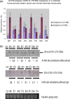
Detection of S and AS RNAs of the U3 ERV-9 LTR (1-2-3-4)1 repeat region by directional RT-PCR. (a) PCR primers used to detect the 200-bp S and AS transcripts from the U3 ERV-9 LTR. (b) Directional RT-PCR analysis of ERV-9 LTR U3 RNAs in nonmalignant primary human cells and in human cancer cell lines. P (positive control) is the U3 ERV-9 LTR directly amplified from K562 genomic DNA, and N (negative control) is the U3 ERV-9 LTR directly amplified from K562 RNA treated with DNase I. ERV-9 LTR U3 products could not be detected in DNase I-treated RNA samples in any cell line (see Fig. S1a in the supplemental material). (c) Real-time PCR analysis of U3 AS and S RNAs in nonmalignant and cancer cell lines. U3 AS RNA was expressed at a significantly higher level than U3 S RNA in normal cells (P < 0.05) but not in cancer cells (P > 0.05). The relative ratio of U3 RNAs between two samples was measured with GAPDH as a cDNA loading control, but the absolute copy numbers of U3 RNAs were not assessed. Relative fold was calculated by setting AS RNA expression level in each cell line as 1-fold. t test was used to evaluate significance.
Fig 2
Fig 2
(A) ERV-9 LTR variants: a and b are LTRs at globin and axin1 genes, c and d are 196- and 155-nt LTRs cloned by a pair of PCR primers shown in Fig. 1, and e and f are 549 and 240 nt LTRs cloned by nested PCR primers (see supplemental material). Sequences were analyzed by NCBI/BLAST. The cDNA sequences of 155-, 196-, 240-, and 549-nt amplicons are shown in Fig. S1 in the supplemental material. (B) The locations of ERV-9 LTRs in introns of different coding genes: a is BRCAA1 transcript, b is a 549-nt LTR embedded in an alternative BRCAA1-012 transcript, c is a 196-nt LTR embedded in MAPK10-018 transcript, and d is a 196-nt LTR embedded in IL-15-008 transcript. Sequences were analyzed by ensemble/blast/blat.
Fig 3
Fig 3
(a) Real-time PCR analysis of U3 AS and S RNAs in matched human primary ductal carcinomas (BT) and adjacent normal mammary tissue pairs (BN). U3 AS RNA was expressed at a significantly higher level than U3 S RNA in normal tissues (P < 0.05) but not in cancer tissues (P > 0.05) within each sample pair. Relative fold was calculated by setting AS RNA expression level in each corresponding normal tissue as 1-fold. ANOVA was used to evaluate significance. (b) High-resolution Northern blot analysis of U3 AS and S RNAs in matched pairs of human ductal carcinomas and adjacent normal mammary tissue. Ductal carcinomas expressed less U3 AS and more U3 S RNAs than adjacent normal tissues. The ∼500-nt AS RNA was detected by nested U3 (1-2-3-4)1 S ODN probes, and the ∼200-nt S RNA was detected by nested U3 (1-2-3-4)1 AS ODN probes. The nested probes were then used as PCR primers to clone these two novel RNAs (Fig. 2).
Fig 4
Fig 4
Analysis of transcription factors assembled with ERV-9 LTR U3 RNAs by RNA-IP. A total of 5 × 10⁷ cells from tumor cell lines and stimulated or unstimulated primary cells were used for RNA-IP (RIP) analysis. The cell lysates were treated with antibodies to each of the following: NF-Y, p53, p300, Ets-1, Sp1, HDAC1, IgG (nonspecific negative control shown in Fig. S2 in the supplemental material), and p21/WAF1 (transcription factor-specific negative control), as well as antibody to PTBP1 as a loading control. The RT-PCR detection of ERV-9 LTR U3 region is described in the legend to Fig. 1b. NF-Y, p53, and sp1 assemble with U3 AS RNAs in HT1080 and MDA231 cells and in quiescent and stimulated human primary T cells.
Fig 5
Fig 5
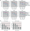
(a) Treatment of human cancer cell lines with ERV-9 LTR U3 S and AS ODNs in vitro. A total of 2.5 × 10⁴ cells was treated with ODNs at 10, 20, or 30 μg/ml for 72 h. The inhibition of proliferation mediated by both U3 S and AS ODNs exceeded those of G3139, GRN163, and MDM2 AS ODNs at all three concentration levels (P < 0.05) in all six cell lines. ANOVA was used to evaluate significance. Relative fold was calculated by setting each cell number in the GFP-treated sample as 1. (b) Treatment of human tumor xenografts with ERV-9 LTR U3 S and AS ODNs in vivo. Animals were inoculated with 2 × 10⁶ tumor cells on day 0. Tumor bearing mice were randomly sorted into 5 groups (n = 8) on day 7. Injection of the tumor site with ODNs was initiated on day 7 and continued daily for 7 days. U3 S and AS ODNs diminished HT1080 and MDA 231 tumor growth to a greater extent than did PBS and GFP controls (P < 0.05). ANOVA was used to evaluate significance. G3139 diminished HT1080 and MDA 231 tumor growth, but the value was not statistically significantly different from that of the negative control by ANOVA.
Fig 6
Fig 6
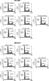
Cell cycle analysis of ODN-treated HT1080 and MDA231 cells. A total of 2.5 × 10⁴ cells was treated with ODNs at 30 μg/ml for 72 h. G3139, GRN163, and MDM2 AS ODNs were positive controls, and GFP ODN was the universal phosphorothioate ODN control. ODN-treated cells were then subjected to flow cytometric cycle analysis. Both U3S and AS ODNs increased the percentage of cells in G0/G1 and reduced the percentage of cells in S/G2 phase.
Fig 7
Fig 7
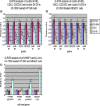
Real-time PCR analysis of cell cycle genes in U3 ODN-treated HT1080 and MDA231 cells. (a and b) U3 ODNs downregulated the mRNA of cyclins B1 and B2, CDC2, and CDC25C (P < 0.05) but not of cyclins D1 and D3 in both cell lines. (c) U3 ODNs upregulated mRNA of p21/WAF1 (P < 0.05). U3 AS ODN also upregulated mRNA of IL-24 (P < 0.05) in MDA231 cells. ANOVA was used to evaluate significance. Relative fold was calculated by setting gene expression levels in GFP ODN-treated cells as 1-fold and defining significance by ANOVA. GFP, U3 Sm, and U3 ASm ODN treatments were used as controls (a).
Fig 8
Fig 8
(a) RNA-IP and real-time PCR analysis of transcription factors assembled with ERV-9 LTR U3 RNAs in MDA231 cell lysates from cells treated with U3 ODNs. U3 ODNs diminished binding of NF-Y, p53, and sp1 with endogenous U3 AS RNAs (P < 0.05). PTBP1 primers were added with ERV-9 LTR primers to amplify PTBP1 RNA as a loading control. Relative fold was calculated by setting the gene expression level in control GFP ODN as 1-fold. ANOVA was used to evaluate significance. (b) Design of a pair of longer U 3 SL and ASL ODNs (158 nt) with 5 tandem repeats of the original 27-nt S or AS RNA in the middle and flanked by a12-nt murine leukemia virus (MLV) PCR linker on both ends. SmL, ASmL, and KL (ERV-K111) are control ODNs which also have 5 tandem repeats of their corresponding sequences (lacking NF-Y binding motifs) in the middle and MLV PCR linker on both ends. (c) Both SL and ASL ODNs bound to NF-Y but not to p53 or sp1, with ASL ODN demonstrating more avid binding to NF-Y. MLV PCR primers detected all constructs with MLV linkers. (d) A total of 2.5 × 10⁴ cells was treated with biotinylated ODNs at 30 μg/ml for 12 h, washed, and lysed. The lysates from each treatment were immunoprecipitated separately with antibodies to NF-Y and biotin. The precipitated pellets were analyzed by Western blotting. IgG and NFKB antibodies were used as negative controls for immunoprecipitation. ERV-K, U3 Sm, and U3 ASm ODNs were used as negative controls for ODN treatment. The ratio of U3 ODN-biotin antibody/NF-Y antibody is the percentage of cellular NF-Y bound by U3 ODNs. The U3 S and AS ODNs bound approximately 10% and 30% total NF-Y, respectively. DR, density ration of bands.
Fig 9
Fig 9
(a) Model depicting the interactions of the U3 AS RNA of ERV-9 LTR with NF-Y, p53, and sp1 as they bear on inhibition of NF-Y binding to promoters of cyclin B1 and B2 in normal cells. (b) Cancer cells express less U3 AS RNA and more NF-Y transcripts, favoring binding to and upregulation of cyclins B1 and B2. Novel U3 miRNA may be present in tumor cells. (c) Exogenous U3 S and AS ODN compete for binding to NF-Y with both endogenous U3 AS RNA of ERV-9 LTR and the promoters of cyclins B1 and B2, thereby inhibiting progression through the cell cycle.

Similar articles

Cited by

References

    1. Subramanian R, Wildschutte J, Russo C, Coffin J. 2011. Identification, characterization, and comparative genomic distribution of the HERV-K (HML-2) group of human endogenous retroviruses. Retrovirology 8:90 doi:10.1186/1742-4690-8-90 - DOI - PMC - PubMed
    1. Stengel A, Roos C, Hunsmann G, Seifarth W, Leib-Mösch C, Greenwood AD. 2006. Expression profiles of endogenous retroviruses in Old World monkeys. J. Virol. 80:4415–4421 - PMC - PubMed
    1. Bannert N, Kurth R. 2006. The evolutionary dynamics of human endogenous retroviral families. Annu. Rev. Genomics Hum. Genet. 7:149–173 - PubMed
    1. Pi W, Yang Z, Wang J, Ruan L, Yu X, Ling J, Krantz S, Isales C, Conway SJ, Lin S, Tuan D. 2004. The LTR enhancer of ERV-9 human endogenous retrovirus is active in oocytes and progenitor cells in transgenic zebrafish and humans. Proc. Natl. Acad. Sci. U. S. A. 101:805–810 - PMC - PubMed
    1. Pi W, Yang Z, Wu M, Wang Y, Fulzele S, Eroglu A, Ling J, Tuan D. 2010. 2010. Long-range function of an intergenic retrotransposon. Proc. Natl. Acad. Sci. U. S. A. 107:12992–12997 - PMC - PubMed

Publication types