. 2012 Dec 7;287(50):42071-83.
doi: 10.1074/jbc.M112.410985. Epub 2012 Oct 18.

Poly-small ubiquitin-like modifier (PolySUMO)-binding proteins identified through a string search

Affiliations

Poly-small ubiquitin-like modifier (PolySUMO)-binding proteins identified through a string search

Huaiyu Sun et al. J Biol Chem. .

Abstract

Polysumoylation is a crucial cellular response to stresses against genomic integrity or proteostasis. Like the small ubiquitin-like modifier (SUMO)-targeted ubiquitin ligase RNF4, proteins with clustered SUMO-interacting motifs (SIMs) can be important signal transducers downstream of polysumoylation. To identify novel polySUMO-binding proteins, we conducted a computational string search with a custom Python script. We found clustered SIMs in another RING domain protein Arkadia/RNF111. Detailed biochemical analysis of the Arkadia SIMs revealed that dominant SIMs in a SIM cluster often contain a pentameric VIDLT ((V/I/L/F/Y)(V/I)DLT) core sequence that is also found in the SIMs in PIAS family E3s and is likely the best-fitted structure for SUMO recognition. This idea led to the identification of additional novel SIM clusters in FLASH/CASP8AP2, C5orf25, and SOBP/JXC1. We suggest that the clustered SIMs in these proteins form distinct SUMO binding domains to recognize diverse forms of protein sumoylation.


Figures

FIGURE 1.
FIGURE 1.
Identification of RNF4-like SUMO-interacting motifs through a computational string search. A diagram describes the computational identification of putative SIM-containing proteins based on SIM2, −3, and −4 in RNF4.
FIGURE 2.
FIGURE 2.
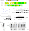
Validation of SUMO-interacting motifs in Arkadia/RNF111. A, shown is the domain structure of Arkadia (top) and an alignment of the predicted SUMO binding regions of Arkadia proteins from mouse (Mus musculus NP_291082), human (Homo sapiens NP_060080), frog (Xenopus tropicalis NP_001072805), chicken (Gallus gallus NP_001186680), and zebrafish (Danio rerio XP_001922708). Solid line rectangles highlight the two motifs (SIM1 and SIM3) matching the initial search string; dashed line rectangles highlight two motifs (SIM0 and SIM2) found through visual inspection. B, shown is a list of Arkadia mutants used for testing SUMO binding. Mutation in each SIM replaced three core hydrophobic residues (Val, Ile, or Leu) with Ala. C, shown is the contribution of Arkadia SIMs to SUMO binding. Various GST fusion proteins as indicated were tested for their association with a FLAG-tagged tri-SUMO2 (FLAG-3xSUMO2) in a GST pulldown assay. The glutathione-agarose bead-bound proteins were detected with anti-GST and anti-FLAG antibodies in an immunoblot. The GST-RNF4 and its sim23Δ, an internal deletion mutant lacking both SIM2 and 3, were used as positive and negative controls, respectively. D, shown is a comparison of the interaction of Arkadia and RNF4 with two major SUMO isoforms. A GST pulldown assay was used to compare the association of Arkadia SBD or RNF4 with either tri-SUMO1 or tri-SUMO2. E, shown is the contribution of Arkadia SIMs to SUMO binding. Various GST fusion proteins as indicated were tested for their association with a FLAG-tagged tri-SUMO1 (FLAG-3xSUMO1) in a GST pulldown assay conducted essentially as in C. IB, immunoblot. F, shown is SIM-dependent association of Arkadia with polySUMO chains. The GST pulldown assay was carried out essentially as in C but against the polySUMO2 ladder. The bead-bound proteins were analyzed with anti-panSUMO or anti-GST immunoblots as indicated. Detectable polySUMO2 species are labeled with the calculated length of SUMO2 units. G, shown is SIM-dependent association between Arkadia and high molecular weight sumoylated proteins in cultured cells detected through coimmunoprecipitation. FLAG-tagged full-length Arkadia in the indicated forms (WT; sim, sim13m; CS, RING domain mutant C971S) were coexpressed with HA-tagged SUMO1 or SUMO2 in 293T cells. Proteins in the anti-FLAG immune complex and in the cell lysate were analyzed in a 4–12% Bis-Tris SDS-PAGE followed by immunoblotting using anti-FLAG and anti-HA antibodies as indicated.
FIGURE 3.
FIGURE 3.
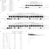
Dominant SIMs are made of VIDLT motif. A, dominant SIMs Arkadia and RNF4 resemble those in PIAS family SUMO ligases. The list shows the alignment of VIDLT-type SIMs in Arkadia, RNF4, PIAS1, PIAS4 (7), Pli1, Rfp2, ATF7IP/MCAF1 (75), CAF1p150 (30), E1A of human adenovirus 5 (76), and USP25 (77). B, all the five core residues in Arkadia SIM3 (VVDLT) are essential for SUMO2 binding. The SIM3 residues Val-382, Val-383, Asp-384, Leu-385, Thr-386, and Val-387 were each replaced with Ala in a sim12m background as indicated. Binding assays were conducted as described in Fig. 2C. IB, immunoblots. C, a rigid structure of VIDLT-type SIMs allowing minimum variations for full strength SUMO1 binding. Additional Arkadia SIM3 mutants, V382Y, V383I, V383L, D384E, D384N, L385I, T386S, and T386V were generated in a sim12m background as indicated. Binding assays were performed as described in Fig. 2C. Examples of purified GST fusion proteins are shown in supplemental Fig. S4. D, a rigid structure of VIDLT-type SIMs allowing minimum variations for full strength SUMO2 binding. Binding assays were performed in parallel to C. E, shown is a summary of single-residue mutations tested in C and D. Mutated residues are indicated in bold; mutations that retained wild-type affinity for SUMO are highlighted. Quantitative densitometry obtained from C and D measures the bound tri-SUMO (FLAG-3xSUMO1 or −3xSUMO2) to the respective GST fusion proteins and is shown as the percentage relative to that of wild-type SIM3. F, shown is a diagram indicating a sequence-dependent SUMO affinity of all putative SIMs (upper panel) and the limited sequence space for the VIDLT-type SIMs (bottom panel).
FIGURE 4.
FIGURE 4.
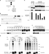
FLASH/CASP8AP2 is a novel SUMO-binding protein with clustered SIMs. A, an alternative string search against (V/I/L/F/Y)(V/I)DLT led to the identification of putative SUMO-binding proteins with clustered VIDLT-type SIMs. B, predicted SIMs in human FLASH/CASP8AP2 (NP_001131140) are shown. Top panel, the relative position of predicted SIMs in FLASH; middle panel, the sequence of the FLASH residues 1661–1815 with the predicted SIMs (1683YVDLT, 1700FIEVT, 1737FIDLT, and 1794YIDLT) highlighted. C, validation of the FLASH SUMO binding domain is shown. FLASH-(1661–1815) and its mutants as indicated were purified as GST fusion proteins and tested for their association with FLAG-3xSUMO2. Analogous to the mutational analysis with Arkadia shown in Fig. 2B, single SIM mutations (sim1m, sim2m, sim3m, sim4m) were generated by replacing all three core hydrophobic residues (Tyr/Phe, Val/Ile, and Leu) with Ala in each of the four putative SIMs. Binding assays were carried out as in Fig. 2C. The GST-Arkadia-(283–415) and its sim123m mutant were used as positive and negative controls, respectively. Dual color immunoblot (anti-GST and anti-FLAG) on a single membrane was processed through the Odyssey Infrared Imaging System. Quantitative densitometry is shown in the bar graph below as the ratio of each FLAG-3xSUMO2 band to its corresponding GST band. IB, immunoblot. D, shown is the contribution of individual SIMs in FLASH SUMO binding domain. The FLASH mutants with only one (sim234m, sim134m, sim124m, sim123m) or none (sim1–4m) of the four SIMs left intact as well as two double SIM mutants (sim12m, sim34m) were tested for SUMO binding as in C. Examples of purified GST-fusion proteins are shown in supplemental Fig. S4. E, shown is the SIM-dependent association between the FLASH SBD and high molecular weight sumoylated proteins in cultured cells. EGFP-tagged fragment of FLASH (residues 1581–1884, either wild-type or sim134m mutant) were coexpressed with HA-tagged SUMO1 or SUMO2 in 293T cells as indicated. Immunoprecipitation (IP) was carried out with anti-GFP antiserum and protein A-agarose beads. Proteins in the precipitated immune complex and in the cell lysate were analyzed in an 8% Tris-glycine SDS-PAGE followed by immunoblotting using anti-GFP and anti-HA antibodies as indicated. The stacking gel was preserved during immunoblotting in order to detect high molecular weight SUMO conjugates that could not enter the resolving gel. vect, EGFP alone; sim, sim134m. *, it is unclear why the sim134m fragment showed a different migration (as also seen with some other SIM mutants in this study); this may reflect a conformational change due to the loss of intact SIMs. F, a precise structure of FLASH SIM3 allows minimum variations for full strength SUMO binding. Single-residue replacements in FLASH SIM3, F1737V, D1739E, L1740I, and T1741S were generated as indicated in a sim1m background. Binding assays were performed as in C and D.
FIGURE 5.
FIGURE 5.
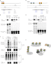
C5orf25 and SOBP each contains an SBD with two VIDLT-type SIMs. A, predicted SIMs in the N terminus of C5orf25 are shown. Top panel, relative position of predicted SIMs in the protein; middle panel, sequence of the N-terminal region (1–74) of C5orf25 with the two SIMs (26FIDLT and 45VIDLT) highlighted; bottom panel, SIM mutants of C5orf25 were generated by replacing a single hydrophobic residue in each or both of the two putative SIMs (sim1m, I27A; sim2m, I46A). B, validation of the C5orf25 SUMO binding domain. C5orf25-(1–74) and its mutants as indicated were purified as GST fusion proteins and tested for their association with FLAG-3xSUMO2. Binding assays were carried out as in Fig. 2C. The GST-FLASH-(1660–1885) and its sim1–4m mutant were used as positive and negative controls, respectively. Examples of purified GST-fusion proteins are shown in supplemental Fig. S4. IB, immunoblots. C, SIM-dependent association between C5orf25 and high molecular weight sumoylated proteins in cultured cells detected through coimmunoprecipitation. FLAG-tagged full-length C5orf25 in indicated forms (WT; sim, sim12m) were coexpressed with HA-tagged SUMO1 or SUMO2 in 293T cells. Immunoprecipitation was carried out with anti-FLAG (M2)-conjugated agarose beads. Proteins in the precipitated immune complex and in the cell lysate were analyzed in an 8% Tris-glycine SDS-PAGE followed by immunoblotting using anti-FLAG and anti-HA antibodies as indicated. The stacking gel was preserved during immunoblotting to detect high molecular weight SUMO conjugates that could not enter the resolving gel. D, shown are predicted SIMs in SOBP. Top panel, the relative position of predicted SIMs in SOBP; middle panel, the sequence of SOBP residues 600–676 with the two SIMs (620VVDLT and 653VIDLT) highlighted; bottom panel, SIM mutants of SOBP were generated by replacing three hydrophobic residue in each or both of the two putative SIMs (sim1m, V620A/V621A/L623A; sim2m, V653A/I654A/L656A). E, SOBP SIMs show differential contributions to SUMO binding. Wild-type and mutant forms of SOBP fragment 600–676 as indicated were purified as GST fusion proteins. The binding assay was performed essentially as described in Fig. 2C. GST-Arkadia-(258–415) and its sim3m mutant (see Fig. 2C) were included as positive and negative controls. F, a diagram illustrating diverse forms of protein sumoylation that can be recognized by SUMO binding domains containing clustered SIMs.

Similar articles

Cited by

References

    1. Creton S., Jentsch S. (2010) SnapShot. The SUMO system. Cell 143, 848–848 - PubMed
    1. Meulmeester E., Melchior F. (2008) Cell biology. SUMO. Nature 452, 709–711 - PubMed
    1. Matunis M. J., Zhang X. D., Ellis N. A. (2006) SUMO. The glue that binds. Dev. Cell 11, 596–597 - PubMed
    1. Minty A., Dumont X., Kaghad M., Caput D. (2000) Covalent modification of p73α by SUMO-1. Two-hybrid screening with p73 identifies novel SUMO-1-interacting proteins and a SUMO-1 interaction motif. J. Biol. Chem. 275, 36316–36323 - PubMed
    1. Hecker C. M., Rabiller M., Haglund K., Bayer P., Dikic I. (2006) Specification of SUMO1- and SUMO2-interacting motifs. J. Biol. Chem. 281, 16117–16127 - PubMed

Publication types