. 2012 Sep;8(9):e1002927.
doi: 10.1371/journal.ppat.1002927. Epub 2012 Sep 27.

Kaposi's sarcoma herpesvirus K15 protein contributes to virus-induced angiogenesis by recruiting PLCγ1 and activating NFAT1-dependent RCAN1 expression

Affiliations

Kaposi's sarcoma herpesvirus K15 protein contributes to virus-induced angiogenesis by recruiting PLCγ1 and activating NFAT1-dependent RCAN1 expression

Kiran Bala et al. PLoS Pathog. 2012 Sep.

Abstract

Kaposi's Sarcoma (KS), caused by Kaposi's Sarcoma Herpesvirus (KSHV), is a highly vascularised angiogenic tumor of endothelial cells, characterized by latently KSHV-infected spindle cells and a pronounced inflammatory infiltrate. Several KSHV proteins, including LANA-1 (ORF73), vCyclin (ORF72), vGPCR (ORF74), vIL6 (ORF-K2), vCCL-1 (ORF-K6), vCCL-2 (ORF-K4) and K1 have been shown to exert effects that can lead to the proliferation and atypical differentiation of endothelial cells and/or the secretion of cytokines with angiogenic and inflammatory properties (VEGF, bFGF, IL6, IL8, GROα, and TNFβ). To investigate a role of the KSHV K15 protein in KSHV-mediated angiogenesis, we carried out a genome wide gene expression analysis on primary endothelial cells infected with KSHV wildtype (KSHVwt) and a KSHV K15 deletion mutant (KSHVΔK15). We found RCAN1/DSCR1 (Regulator of Calcineurin 1/Down Syndrome critical region 1), a cellular gene involved in angiogenesis, to be differentially expressed in KSHVwt- vs KSHVΔK15-infected cells. During physiological angiogenesis, expression of RCAN1 in endothelial cells is regulated by VEGF (vascular endothelial growth factor) through a pathway involving the activation of PLCγ1, Calcineurin and NFAT1. We found that K15 directly recruits PLCγ1, and thereby activates Calcineurin/NFAT1-dependent RCAN1 expression which results in the formation of angiogenic tubes. Primary endothelial cells infected with KSHVwt form angiogenic tubes upon activation of the lytic replication cycle. This effect is abrogated when K15 is deleted (KSHVΔK15) or silenced by an siRNA targeting the K15 expression. Our study establishes K15 as one of the KSHV proteins that contribute to KSHV-induced angiogenesis.


Conflict of interest statement

The authors have declared that no competing interests exist.

Figures
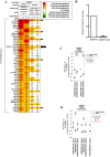
Figure 1
Figure 1. K15 dependent cellular gene expression in HUVECs.
(A) Microarray analysis of cellular mRNAs upregulated in HUVECs by K15 overexpression or after KSHV infection. HUVECs were transduced with a K15-expression vector (lanes 1–2) or infected with either KSHVwt or KSHVΔK15 (lanes 3–4). Depicted are the top-ranking 50 transcripts most strongly upregulated by K15 overexpression, showing an at least two-fold induction by K15 overexpression in each of two experiments performed using cells from two healthy donors (lanes 1–2). Asterisks next to fold change values within heatmap lanes 3–4 indicate genes that were induced more than two-fold in KSHVwt-infected cells compared to cells mock infected with heat-inactivated virus. Empty cells in the heatmap correspond to undetectable mRNA levels in both of the two samples compared in each lane. Arrows indicate cellular genes, inducible upon KSHVwt infection with more than two-fold elevated expression in KSHVwt- relative to KSHVΔK15-infected cells in each of the two experiments performed. (B) Quantitative PCR analysis of K15 transcript in mRNA derived from KSHVwt- or KSHVΔK15-infected cells used for the microarray experiment. (C) Quantitative PCR analysis of RCAN1/DSCR1 and VEGFA transcripts in HUVECs transduced with a retroviral K15 expression vector or with an empty control vector. (D) HUVECs from two healthy donors (Exp1–2) were infected with heat-inactivated, KSHVwt, or KSHVΔK15 and were lysed 4 days after infection. RNA was extracted, reversely transcribed and subjected to quantitative TaqMan-based PCR analysis. mRNA fold change values of KSHVwt-infected versus mock-infected (with heat-inactivated KSHVwt = HI) cells were depicted in black, values of KSHVΔK15- infected versus mock-infected cells were depicted in red. Input mRNA samples are identical to samples used in microarray experiments (compare panel A lanes 3–4).
Figure 2
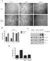
Figure 2. K15 induces capillary tube formation and increases RCAN1.1 and RCAN1.4 protein expression.
(A) HUVECs transduced with pSF91 or pSF91K15 were plated on matrigel with medium (EBM2+2%FBS) with or without VEGF (50 ng/ml) 30 hours after transduction and were assessed 6 hours later for their ability to form capillary tubes. Arrows indicate cellular junctions counted to calculate the angiogenic index. (B) Angiogenic index (number of cellular junctions) in pSF91 and pSF91K15 transduced cells. Significance levels for the indicated comparisons are marked with ‘**’ (p<0.01) (see Material and Methods). (C) Western blot showing the increased expression of both RCAN1.1 and RCAN1.4 in K15 transduced cells in medium with and without VEGF. (D) HUVECs were resuspended in supernatant collected from cells transduced with K15 or vector control for 48 hours and then were plated onto matrigel for 6 hours to score for tube formation. Medium (EBM2+2%FBS) with VEGF (50 ng/ml) was used as a positive control.
Figure 3
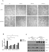
Figure 3. K15-induced capillary tube formation depends on RCAN1.
HUVECs were transduced with pSF91K15 or the pSF91 vector and 30 hours later siRNAs against RCAN1, RCAN1.1 and RCAN 1.4 were transfected. 36 hours after transfection, cells were plated on matrigel to score for capillary tube formation (A, B) or analysed by Western blot to verify silencing of individual RCAN1 isoforms (C). Statistical significance levels for the comparisons of indicated samples are marked with ‘*’ (p<0.05) and ‘**’ (p<0.01).
Figure 4
Figure 4. K15 dependent capillary tube formation involves the PLCγ1-Calcineurin-NFAT pathway.
(A, B) HUVECs were treated with a Calcineurin inhibitor (Cyclosporin A; 1 µM), or a PLCγ inhibitor (U73122; 20 µM) for 6 hours and were then plated on matrigel in the presence of VEGF-A to score for capillary tube formation (A). Parallel samples were analysed on Western blots to show the phosphorylation of NFAT1 and PLCγ1 and the levels of total PLCγ1, RCAN1.1 and RCAN 1.4 (B). (C, D) HUVECs were transduced with pSF91 or pSF91K15 and after 30 hours were treated with CsA or U73122 and tube formation was analysed as in figure 2 (C). Phosphorylation of NFAT1, PLCγ1, and expression of RCAN1 isoforms and K15 was measured by Western blot (D).
Figure 5
Figure 5. Silencing of Calcineurin, NFAT1 and PLCγ1 inhibits K15-induced capillary tube formation.
HUVECs were transduced with pSF91 or pSF91K15 and (A) changes in the angiogenic index in response to K15 expression and silencing of NFAT1 or Calcineurin were analysed. (B) NFAT1 phosphorylation and expression of NFAT1, Calcineurin, RCAN1 in response to K15 overexpression and silencing of NFAT1 and Calcineurin in the absence of VEGF were analysed by Western blot. (C) Changes in the angiogenic index in response to K15 expression and silencing of PLCγ1 and NFκB p65. (D) Expression of RCAN1, PLCγ1, p65 in response to K15 overexpression and silencing of PLCγ1 or p65 in the absence of VEGF were analysed by Western blot. Transduction with pSF91, pSF91K15 and transfection with siRNA was performed as in figure 3.
Figure 6
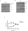
Figure 6. K15 activates PLCγ1 in a protracted manner via its SH2 binding domain.
(A) HUVECs were treated with 50 ng/ml VEGF in 12 well plates for the indicated time points and analysed by Western blots to investigate the effects on the phosphorylation of PLCγ1 and expression of RCAN1 isoforms. (B) HUVECs were transduced with a retroviral vector expressing K15 (pSF91K15), plated in 12 well plates for the indicated time points and the phosphorylation of PLCγ1 and expression of RCAN1 isoforms was monitored by Western blot. (C) HUVECs were transduced with pSF91K15 or control vector and the angiogenic index was measured after treating the cells with VEGF at the indicated concentrations and plating them on matrigel.
Figure 7
Figure 7. K15 activates PLCγ1 via its SH2 binding domain.
(A) Schematic representation of K15 showing the SH2 and SH3 binding sites in the cytoplasmic domain along with corresponding mutants. (B) HUVECs were transduced with retroviral vectors expressing K15 and the indicated mutants of the K15 SH2 and SH3 binding sites or the control vector (pSF91). Thirty hours after transduction cells were lysed and analysed by Western blot for PLCγ1 phosphorylation and expression of K15 and RCAN1 isoforms. (C) Parallel samples from the experiment shown in (B) were plated on matrigel in the absence of VEGF and scored for capillary tube formation as in figure 2.
Figure 8
Figure 8. K15P and K15M recruit and activate PLCγ1.
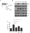
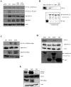
(A) Co-immunoprecipitation of K15-P with PLCγ1: pSF91K15, the mutants (YF, ΔSH3, YF/ΔSH3) and control vector (pSF91) were used to transiently transfect HEK293. After immunoprecipitation of endogenous PLCγ1, co-immunoprecipitated K15-P was visualised on Western blots using a polyclonal antibody to K15. (B) Purified GST or GST fusion proteins containing the cytoplasmic domain of K15-P wt and mutants YF, ΔSH3, YF/ΔSH3 were used in a GST pulldown assay with lysates of HEK293T cells. Endogenous PLCγ1 bound to GST-K15 beads was visualised with an antibody against PLCγ1. Purified GST or GST-K15 fusion proteins were visualized by Coomassie blue staining to show that equal amounts of fusion protein were used for GST pulldown experiments (bottom). (C) Co-immunoprecipitation of K15-P and K15-M with PLCγ1: HEK293T cells were transiently transfected with a vector containing a FLAG-tagged K15P or K15M, or the control vector (pFJ) and endogenous PLCγ1 was immunoprecipitated. Co-immunoprecipitated K15-P or K15-M was visualised by an antibody to the FLAG tag. (D) Lysates of HEK293T cells were incubated with beads coated with GST, GST-K15-P or GST-K15-M and bound endogenous PLCγ1 was detected on Western blots. (E) Lysates of HEK293T cells transfected with K15-P or K15-M expression vector or control vector (pFJ) were analysed on Western blots using an antibody to the PLCγ1 tyrosine 783 phosphorylation site.
Figure 9
Figure 9. K15 induced angiogenesis does not involve VEGF receptors.
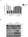
(A) HUVECs were transduced with pSF91 or pSF91K15 and 30 hours later transfected with siRNA to VEGFR1 (FLT1), VEGFR2 (KDR) and VEGFR3 (FLT4). Thirty-six hours after transfection, cells were plated on matrigel in the absence or presence of VEGF and scored for capillary tube formation. (B) Parallel samples were analysed by Western blot to verify silencing of VEGF receptors and K15 expression.
Figure 10
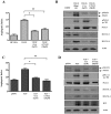
Figure 10. Lack of K15 abrogates angiogenic tube formation induced by KSHV infection of HUVECs.
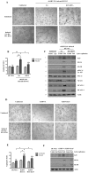
(A–C) Capillary tube formation in HUVECs infected with rKSHV.219. Seventy-two hours after infection of HUVECs, siRNA against K15 or control siRNA was microporated. Twenty-four hours after transfection the lytic cycle was induced with sodium butyrate (Na-Bu) and 10% RTA. Forty-eight hours later, cells were plated on matrigel and scored for tube formation (A). (B) Angiogenic index for the experiment shown in (A). Significance levels for the indicated comparisons of different samples are marked with ‘*’ (p<0.05) and ‘**’ (p<0.01). (C) Western blot analysis for the experiment shown in (A) to confirm the silencing of K15 expression and to quantify the expression of K8.1 (a late lytic glycoprotein), RCAN1.1/1.4, and phosphorylated PLCγ1 and phosphorylated NFAT1. (D–F) HUVEC were infected with BAC-derived KSHVwt or KSHVΔK15 for 72 hours. Following activation of the lytic cycle as in (A–C), cells were plated on matrigel for angiogenic tube formation (D). (E) Angiogenic index for the experiment shown in (D). Significance levels for the indicated comparisons are marked with ‘*’ (p<0.05). (F) Parallel samples were analysed by Western blot for the expression of K15 and K8.1 A/B.
Figure 11
Figure 11. Schematic diagram showing the likely involvement of K15 in the PLCγ1-Calcineurin-NFAT pathway.
As shown in this report, K15 directly recruits PLCγ1 and induces its phosphorylation by an unidentified kinase. The PLCγ1 dependent production of IP3 leads to increased calcium influx, Calcineurin activity, NFAT dephosphorylation and ultimately increased expression of NFAT-dependent genes, including RCAN1.1/1.4 and angiogenesis. CsA: Cyclosorin A, U73122: PLCγ1 inhibitor.

Similar articles

Cited by

References

    1. Chang Y, Cesarman E, Pessin MS, Lee F, Culpepper J, et al. (1994) Identification of herpesvirus-like DNA sequences in AIDS-associated Kaposi's sarcoma. Science 266: 1865–1869. - PubMed
    1. Cesarman E, Chang Y, Moore PS, Said JW, Knowles DM (1995) Kaposi's sarcoma-associated herpesvirus-like DNA sequences in AIDS-related body-cavity-based lymphomas. N Engl J Med 332: 1186–1191. - PubMed
    1. Soulier J, Grollet L, Oksenhendler E, Cacoub P, Cazals-Hatem D, et al. (1995) Kaposi's sarcoma-associated herpesvirus-like DNA sequences in multicentric Castleman's disease. Blood 86: 1276–1280. - PubMed
    1. Ganem D (2010) KSHV and the pathogenesis of Kaposi sarcoma: listening to human biology and medicine. J Clin Invest 120: 939–949. - PMC - PubMed
    1. Cai Q, Verma S, Lu J, Robertson E (2010) Molecular biology of Kaposi's sarcoma-associated herpesvirus and related oncogenesis. Adv Virus Res 78: 87–142. - PMC - PubMed

Publication types

MeSH terms

Associated data

Grants and funding

This work was supported by Collaborative Research Centre (CRC 566 of the Deutsche Forschungsgemeinschaft), the European Union Integrated Project INCA (The role of chronic infections in the development of cancer; LSHC-CT-2005-018704) and the Molecular Medicine PhD Program of Hannover Medical School. The funders had no role in study design, data collection and analysis, decision to publish, or preparation of the manuscript.