

Review
doi: 10.1083/jcb.201201041.
Tracing epithelial stem cells during development, homeostasis, and repair
Affiliations
- PMID: 22641343
- PMCID: PMC3365500
- DOI: 10.1083/jcb.201201041
Item in Clipboard
Review
Tracing epithelial stem cells during development, homeostasis, and repair
J Cell Biol.
.
Abstract
Epithelia ensure many critical functions of the body, including protection against the external environment, nutrition, respiration, and reproduction. Stem cells (SCs) located in the various epithelia ensure the homeostasis and repair of these tissues throughout the lifetime of the animal. Genetic lineage tracing in mice has allowed the labeling of SCs and their progeny. This technique has been instrumental in characterizing the origin and heterogeneity of epithelial SCs, their tissue location, and their differentiation potential under physiological conditions and during tissue regeneration.
Figures
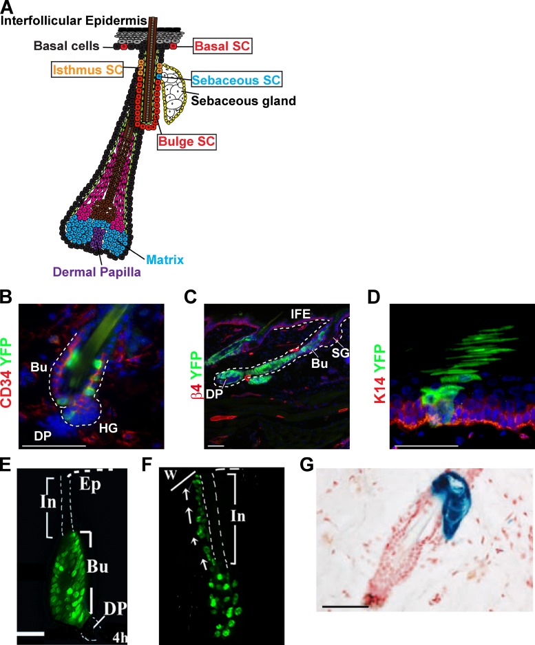
Lineage tracing of the skin epidermis. (A) Schematic representation of the skin epidermis and the different epidermal SCs: the interfollicular epidermis (IFE) SCs, the isthmus SCs, and the bulge SCs. (B and C) Lineage tracing of bulge stem cells (green) using K19CREER-RosaYFP mice induced with 10 mg tamoxifen between d 21 and 25, and analyzed 1 wk (B) or 5 wk (C) after induction. Immunostaining of CD34 (B) or integrin β4 (C) and YFP show the initial labeling of CD34+ bulge SC (B) and the presence of YFP cells in the newly formed hair follicle 5 wk after (C). (D) Lineage tracing of basal cells (green) from the interfollicular epidermis using K14CREER-RosaYFP mice induced with 1 mg tamoxifen and analyzed 5 wk later. Immunostaining of K14 (red) and YFP (green) shows the presence of a column of YFP-marked cells spanning from the basal layer (red) to the cornified layer, corresponding to a unit of interfollicular epidermis maintained by a single SC. (E and F) Migration of H2B-GFP label-retaining bulge SCs to the interfollicular epidermis after wounding, showing the early contribution of bulge SC to the wound repair. Adapted from Tumbar et al. (2004) with permission from AAAS. (G) Lineage tracing using Lgr6-GFP-IresCREER/Rosa-LacZ mice induced at d 20 and analyzed 1 yr later, showing the labeling of the sebaceous gland, and demonstrating the existence of long-lived SC of the sebaceous gland. Adapted from Snippert et al. (2010a) with permission from AAAS. Bu, bulge; DP, dermal papilla; HG, hair germ; IFE, interfollicular epidermis; SG, sebaceous gland; In, infudibulum; Ep, epidermis; w, wound. Bars, 50 µM.
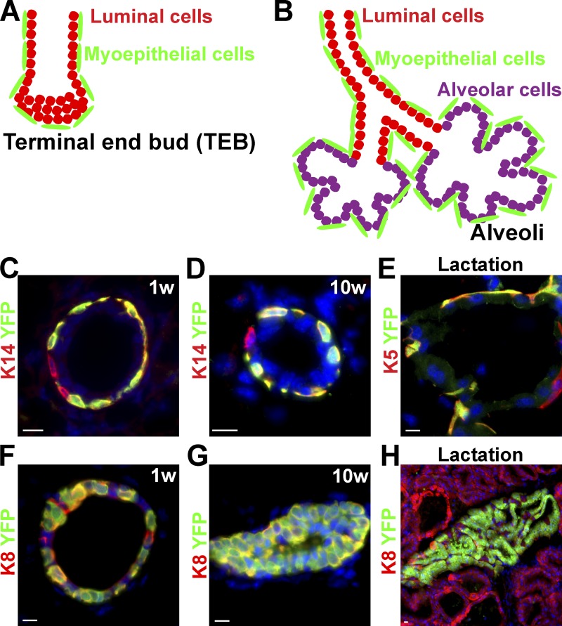
Lineage tracing of the mammary gland. (A and B) Schematic representation of the main epithelial cell types (myoepithelial, luminal, and alveolar cells) of the breast during post-natal development (A) and pregnancy (B). (C–E) Lineage tracing of myoepithelial cells (green) using K14rtTA/TetOCre/RosaYFP mice induced at the onset of puberty (4 wk old) and analyzed 1 wk (C) or 10 wk (D) later or during lactation (E). Immunostaining of K14 (C and D) or K5 (E) (red) and YFP (green) demonstrates the labeling of unipotent SCs that ensure myoepithelial lineage expansion during puberty and pregnancy. (F–H) Lineage tracing of luminal cells (green) using K8CREER/RosaYFP mice induced at the onset of puberty (4 wk old) and analyzed 1 wk (F) or 10 wk later (G), or during lactation (H) shows the labeling of unipotent SCs that ensure luminal lineage expansion during puberty and pregnancy. Adapted from Van Keymeulen et al. (2011) with permission from Nature Publishing Group. Bars, 10 µM.
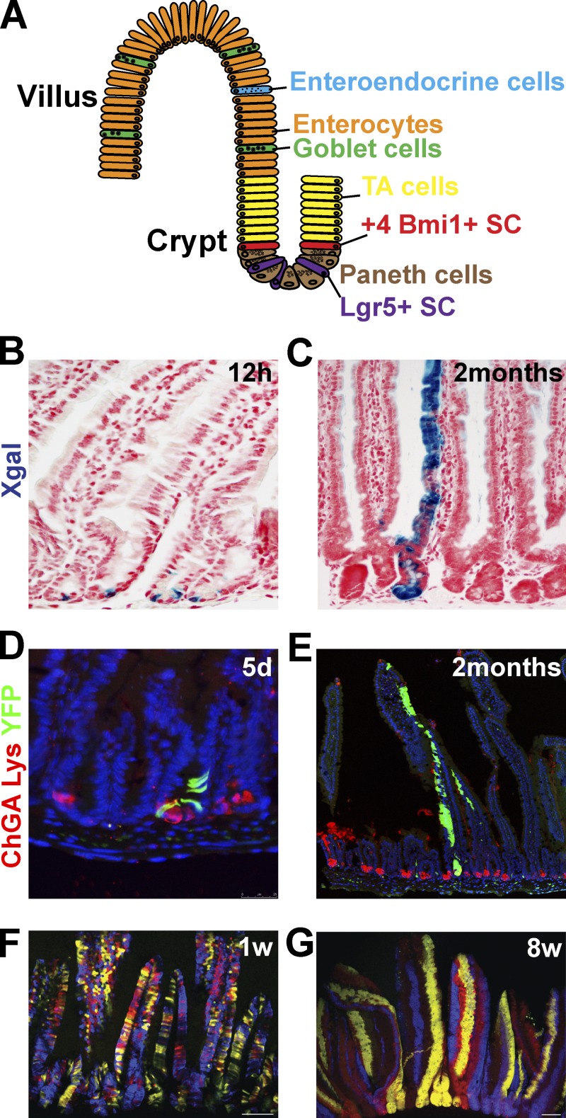
Lineage tracing of the intestine. (A) Schematic representation of the intestinal epithelium lineages (enterocytes, enteroendocrine, goblet, and paneth cells) and its SCs. (B and C) Lineage tracing of Lgr5+ SCs (blue) using Lgr5-GFP-IresCREER/RosaLacZ mice analyzed 12 h (B) or 60 d (C) after induction demonstrates the initial labeling of columnar basal cells (B) that give rise to the differentiated cells of a villus (C). Adapted from Barker et al. (2007) with permission from Nature Publishing Group. (D and E) Lineage tracing of Bmi1+ SCs (green) using Bmi1CREER/Rosa YFP mice analyzed 5 d (D) or 2 mo (E) after induction. Staining of YFP (green) and of chromogranin A (ChGA) labeling enteroendocrine cells (red); and lysozyme (Lys) antibody, labeling Paneth cells (red), showing the initial labeling of cells above the paneth cells (D) that give rise to the differentiated cells of a villus (E). Images courtesy of E. Sangiorgi and M. Capecchi. (F and G) Lineage tracing of crypts using Ah-CREER/RosaConfetti mice analyzed 1 wk (F) or 8 wk (G) after induction, showing the initial multicolor labeling of the crypt and villus unit that progressively become monoclonal (one color per crypt) over time. Adapted from Snippert et al. (2010b) with permission from Elsevier.
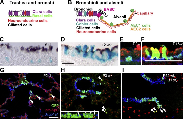
Lineage tracing of the airway epithelium. (A and B) Schematic representation of the airway epithelium that can be divided into trachea and bronchi, bronchioli, and alveoli. (C–F) Lineage tracing of tracheal basal cells using K5CREER/Rosa-LacZ and analyzed 6 d (C and E), 3 wk (F), or 12 wk (D) after TAM administration. Paraffin sections of X-gal–stained (blue) (K5-labeled cells and their progeny) and anti-acetylated tubulin (brown, cilia), showing the initial labeling of basal cells (C) that give rise to luminal cells during postnatal development (D). (E and F) Lineage tracing of tracheal basal cells using K5CREER/Rosa-YFP and analyzed 3 and 15 wk after TAM administration showing the initial labeling of basal cells (green) that give rise to Clara cells (red). Adapted from Rock et al. (2009) with permission from Proc. Natl. Acad. Sci. USA. (G–I) Lineage tracing of the Clara cells in the bronchioli using Scgb1a1CREER/RosaYFP mice induced at E18.5 and analyzed 2 d (G), 3 wk (H), or 1 yr (I) after induction. Immunostaining of GFP (green), pro-SPC (AEC2) (red), and Scgb1a1 (Clara cells) (blue) shows the long-term renewal of Clara cells and the expansion of ciliated cells over time. Arrowheads represent lineage-labeled AEC2 cells. Arrows represent lineage-labeled putative BASCs. Inset in H shows labeled ciliated cells (arrowheads), but no neuroendocrine cells (red, arrow). The stable frequency of lineage-labeled AEC2 cells over time (1–3%) suggests that BASC cells do not contribute to alveoli expansion during postnatal growth. Adapted from Rawlins et al. (2009) with permission from Elsevier. AEC1, alveolar type I; AEC2, alveolar type II; BADJ, bronchioalveolar duct junction; BASC, bronchioalveolar stem cell. Bars: (C and D) 20 µM; (E and F) 25 µM; (G) 50 µM.
Similar articles
-
Lineage-Restricted Mammary Stem Cells Sustain the Development, Homeostasis, and Regeneration of the Estrogen Receptor Positive Lineage.Cell Rep. 2017 Aug 15;20(7):1525-1532. doi: 10.1016/j.celrep.2017.07.066. Cell Rep. 2017. PMID: 28813665 Free PMC article.
-
Bipotent stem cells support the cyclical regeneration of endometrial epithelium of the murine uterus.Proc Natl Acad Sci U S A. 2019 Apr 2;116(14):6848-6857. doi: 10.1073/pnas.1814597116. Epub 2019 Mar 14. Proc Natl Acad Sci U S A. 2019. PMID: 30872480 Free PMC article.
-
Plasticity within stem cell hierarchies in mammalian epithelia.Trends Cell Biol. 2015 Feb;25(2):100-8. doi: 10.1016/j.tcb.2014.09.003. Epub 2014 Oct 9. Trends Cell Biol. 2015. PMID: 25308311 Review.
-
Lineage enforcement by inductive mesenchyme on adult epithelial stem cells across developmental germ layers.Stem Cells. 2009 Dec;27(12):3032-42. doi: 10.1002/stem.244. Stem Cells. 2009. PMID: 19862839
-
Hippo signaling in epithelial stem cells.Acta Biochim Biophys Sin (Shanghai). 2015 Jan;47(1):39-45. doi: 10.1093/abbs/gmu111. Epub 2014 Dec 4. Acta Biochim Biophys Sin (Shanghai). 2015. PMID: 25476205 Review.
Cited by
-
Cancer stem cell definitions and terminology: the devil is in the details.Nat Rev Cancer. 2012 Nov;12(11):767-75. doi: 10.1038/nrc3368. Epub 2012 Oct 11. Nat Rev Cancer. 2012. PMID: 23051844 Review.
-
Unravelling cancer stem cell potential.Nat Rev Cancer. 2013 Oct;13(10):727-38. doi: 10.1038/nrc3597. Nat Rev Cancer. 2013. PMID: 24060864 Review.
-
Spatial organization within a niche as a determinant of stem-cell fate.Nature. 2013 Oct 24;502(7472):513-8. doi: 10.1038/nature12602. Epub 2013 Oct 6. Nature. 2013. PMID: 24097351 Free PMC article.
-
Generation of Spheres from Dental Epithelial Stem Cells.Front Physiol. 2017 Jan 19;8:7. doi: 10.3389/fphys.2017.00007. eCollection 2017. Front Physiol. 2017. PMID: 28154538 Free PMC article.
-
Cancer stem cells: biological functions and therapeutically targeting.Int J Mol Sci. 2014 May 9;15(5):8169-85. doi: 10.3390/ijms15058169. Int J Mol Sci. 2014. PMID: 24821540 Free PMC article. Review.
References
-
- Adamson I.Y., Bowden D.H. 1975. Derivation of type 1 epithelium from type 2 cells in the developing rat lung. Lab. Invest. 32:736–745 - PubMed
-
- Barker N., Huch M., Kujala P., van de Wetering M., Snippert H.J., van Es J.H., Sato T., Stange D.E., Begthel H., van den Born M., et al. 2010b. Lgr5(+ve) stem cells drive self-renewal in the stomach and build long-lived gastric units in vitro. Cell Stem Cell. 6:25–36 10.1016/j.stem.2009.11.013 - DOI - PubMed
Publication types
MeSH terms
LinkOut - more resources
Full Text Sources
Medical
