. 2012 Jun;18(6):961-6.
doi: 10.1038/nm.2805.

The ephrin receptor tyrosine kinase A2 is a cellular receptor for Kaposi's sarcoma–associated herpesvirus

Affiliations

The ephrin receptor tyrosine kinase A2 is a cellular receptor for Kaposi's sarcoma–associated herpesvirus

Alexander S Hahn et al. Nat Med. 2012 Jun.

Abstract

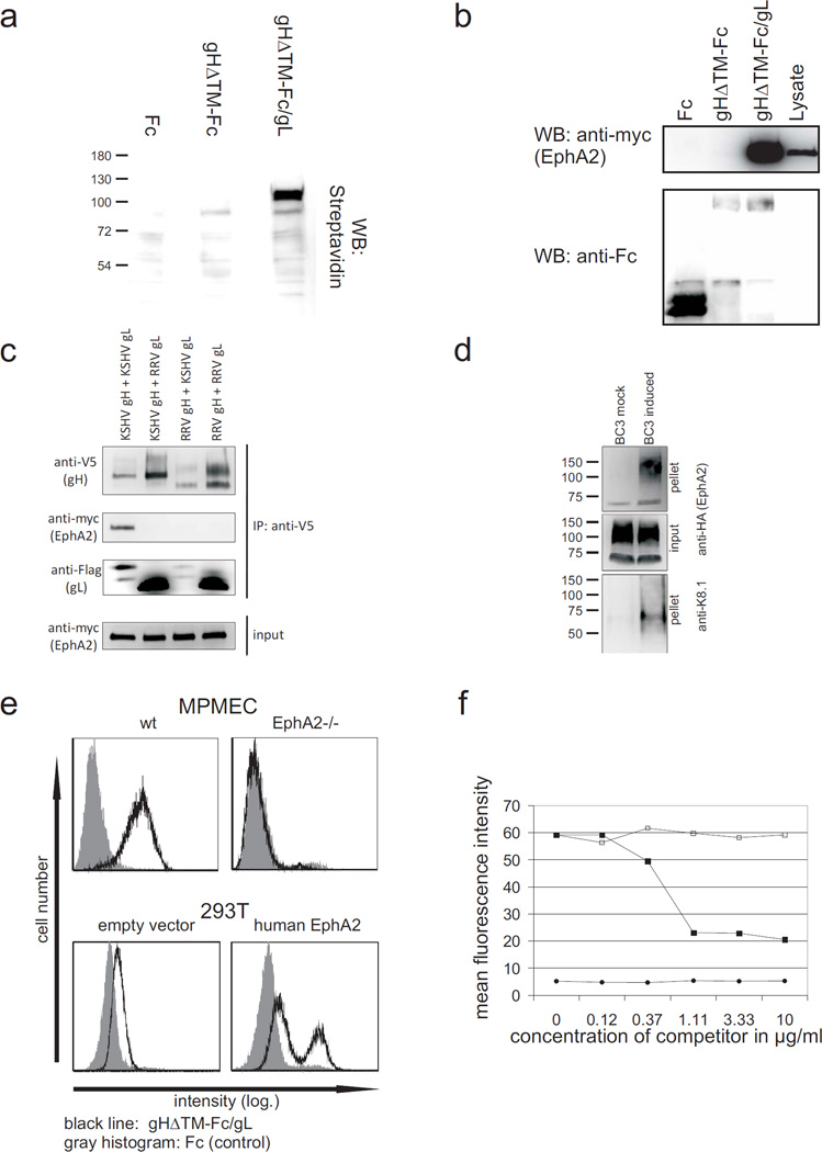
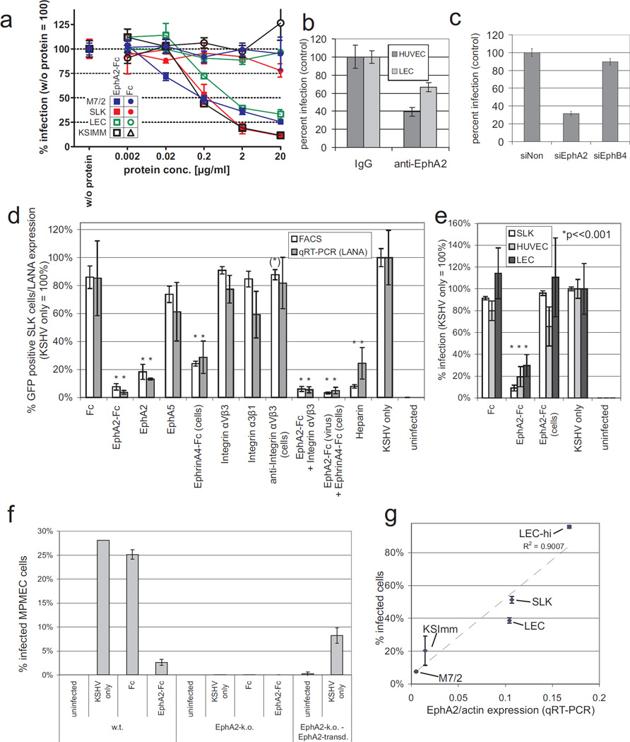
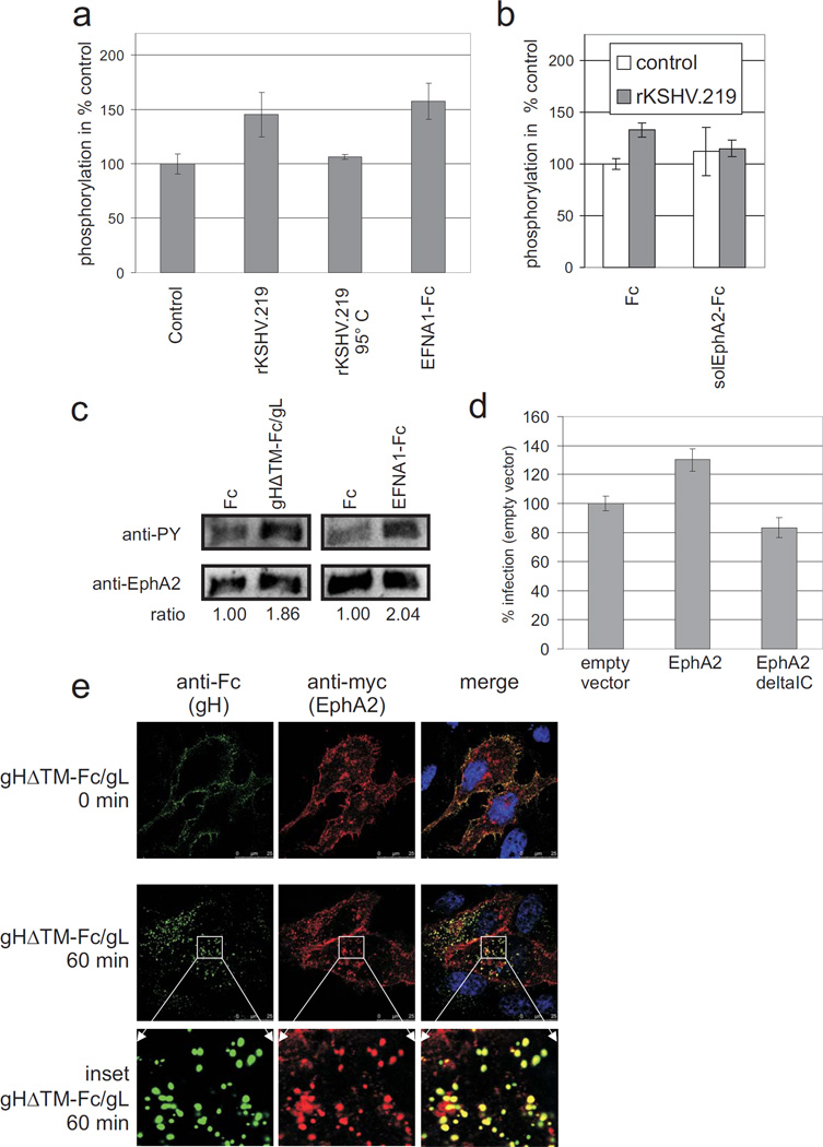
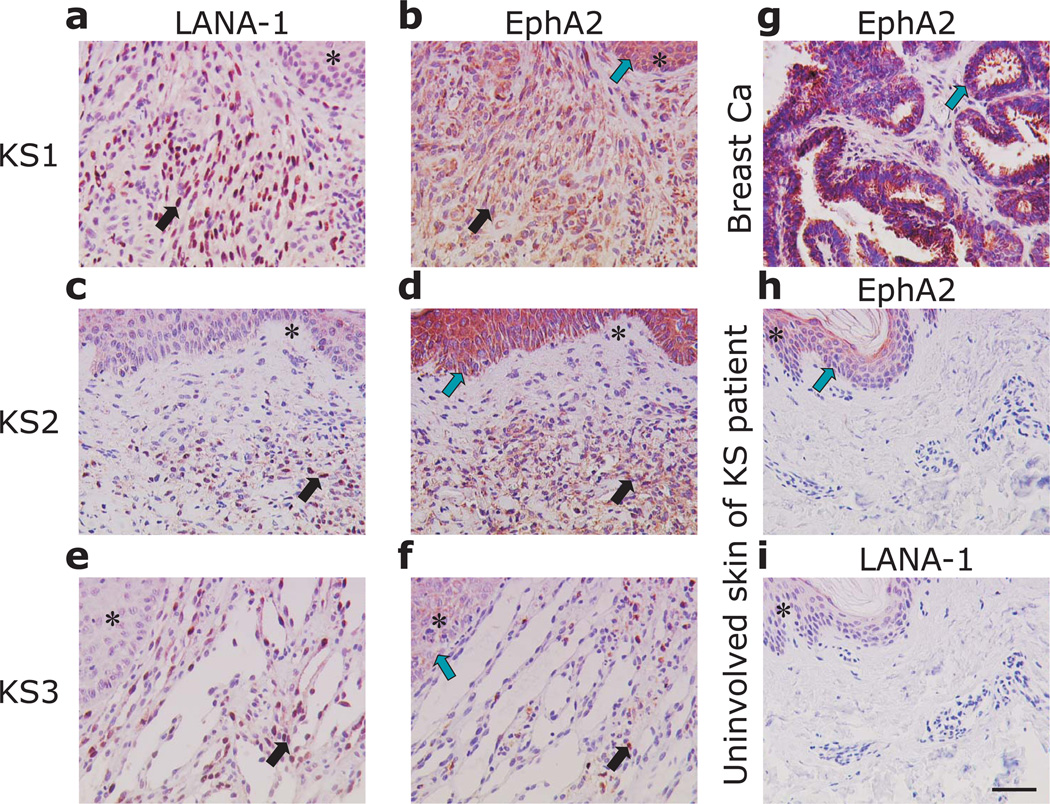
Kaposi's sarcoma-associated herpesvirus (KSHV) is the causative agent of Kaposi's sarcoma(1), a highly vascularized tumor originating from lymphatic endothelial cells, and of at least two different B cell malignancies(2,3). A dimeric complex formed by the envelope glycoproteins H and L (gH-gL) is required for entry of herpesviruses into host cells(4). We show that the ephrin receptor tyrosine kinase A2 (EphA2) is a cellular receptor for KSHV gH-gL. EphA2 co-precipitated with both gH-gL and KSHV virions. Infection of human epithelial cells with a GFP-expressing recombinant KSHV strain, as measured by FACS analysis, was increased upon overexpression of EphA2. Antibodies against EphA(2) and siRNAs directed against EphA2 inhibited infection of endothelial cells. Pretreatment of KSHV with soluble EphA2 resulted in inhibition of KSHV infection by up to 90%. This marked reduction of KSHV infection was seen with all the different epithelial and endothelial cells used in this study. Similarly, pretreating epithelial or endothelial cells with the soluble EphA2 ligand ephrinA4 impaired KSHV infection. Deletion of the gene encoding EphA2 essentially abolished KSHV infection of mouse endothelial cells. Binding of gH-gL to EphA2 triggered EphA2 phosphorylation and endocytosis, a major pathway of KSHV entry(5,6). Quantitative RT-PCR and in situ histochemistry revealed a close correlation between KSHV infection and EphA2 expression both in cultured cells derived from human Kaposi's sarcoma lesions or unaffected human lymphatic endothelium, and in situ in Kaposi's sarcoma specimens, respectively. Taken together, our results identify EphA2, a tyrosine kinase with known functions in neovascularization and oncogenesis, as an entry receptor for KSHV.


Figures

Fig. 1
Fig. 1. KSHV gH/gL binds to human EphA2
(a) KSHV gH/gL precipitates a membrane protein of approximately 110 kDa. Membrane proteins of sog9 mouse fibroblasts were biotinylated and precipitated with the denoted proteins bound to protein-A sepharose. Horseradish peroxidase coupled to streptavidin was used to detect precipitated membrane proteins. (b, c) KSHV gH/gL precipitates EphA2. 293T cells were transfected with an expression plasmid for myc-tagged human EphA2. The cells were lysed and precipitated with (b) protein-A-immobilized Fc, gHΔTM-Fc or gHΔTM-Fc/gL or (c) V5 tagged KSHV or RRV 26–95 gH in combination with KSHV or RRV 26–95 gL (Flag tag) immoblised to protein-G via anti-V5 antibodies. Precipitates were washed and subjected to Western blot analysis. (d) EphA2 co-sediments with KSHV. Pre-cleared and 10× concentrated supernatant from either lytically induced or mock-treated BC3 cells was incubated with supernatant from 293T cells expressing the HA-tagged soluble ectodomain of EphA2. KSHV virions were then pelleted by centrifugation, washed twice with PBS and subjected to PAGE and immunoblot with either anti-HA to detect EphA2 or anti-K8.1 to detect KSHV. (e) KSHV gH/gL binding to cells is mediated by EphA2. Cells were incubated with either Fc-protein (grey histogram) or gHΔTM-Fc/gL (black line). Heparin was added at 100 units/ml to inhibit binding of gH/gL to heparan sulphate. Anti-human IgG coupled to FITC was used as a secondary antibody and detected by FACS. Upper panel: MPMECs from EphA2−/− mice or the parental strain (wt) that were detached by brief treatment with trypsin. lower panel: 293T cells either transfected with an EphA2 expression construct (right) or empty vector (left). (f) Binding of soluble gHΔTM-Fc/gL to 293T cells was blocked with soluble EphA2-Fc. 293T cells were incubated with supernatants from cells expressing either soluble gHΔTM-FcStrep/gL (squares) or FcStrep-protein (circles). Either solEphA2-Fc (filled symbols) or Fc (unfilled squares) was added to the protein solutions in increasing concentration 30 min before incubation with 293T cells. Binding of the Strep-tagged proteins was determined by FACS.
Fig. 2
Fig. 2. Permissiveness for KSHV infection correlates with EphA2 expression in cell culture
(a) Dose-dependent inhibition of KSHV infection by soluble EphA2. Primary LEC (green), two cell lines derived from KS (SLK:red, KSImm:black) and spindle cells derived from a KS biopsy (M7/2: blue) were infected with KSHV.219 at an SLK-MOI of 0.5 (SLK, KSImm, M7/2) or 0.2 (LEC). Prior to infection, each virus was incubated with either solEphA2-Fc (squares) or Fc (control, circles) for 30 min at the indicated concentrations. GFP-positive cells were detected by FACS 3 days after infection. The infection rate achieved in the absence of either protein (solEphA2-Fc or Fc) was set to 100 which corresponded to approx 15% - 40% infected cells depending on cell type (n=6 for SLK and KSImm, n=3 for LEC and M7/2, error bars represent sd). (b) Antibodies to EphA2 inhibit KSHV infection in primary HUVEC and LEC. HUVEC (dark grey) and LEC (light grey) cells were incubated with antibody to EphA2 or rabbit IgG at 1 mg/ml and infected as described in (b) (n=7, error bars represent sem). (c) siRNAs against EphA2 inhibit KSHV infection of LECs. LECs were transfected with either a non-targeting siRNA (siNon), an siRNA pool against EphA2 (siEphA2) or against the related EphB4 (siEphB4) two days prior to infection with rKSHV.219. The relative percentage of GFP-expressing cells is shown (siNon = 100%, n=4, error bars represent sem). (d) Effect of soluble proteins and heparin on KSHV infection of SLK cells. SLK cells were infected with KSHV.219 at an MOI of 0.5. Prior to infection, virus or – where indicated – the cells were incubated with the indicated protein(s). A concentration of 2 µg/ml was used except for integrins and anti-integrin αVβ3 (10µg/ml). Heparin was used at a concentration of 166 IU/ml. KSHV infection was determined on day 3 p.i. by both FACS for GFP-positive cells and qRT-PCR for LANA expression normalized to GAPDH. Values achieved in the presence of KSHV only were set to 100 % (infection rate by FACS: 44.3%). Mean and sd from 6 (FACS) or 3 (qPCR) independent experiments are shown. Error bars represent sd, asterisks indicate statistically significant differences to KSHV only (*:p < 0.001; (*): p < 0.01). (e) Inhibition of KSHV by EphA2 requires contact of EphA2 with the virus. Cells of endothelial origin (SLK, HUVEC, LEC) were infected with KSHV.219 at an SLK-MOI of 0.5. Prior to infection, either the viral inoculum or the cells were incubated with EphA2-Fc (2 µg/ml, 30 min, 37°C) or left untreated. Fc-protein was used as a control. KSHV infection was determined on day 3 by FACS. Mean and sd of relative infection (KSHV only = 100%) from 3 independent experiments are shown. Error bars represent sd, asterisks indicate statistically significant differences to KSHV only (p < 0.001). (f) Loss of EphA2 expression renders MPMEC resistant to KSHV infection. MPMEC cells from either wild type (w.t) or an EphA2 knock-out (EphA2-k.o.) mouse were infected with KSHV (SLK-MOI: 2). EphA2-k.o. cells transduced with a retroviral vector expressing murine EphA2 were infected as a control (right). Where indicated, KSHV was incubated with EphA2-Fc or control protein prior to infection (2 µg/ml, 30min). Mean and sd from 3 independent experiments are shown. (g) Susceptibility for KSHV infection correlates with EphA2 expression in KS cells and lymphatic endothelium. Human cells and cell lines derived from KS (M7/2, KSImm, SLK) or two different batches of human lymphatic endothelial cells (LEC, LEC-hi) were infected with KSHV at constant cell/virus ratio corresponding to an MOI of approx. 0.5 on SLK cells. Cells were harvested 2 days p.i., infection was determined by FACS and both EphA2 and actin mRNA were quantified by qRT-PCR. The quotient of mRNA expressions (EphA2/actin) is plotted on the x-axis against the percentage of KSHV infected cells on the y-axis (R2 = square correlation coefficient, n=6 for SLK and KSImm, 3 for LEC, LEC-hi and M7/2).
Fig. 3
Fig. 3. KSHV or gH/gL trigger tyrosine phosphorylation and endocytosis of EphA2
(a,b) KSHV triggers tyrosine phosphorylation of EphA2. (a) 293T cells were incubated for 10 min with concentrated rKSHV.219 (n = 5), heat-inactivated rKSHV.219 (5 min 95°C, n = 2) or a negative control prepared identically from the supernatant of KSHV-negative 293 cells (n = 5). EFNA1-Fc at a final concentration of 5 µg/ml served as a positive control (n = 5). EphA2 phosphorylation was determined by phospho-EphA2 ELISA (R&D Systems). Optical densities obtained after incubation with the KSHV-negative control preparation were set to 100%. Arithmetic mean and standard deviation are shown. Only the differences between control and rKSHV.219 (p < 0.005) or EFNA1-Fc (p < 0.005) were statistically significant (Student’s t-test). (b) 293T cells were stimulated with either rKSHV.219 (grey bars) or a KSHV-negative control preparation (white bars) as in (a). In addition, Fc or solEphA2-Fc were added to the preparations at a final concentration of 5 µg/ml 30 min prior to incubation with the cells. EphA2 phosphorylation was determined by ELISA. Relative mean values (control preparation with Fc-protein = 100%) and standard deviation from three independent experiments are shown. Differences in EphA2 phosphorylation between control and rKSHV.219 were significant for Fc (p < 0.005), but not for solEphA2-Fc (p = 0.85). Reduction of KSHV-induced EphA2 phosphorylation by pre-incubation of KSHV with solEphA2-Fc was significant (p < 0.05). (c) gH/gL triggers tyrosine phosphorylation of EphA2. Serum starved 293T cells were stimulated for 10 min with gHΔTM-Fc/gL or Fc (1.5 µg/ml) cross-linked via anti-Fc monoclonal antibody. EphA2 was immunoprecipitated and assayed for tyrosine phosphorylation by western blotting and detection with anti-phosphotyrosine monoclonal antibody followed by detection of EphA2 for normalization. (d) The intracellular domain of EphA2 is required to enhance KSHV infection of 293T cells. 293T cells were transiently transfected with either empty vector, an expression plasmid for EphA2 or an EphA2 variant lacking the intracellular domain (EphA2deltaIC). The cells were then infected with rKSHV.219 and the rate of infection was quantified and normalized to empty vector control (n=3, error bars represent sd). (e) gH/gL and EphA2 colocalise after endocytosis. gHΔTM-Fc/gL was first preincubated and crosslinked for 30 min with anti-human-Fc-Alexa488 antibody. HELA cells that were transfected with an EphA2myc expression plasmid two days before were then incubated with the crosslinked gHΔTM-Fc/gL for 1h at 4°C, then washed, shifted to 37°C and fixed with 3% PFA after 0 min (first row) or 60 min at 37°C (2nd row, enlarged insets are shown in row 3). EphA2myc was detected with primary monoclonal 9E10 anti-myc-epitope antibody and anti-mouse-Cy3 secondary antibody. Cells were visualized by sequential confocal laser scanning microscopy.
Fig. 4
Fig. 4. EphA2 is expressed in KSHV-infected spindle cells of Kaposi’s sarcoma
(a – f) Immunohistochemical detection of LANA-1 and EphA2 in two different KS skin lesions (KS1, KS2) and one lung KS (KS3). (g) EphA2 expression in breast cancer was used as a positive control. (h, i) EphA2 and LNA-1 expression in uninvolved tissue sections of the skin of a KS patient. Similar tissue areas of consecutive sections are indicated by asterisks. Areas with KS spindle cells, exhibiting prominent nuclear LNA-1 staining and cytoplasmic EphA2 staining in consecutive sections are indicated by black arrows. Green arrows indicate EphA2 expression in the epidermis overlaying KS (b, d), in the tumour cells of breast cancer (g) or the epidermal layer of uninvolved skin (h), respectively. The magnification bar in panel e is 50 µm and is identical for all other panels.

Comment in

Similar articles

Cited by

References

    1. Chang Y, et al. Identification of herpesvirus-like DNA sequences in AIDS-associated Kaposi's sarcoma. Science. 1994;266:1865–1869. - PubMed
    1. Cesarman E, Chang Y, Moore PS, Said JW, Knowles DM. Kaposi's sarcoma-associated herpesvirus-like DNA sequences in AIDS-related body-cavity-based lymphomas. N. Engl. J. Med. 1995;332:1186–1191. - PubMed
    1. Soulier J, et al. Kaposi's sarcoma-associated herpesvirus-like DNA sequences in multicentric Castleman's disease. Blood. 1995;86:1276–1280. - PubMed
    1. Campadelli-Fiume G, et al. The multipartite system that mediates entry of herpes simplex virus into the cell. Rev. Med. Virol. 2007 - PubMed
    1. Chandran B. Early Events in Kaposi's Sarcoma-Associated Herpesvirus Infection of Target Cells. J. Virol. 2010;84:2188–2199. - PMC - PubMed

Publication types