. 2012;8(4):e1002660.
doi: 10.1371/journal.ppat.1002660. Epub 2012 Apr 19.

T regulatory cells control susceptibility to invasive pneumococcal pneumonia in mice

Affiliations

T regulatory cells control susceptibility to invasive pneumococcal pneumonia in mice

Daniel R Neill et al. PLoS Pathog. 2012.

Abstract

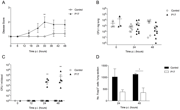
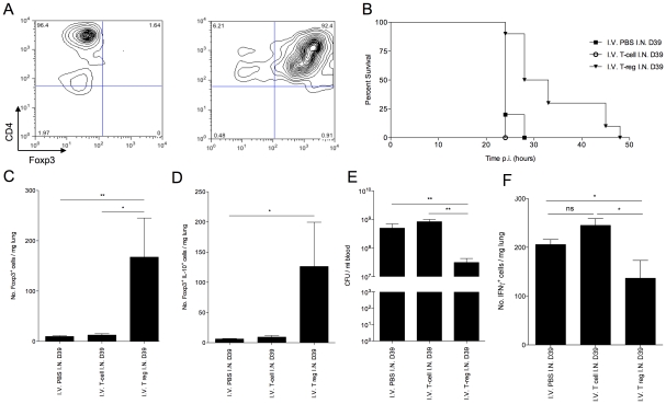
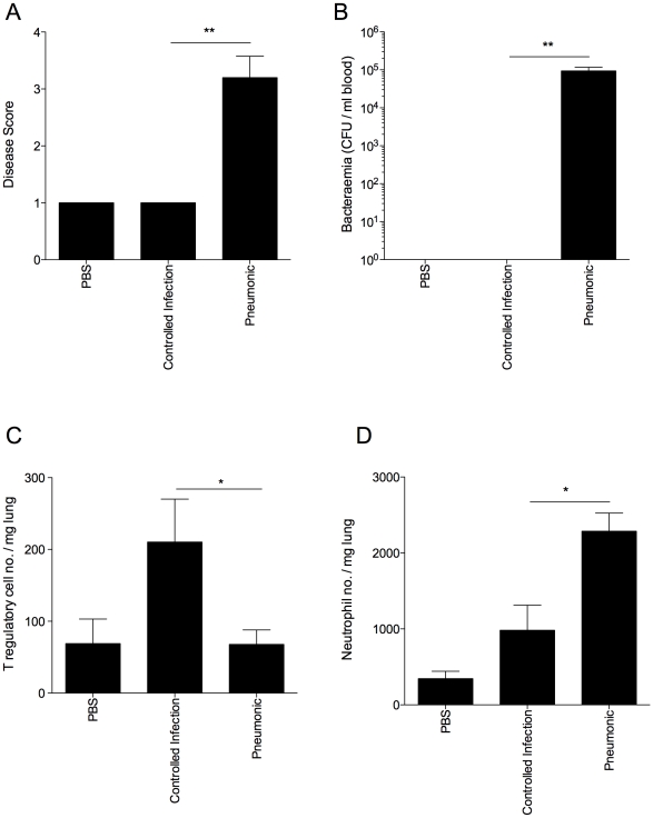
Streptococcus pneumoniae is an important human pathogen responsible for a spectrum of diseases including pneumonia. Immunological and pro-inflammatory processes induced in the lung during pneumococcal infection are well documented, but little is known about the role played by immunoregulatory cells and cytokines in the control of such responses. We demonstrate considerable differences in the immunomodulatory cytokine transforming growth factor (TGF)-β between the pneumococcal pneumonia resistant BALB/c and susceptible CBA/Ca mouse strains. Immunohistochemistry and flow cytometry reveal higher levels of TGF-β protein in BALB/c lungs during pneumococcal pneumonia that correlates with a rapid rise in lung Foxp3(+)Helios(+) T regulatory cells. These cells have protective functions during pneumococcal pneumonia, because blocking their induction with an inhibitor of TGF-β impairs BALB/c resistance to infection and aids bacterial dissemination from lungs. Conversely, adoptive transfer of T regulatory cells to CBA/Ca mice, prior to infection, prolongs survival and decreases bacterial dissemination from lungs to blood. Importantly, strong T regulatory cell responses also correlate with disease-resistance in outbred MF1 mice, confirming the importance of immunoregulatory cells in controlling protective responses to the pneumococcus. This study provides exciting new evidence for the importance of immunomodulation during pulmonary pneumococcal infection and suggests that TGF-β signalling is a potential target for immunotherapy or drug design.


Conflict of interest statement

The authors have declared that no competing interests exist.

Figures

Figure 1
Figure 1. Many Tgfβ1 interaction partners show opposing expression profiles in BALB/c and CBA/Ca mice at 6 hours p.i.
Ingenuity pathway analysis of microarray data from 6 hours p.i. Red=Gene significantly upregulated as compared to t=0 levels. Green=Down-regulated. BALB/c levels are compared to BALB/c t=0 expression and CBA/Ca levels are compared to CBA/Ca t=0 expression. Orange outline=Gene found within Spir1 susceptibility-determining locus. Solid lines indicate direct interactions, dotted lines indicate indirect interactions.
Figure 2
Figure 2. tgfb1 expression and TGF-β production is upregulated in BALB/c lungs in contrast to CBA/Ca lungs during pneumococcal infection.
Relative gene expression of tgfβ1 in lungs of BALB/c and CBA/Ca mice before (A) and during (B) pneumococcal infection. Data were normalised against a housekeeping gene (Hprt1) and are presented using BALB/c results as a calibrator (A) or else using BALB/c or CBA/Ca sham-infection data from the appropriate time-point as calibrators (B). For 6 and 12 hours p.i. data for each strain was calibrated against sham-infection data for that strain at the same time-point. i.e. BALB/c to BALB/c sham and CBA/Ca to CBA/Ca sham. (C) Antibody staining of TGF-β in airway epithelial cells from BALB/c and CBA/Ca mice during pneumococcal infection. *=p<0.05, ***=p<0.005. For all figures data represent mean +/− SEM. Data are representative of two independent experiments with 5 mice per time-point per strain and per treatment group.
Figure 3
Figure 3. IL-10-producing natural T regulatory cells are rapidly recruited to the lung in S. pneumoniae-infected BALB/c mice.
All results are from intranasal-infection of mice with wild-type S. pneumoniae D39. (A) Foxp3 immunostaining of lung sections taken from BALB/c and CBA/Ca mice during pneumococcal infection. Inset shows example staining from 12 hour p.i. BALB/c mouse. Foxp3+ cells stain brown. (B) Number of Foxp3+ T regulatory cells, (C) Number of Foxp3+Helios+ cells, (D) number of Foxp3+IL-10+ cells and (E) number of Foxp3+CTLA-4+ cells per mg lung in S. pneumoniae-infected BALB/c and CBA/Ca mice, as identified by flow cytometry. Data in (D) are from 24 hrs p.i., PBS group contains both BALB/c and CBA/Ca mice. White bars=BALB/c, black bars=CBA/Ca. *'s indicate significant difference, where *=p<0.05, **=p<0.01, and ***=p<0.005. For all graphs data represent mean +/− SEM. All results are representative of between 2–4 independent experiments with >4 mice per group.
Figure 4
Figure 4. CBA/Ca mice display uncontrolled lung inflammation and associated apoptosis following S. pneumoniae infection.
All results are from intranasal-infection of mice with wild-type S. pneumoniae D39. (A) Number of IFNγ+NKp46+CD4 cells per mg lung in PBS-treated or S. pneumoniae-infected BALB/c and CBA/Ca mice at 24 hours p.i. (B) Top left shows gating of NK cells in lung homogenate. Remaining panels show example IFNγ-staining from PBS-treated CBA/Ca or S. pneumoniae-infected BALB/c and CBA/Ca mice at 24 hours p.i. (C) Proportion (%) of apoptotic cells in areas of inflammation within lungs of S. pneumoniae-infected BALB/c and CBA/Ca mice. Inset shows apoptotic cell (arrow) within area of inflammation as defined by presence of inflammatory infiltrate. White bars=BALB/c, black bars=CBA/Ca. *'s indicate significant difference, where *=p<0.05 and ***=p<0.005. For all graphs data represent mean +/− SEM. Results in (A and B) are representative of 3 independent experiments with >4 mice per group. Results in (C) are from a single experiment with 5 mice per group. PBS-treated groups contained both BALB/c and CBA/Ca mice.
Figure 5
Figure 5. Inhibition of TGF-β activity impairs BALB/c resistance to pneumococcal infection.
All results are from intranasal-infection of mice with wild-type S. pneumoniae D39. (A) Mean animal disease score in S. pneumoniae-infected PBS-treated or P17-treated BALB/c mice over a 48 hour time-course. Normal mice score 1. (B) Number of bacteria per mg lung tissue and (C) per ml blood in P17- (▴) or PBS-treated (○) mice. CFU=colony forming units. (D) Quantification of Foxp3+ immunostaining in antibody-stained lung sections from P17- (black bars) or PBS-treated (white bars) S. pneumoniae-infected mice. *'s indicate significant difference, where *=p<0.05 and **=p<0.01. For all graphs data represent mean +/− SEM. Results in (A–D) are representative of 2 independent experiments with >5 mice per group.
Figure 6
Figure 6. Adoptive transfer of in vitro generated Foxp3+ T regulatory cells improves CBA/Ca survival in pneumococcal infection.
(A) CD4 and Foxp3 FACS staining of isolated CBA/Ca CD4+, CD25 splenocytes before (left column) and after (right column) 5 days culture with anti-CD3, anti-CD28 and TGF-β. 5 day cultured cells were used for adoptive transfer. All results in (B–D) are from intranasal-infection of mice with wild-type D39 S. pneumoniae. (B) Survival of S. pneumoniae-infected CBA/Ca mice that had received PBS (▪), CD4+ T cells (○), or T regulatory cells (▾) i.v. prior to intranasal infection with S. pneumoniae. Survival of T regulatory cell treatment group is significantly different to both CD4+ T cell treatment group (p<0.01) and PBS treatment group (p<0.01) (C) Number of Foxp3+ cells and (D) number of IL-10+Foxp3+ cells per mg lung at 24 hours p.i. (E) Number of bacteria/ml blood at 24 hour post-infection. (F) Number of IFN-γ+ cells per mg lung at 24 hours p.i.. CFU=colony forming units. I.N.=Intranasal. I.V.=Intravenous. *'s indicate significant difference, where *=p<0.05 and ***=p<0.001. For all graphs data represent mean +/− SEM. Results in (A) are representative of 5 independent experiments. Results in (B–F) are representative of two independent experiments with >6 mice per group.
Figure 7
Figure 7. Disease resistance in outbred MF1 mice correlates with strong T regulatory cell responses in the lung.
All results are from intranasal-infection of outbred MF1 mice with wild-type S. pneumoniae D39. (A) Animal disease score at 48 hours p.i. A score of 1 is a normal mouse. Any infected mouse scoring 1 at 48 hours p.i. was considered to have a controlled infection. (B) Number of bacteria/ml blood at 48 hours p.i. CFU=colony forming units. Number of Foxp3+ T regulatory cells (C) and Gr-1hiF4/80lo neutrophils (D) per mg lung in PBS-treated or S. pneumoniae-infected MF1 mice at 48 hours p.i., as identified in flow cytometry. *'s indicate significant difference, where *=p<0.05 and **=p<0.01. For all graphs data represent mean +/− SEM. Results are representative of 2 independent experiments, each with 3 PBS-treated and >10 infected mice.

Similar articles

Cited by

References

    1. Paterson GK, Orihuela CJ. Pneumococci: immunology of the innate host response. Respirology. 2010;15:1057–1063. - PMC - PubMed
    1. Buttery J, Moxon ER. Capsulate bacteria and the lung. Br Med Bull. 2002;61:63–80. - PubMed
    1. Kaplanski G, Marin V, Montero-Julian F, Mantovani A, Farnarier C. IL-6: a regulator of the transition from neutrophil to monocyte recruitment during inflammation. Trends Immunol. 2003;24:25–29. - PubMed
    1. Kadioglu A, Gingles NA, Grattan K, Kerr A, Mitchell TJ, et al. Host cellular immune response to pneumococcal lung infection in mice. Infect Immun. 2000;68:492–501. - PMC - PubMed
    1. Kadioglu A, Coward W, Colston MJ, Hewitt CR, Andrew PW. CD4-T-lymphocyte interactions with pneumolysin and pneumococci suggest a crucial protective role in the host response to pneumococcal infection. Infect Immun. 2004;72:2689–2697. - PMC - PubMed

Publication types

MeSH terms

LinkOut - more resources