. 2012;8(3):e1002566.
doi: 10.1371/journal.ppat.1002566. Epub 2012 Mar 1.

Kaposi's sarcoma herpesvirus upregulates Aurora A expression to promote p53 phosphorylation and ubiquitylation

Affiliations

Kaposi's sarcoma herpesvirus upregulates Aurora A expression to promote p53 phosphorylation and ubiquitylation

Qiliang Cai et al. PLoS Pathog. 2012.

Abstract

Aberrant expression of Aurora A kinase has been frequently implicated in many cancers and contributes to chromosome instability and phosphorylation-mediated ubiquitylation and degradation of p53 for tumorigenesis. Previous studies showed that p53 is degraded by Kaposi's sarcoma herpesvirus (KSHV) encoded latency-associated nuclear antigen (LANA) through its SOCS-box (suppressor of cytokine signaling, LANA(SOCS)) motif-mediated recruitment of the EC(5)S ubiquitin complex. Here we demonstrate that Aurora A transcriptional expression is upregulated by LANA and markedly elevated in both Kaposi's sarcoma tissue and human primary cells infected with KSHV. Moreover, reintroduction of Aurora A dramatically enhances the binding affinity of p53 with LANA and LANA(SOCS)-mediated ubiquitylation of p53 which requires phosphorylation on Ser215 and Ser315. Small hairpin RNA or a dominant negative mutant of Aurora A kinase efficiently disrupts LANA-induced p53 ubiquitylation and degradation, and leads to induction of p53 transcriptional and apoptotic activities. These studies provide new insights into the mechanisms by which LANA can upregulate expression of a cellular oncogene and simultaneously destabilize the activities of the p53 tumor suppressor in KSHV-associated human cancers.


Conflict of interest statement

The authors have declared that no competing interests exist.

Figures
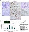
Figure 1
Figure 1. Aurora A expression is up-regulated in KSHV latently infected cells.
(A) The protein levels of Aurora A were enhanced in KSHV-associated KS patient skin tissue. Kaposi's sarcoma tissue specimens from 3 patients were analyzed for both LANA and Aurora A expression by immunohistochemistry. A representative sample of Kaposi's sarcoma patient (middle and right panels) and the adjacent normal tissue (left panel) are shown. (B) Quantitative real-time PCR analysis of Aurora A and LANA transcripts in PBMC with GFP-KSHV infection. Total RNA was isolated from cells with GFP-KSHV infection for 0, 1, 2, 4 and 7 days. Real-time PCR was performed as described in Materials and Methods. The relative levels of Aurora A and LANA transcript were calculated by the cycle threshold (ΔΔCt) values and shown by the fold change compared to mock (day 0). All samples were tested in triplicate and the calculation of the mean and standard deviation from two separate experiments. The mock cells without or with GFP-KSHV infection for 4 days were shown on the top. (C) Immunoblotting analysis. Whole cell lysate of human PBMC cells with GFP-KSHV infection for 4 days or mock, were subjected to immunoblotting with antibodies against LANA, Aurora A and GAPDH. The relative instensity (RI) of Aurora A is quantified and shown in the figure.
Figure 2
Figure 2. Aurora A transcript is dramatically enhanced in a LANA-dependent manner.
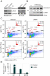
(A) The level of Aurora A expression was decreased in BC3 cells with LANA knockdown. Cell lysates from BC3 cells with LANA (shLANA) or control firefly luciferase knockdown (shLuc) were subjected to immunoblotting against LANA, Aurora A and GAPDH, or total RNA were extracted for RT-PCR analysis. (B) LANA increases endogenous Aurora A expression in a dose-dependent manner. Fifteen million 293 cells were transiently transfected either with empty vector or increasing amounts of pA3M-LANA. At 36 hours post-transfection, the cells were harvested, lysed and individually subjected to western blot with antibodies against Aurora A, LANA and Actin. (C) Cytometric profile of Aurora A expression in HeLa cells in the presence or absence of LANA. HeLa cells transfected with pA3F-LANA or pA3F vector alone were individually divided and subjected to propidium iodide (PI)-staining cell cycle analysis (inset) combined with anti-Aurora A (FITC) or normal IgG (FITC) staining. A representative of double staining with the gating percentage of Aurora (IgG) expression at G1, S or G2/M phase of cell cycle was shown. (D) The relative fold of Aurora expression in each phase of HeLa cells with LANA expression or vector alone from panel C was presented and normalized by IgG staining control. Data from two repeat experiments.
Figure 3
Figure 3. The Sp1 cis-acting element is critical for LANA-mediated enhancement of Aurora A transcription.
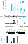
(A) HEK293 cells were co-transfected full length Aurora A promoter or its mutants driving reporter plasmid with either pA3M-LANA or pA3M vector. At 24-hrs post-transfection, cells were harvested and subjected to reporter assay. The schematic representation of full length Aurora A gene promoter or its mutants-driven luciferase constructs is shown at the bottom panel. The results were presented by the RLU (relative luciferase unit) fold compared to pGL3-basic with vector alone. Data is presented as means±SD of three independent experiments. (B) ChIP analysis of endogenous Aurora A promoter in BC3 cells with or without LANA knockdown. ChIP analysis was conducted using normal mouse IgG, α-E2F, α-Sp1 agarose, and subjected to PCR analysis using primer F(−400) and R(−60) indicated in A. The results of quantitative real-time PCR are shown at the bottom panel.
Figure 4
Figure 4. Coexpression of Aurora A enhances LANA-mediated repression of p53 transcriptional activity which depends on its kinase activity.
Ten million of Saos-2 (A), MEF (B), or 293 (C) cells were co-transfected 3 µg p53 reporter plasmid with construct expressing LANA, Aurora A (WT or KR) or both. At 24 hr post-transfection, cells were harvested and lysed for luciferase assays. The expression levels of each target proteins were detected by westernblotting and shown at the bottom panels. β-actin blot was used as loading control.
Figure 5
Figure 5. The active form of Aurora A kinase enhances LANASOCS-mediated ubiquitylation of p53.
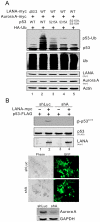
(A) Active form of Aurora A kinase enhanced the interaction of p53 with LANA. Saos-2 or MEF cells were co-transfected p53-FLAG with pA3M-LANA plus either vector control, wild type (WT) Aurora A or its kinase inactive mutant (KR). At 36 hr post-transfection, the cells lysates were immunoprecipitated with anti-FLAG (M2) antibody, followed by immunoblotting with indicated antibodies. The relative intensity (RI) of p53-interacting LANA was presented by normalization with the protein amounts of both immunoprecipitated p53 and input LANA. (B) The LANASOCS–mediated ubiquitylation of p53 was enhanced by Aurora A coexpression in a kinase activity-dependent manner. MEF cells were cotransfected with the plasmids expressing HA-Ub and p53-FLAG plus LANA-myc (WT or ΔSOCS) and Aurora A-myc (WT or KR). At 36 hr post-transfection with additional 6 hr MG132 treatment before harvest, cell lysates were analyzed by immunoblotting with as indicated antibodies.
Figure 6
Figure 6. Aurora A-induced phosphorylation of p53 is a target for LANASOCS-mediated ubiquitylation.
(A) The Aurora A-induced phosphorylation on Ser 215 and Ser 315 was critical for LANA-mediated ubiquitylation of p53. MEF cells were cotransfected with different combinations of HA-Ub, HA-p53 (WT, S215A, S315A or S215A/S315A), LANA-myc (WT or ΔSOCS) and Aurora A-myc (WT or KR) as indicated in the figure. At 36 hr post-transfection with additional 6 hr MG132 treatment before harvest, cell lysates were analyzed by immunoblotting with as indicated antibodies. (B) LANA upregulated Aurora A-mediated phosphorylation of p53. MEF cells were cotransfected p53-FLAG with either plasmid expressing shAurora A or shLuc in the presence or absence of LANA-myc as indicated in the figure. At 36 hr post-transfection with additional 6 hr MG132 treatment before harvest, cell lysates were analyzed by immunoblotting with specific antibodies. The knockdown efficiency of shRNA against Aurora A is shown at the bottom panel.
Figure 7
Figure 7. Aurora A is important for LANA to inhibit p53-induced apoptosis.
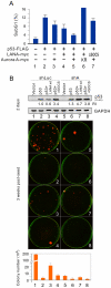
(A) Saos-2 cells were cotransfected with different combination of plasmids expressing p53-FLAG, LANA-myc (wt or ΔSOCS), Aurora A-myc (wt or KR) or empty vectors as indicated in the figure. At 36 hr post-transfection, cell cycle profiles were analyzed with Flow cytometry. The percentage of subG1 population cells is presented. (B) Knockdown of Aurora A blocks LANA-mediated inhibition of p53 suppression function in colony formation. Saos-2 cells were cotransfected with different combination of plasmids expressing p53-FLAG, LANA-myc (wt or ΔSOCS) or empty vectors in the presence of shAurora A or shluc control as indicated in figure with number 1 to 8. Upper panel, Immunoblotting analysis of p53-FLAG expression at 48-hr posttransfection. Middle panel, a representative of colony formation after 3-week culture was shown. Lower panel, the data are presented as the average from two independent experiments.
Figure 8
Figure 8. Downregulation of Aurora A induces cell cycle arrest and apoptosis in PEL cells.
(A) Induction of PARP1 cleavage and increased p53 expression in cells with Aurora A knockdown. Parental BC3 cells and BC3 cells transduced by lentivirus expressing small hairpin against Aurora A (shA#1, shA#2) or luciferase (shLuc), were exposed to 0.1% sera for 18 hr. Cell lysates were subjected to immunoblotting as indicated antibodies in the figure. The relative instensity (RI) of p53 protein level is shown. (B) FACS analysis of BC3 cells with or without Aurora knockdown. Aurora A depletion induced cells with greater than 4N DNA content (Propidium iodide staining). (C) Fold changes in the subG1 population of cells with Aurora A knockdown, as analyzed by FACS. (D) DAPI staining of representative fields show multinucleation (white arrows), and inset fields show corresponding photographs to the white rectangles in the upper/lower panels. (E) Quantification of the multinucleated (M) and fragmented nuclei (F) in BC3 cells with or without Aurora A knockdown. (F) Aurora A knockdown dramatically increased p53 accumulation and subG1 population in KSHV-infected not uninfected cells. 293 (Mock) and 293-Bac36 (KSHV) cells were individually transduced by lentivirus expressing small hairpin against Aurora A (shA) and luciferase (shLuc) followed by immunoblotting and subG1 population analysis as described in panels A and C.
Figure 9
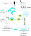
Figure 9. A model depicting the role of Aurora A targeted by LANA.
In the KSHV latently infected cells, LANA upregulates Aurora A expression through targeting Sp1-binding site. The elevated Aurora A induced phosphorylation of p53 on Ser 215 and Ser315 which enhanced the binding affinity of p53 with LANA and promoted LANASOCS-mediated ubiquitylation and degradation of p53, and thus inhibited p53 transcriptional and apoptotic activities. Loss of p53 function and aberrant expression of Aurora A trigger chromosome instability for cell survival and growth.

Similar articles

Cited by

References

    1. Dupin N, Diss TL, Kellam P, Tulliez M, Du MQ, et al. HHV-8 is associated with a plasmablastic variant of Castleman disease that is linked to HHV-8-positive plasmablastic lymphoma. Blood. 2000;95:1406–1412. - PubMed
    1. Chang Y, Cesarman E, Pessin MS, Lee F, Culpepper J, et al. Identification of herpesvirus-like DNA sequences in AIDS-associated Kaposi's sarcoma. Science. 1994;266:1865–1869. - PubMed
    1. Cesarman E, Chang Y, Moore PS, Said JW, Knowles DM. Kaposi's sarcoma-associated herpesvirus-like DNA sequences in AIDS-related body-cavity-based lymphomas. N Engl J Med. 1995;332:1186–1191. - PubMed
    1. Soulier J, Grollet L, Oksenhendler E, Cacoub P, Cazals-Hatem D, et al. Kaposi's sarcoma-associated herpesvirus-like DNA sequences in multicentric Castleman's disease. Blood. 1995;86:1276–1280. - PubMed
    1. Godfrey A, Anderson J, Papanastasiou A, Takeuchi Y, Boshoff C. Inhibiting primary effusion lymphoma by lentiviral vectors encoding short hairpin RNA. Blood. 2005;105:2510–2518. - PubMed

Publication types

MeSH terms