. 2012 Mar 21;31(6):1440-52.
doi: 10.1038/emboj.2011.501. Epub 2012 Feb 14.

PP4 dephosphorylates Maf1 to couple multiple stress conditions to RNA polymerase III repression

Affiliations

PP4 dephosphorylates Maf1 to couple multiple stress conditions to RNA polymerase III repression

Andrew J Oler et al. EMBO J. .

Abstract

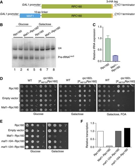
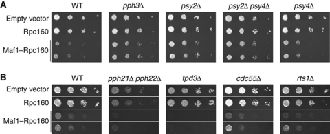
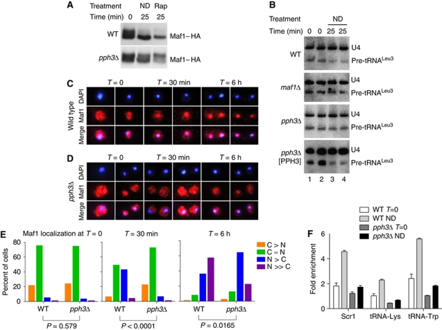
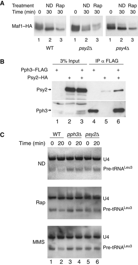
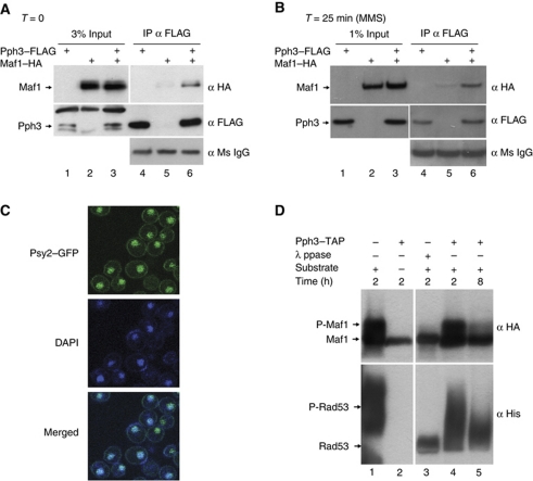
Maf1 is the 'master' repressor of RNA polymerase III (Pol III) transcription in yeast, and is conserved in eukaryotes. Maf1 is a phospho-integrator, with unfavourable growth conditions leading to rapid Maf1 dephosphorylation, nuclear accumulation, binding to RNA Pol III at Pol III genes and transcriptional repression. Here, we establish the protein phosphatase 4 (PP4) complex as the main Maf1 phosphatase, and define the involved catalytic (Pph3), scaffold (Psy2) and regulatory subunits (Rrd1, Tip41), as well as uninvolved subunits (Psy4, Rrd2). Multiple approaches support a central role for PP4 in Maf1 dephosphorylation, Maf1 nuclear localization and the rapid repression of Pol III in the nucleus. PP4 action is likely direct, as a portion of PP4 co-precipitates with Maf1, and purified PP4 dephosphorylates Maf1 in vitro. Furthermore, Pph3 mediates (either largely or fully) rapid Maf1 dephosphorylation in response to diverse stresses, suggesting PP4 plays a key role in the integration of cell nutrition and stress conditions by Maf1 to enable Pol III regulation.


Conflict of interest statement

The authors declare that they have no conflict of interest.

Figures

Figure 1
Figure 1
A Maf1–Rpc160 fusion functionally represses Pol III transcription. (A) Constructs of galactose-inducible Rpc160 or Maf1–Rpc160 fusion. (B) Northern blot showing levels of pre-tRNALeu3 and U4 in strains with Maf1–Rpc160 or Rpc160 constructs, under conditions of repression by glucose (lanes 1–4) or induction by galactose (lanes 5–8). Expression of the Maf1–Rpc160 fusion but not Rpc160 represses pre-tRNALeu3 transcription. (C) Quantification of lanes 5–8 of (B) as a ratio of tRNA/U4 band intensity with Rpc160 set to 1. Error bars represent s.d. (D) Expression of the Maf1–Rpc160 fusion but not Rpc160 confers a growth defect in both RPC160+ and rpc160Δ cells (right and left half of each plate, respectively). Cells were grown on plates containing glucose, galactose or galactose + FOA. Note that FOA only permits growth of cells lacking PMET25Rpc160 as this vector carries a URA3 marker. Two biological replicates are shown for each strain. See Materials and methods for more details. (E) The fusion growth defect is dependent on a functional Maf1. Strong, dephosphorylation-resistant missense mutations—maf1-104 or maf1-124—in the Maf1 portion of the fusion partially suppress the fusion growth defect. (F) Yeast transformed with plasmids for galactose-inducible Rpc160 (p2266), Maf1–Rpc160 (p2268), maf1-124–Rpc160 (p2424) or maf1-104–Rpc160 (p2425) were grown in galactose for 4 h and RNA was isolated immediately, followed by northern blot. Relative transcription is expressed as a ratio of the pre-tRNALeu3 band to U4 band, with Rpc160 set to 1. Data are from a single replicate.
Figure 2
Figure 2
Maf1–Rpc160 fusion identifies Pph3 and Psy2 as Maf1-dependent repressors of Pol III. (A) WT and various PP4 mutants harbouring plasmids for empty vector, Rpc160 or Maf1–Rpc160 fusion were grown on plates containing galactose. The fusion growth defect is partially rescued in PP4 mutant strains pph3Δ and psy2Δ (two biological replicates in the bottom two rows of spot dilutions) compared with controls (top two rows). Little effect on the fusion growth defect is seen in psy4Δ strain. See Materials and methods for details. (B) The fusion growth defect remains (cdc55Δ, rts1Δ) or is intensified (tpd3Δ, pph21Δpph22Δ) in PP2A mutant strains. Note that rows in (B) for each strain are from the same plate image.
Figure 3
Figure 3
Pph3 mediates Pol III repression. (A) Western blot of WT and pph3Δ yeast treated with ND or rapamycin (Rap). ND and Rap treatment cause dephosphorylation of Maf1 in WT but not pph3Δ mutant strain. (B) Northern blot showing repression of pre-tRNALeu3 transcription in WT strain (top panel) in response to ND. Repression is attenuated in pph3Δ strain (third panel) and repression is restored by complementing with Pph3 on a plasmid (bottom panel). Lack of Pol III repression in maf1Δ strain is shown for comparison (second panel). Two biological replicates are shown. (C, D) Nuclear localization of Maf1–HA in response to ND in (C) WT and (D) pph3Δ. Maf1 is stained red while nuclear DNA is stained blue (DAPI). Localization of Maf1 to the nucleus is delayed in pph3Δ compared with WT. (E) Distribution of Maf1 localization in WT and pph3Δ strains at T=0 (left panel; n=230 for WT, n=150 for pph3Δ), T=30 min (middle panel; n=470 for WT, n=986 for pph3Δ) and T=6 h (right panel; n=239 for WT, n=341 for pph3Δ). Significance values calculated as difference between nuclear (N≫C, N>C) and cytoplasmic (C>N, C≈N) Maf1 in WT and pph3Δ using Fisher's exact test. (F) ChIP of Maf1 in WT or pph3Δ cells at T=0 or 25 min ND treatment. ChIP values are expressed as enrichment over a control locus, the Tra1 gene.
Figure 4
Figure 4
PP4 complex members play a role in steps of Pol III repression. (A) Western blot of WT, psy2Δ and psy4Δ yeast treated with ND or rapamycin (Rap). Maf1 is dephosphorylated in WT and the psy4Δ mutant, but not in the psy2Δ mutant in response to ND and Rap treatment. (B) Co-IP of tagged Pph3–FLAG and Psy2–HA in psy4Δ mutant strain, revealed by western blot. Psy2 and Pph3 can interact in the absence of Psy4. (C) Northern blot of WT, pph3Δ and psy2Δ strains treated with ND, Rap or MMS. Pph3 and Psy2 are required for reduction of nascent tRNA levels in ND, Rap and MMS treatments.
Figure 5
Figure 5
Pol III repression requires PP4 accessory factors Rrd1 and Tip41. (A) The fusion growth defect is rescued in rrd1Δ and tip41Δ mutants, but not rrd2Δ. See Materials and methods for details. (B) Western blot showing PP4-associated factors Tip41 and Rrd1 are required for dephosphorylation of Maf1 during ND or exposure to rapamycin, whereas PP2A-associated Rrd2 is not required, correlating to the fusion growth data in (A).
Figure 6
Figure 6
Pph3 directly dephosphorylates Maf1 in the nucleus. (A, B) Co-precipitation of Maf1 and Pph3 in (A) unstressed cells and (B) MMS-treated cells. Cells transformed with tagged Maf1 and/or Pph3 were isolated after brief (15 min) formaldehyde crosslinking. Extracts were incubated with FLAG beads to immunoprecipitate Pph3 and bound Maf1. Maf1 is enriched in Pph3–FLAG-containing extracts (lane 6) over background (lane 5). (C) Psy2–GFP localizes largely to the nucleus (and partially to the cell membrane) in unstressed cells. (D) Pph3–TAP complexes are active on purified phosphorylated Maf1 (top panels) and Rad53 (bottom panels). Left and right panels are from the same gel. The band seen in the top panel, lane 2 is likely crossreacting Pph3–TAP, due to the protein A epitope present on Pph3–TAP, which runs near dephosphorylated Maf1. See text and Supplementary data for details.
Figure 7
Figure 7
Pph3 is required for dephosphorylation of Maf1 in multiple stress conditions. (AC) Involvement of Pph3 in Maf1 dephosphorylation in (A) MMS, CPZ, DTT, (B) phosphorus deprivation (–P) and (C) extended CHX treatment. See Materials and methods for details on growth conditions. (D) Model for PP4-dependent regulation of Maf1 action in repression of Pol III. In favourable growth conditions (left panel), activity of Maf1 kinases predominates over activity of PP4, inhibiting Maf1–Pol III interaction and allowing Pol III to remain active. Phosphorylated Maf1 is exported from the nucleus in an Msn5-dependent manner in S288C background yeast. Maf1 is shuttled into the nucleus due to nuclear localization sequences. Hyperphosphorylated Maf1 is represented with multiple P's, while hypophosphorylated Maf1 is represented by Maf1 lacking P's. Darkly coloured Maf1 illustrates the predominating species of Maf1: hyperphosphorylated form in favourable growth (left), and hypophosphorylated form in unfavourable growth (right). In conditions where Maf1 is predominately phosphorylated (left panel), the rate of export by Msn5 is high, leading to mostly cytoplasmic localization. Lightly coloured Maf1 represents a small or diminishing pool of Maf1, due to action by kinases in favourable growth (left panel) or by the PP4 phosphatase in unfavourable growth (right panel). In unfavourable growth conditions (right panel), activity of PP4 phosphatase predominates over kinase activity, producing hypophosphorylated Maf1, which binds tightly to Pol III and inhibits its activity. Double arrows between Rrd1, Tip41 and components of PP4 represent physical interaction based on experimental evidence (Gingras et al, 2005; Van Hoof et al, 2005; Krogan et al, 2006; Collins et al, 2007). Core and noncore subunits of PP4 are coloured based on their requirement for Maf1 phosphatase activity: green-coloured subunits are required, while the black-coloured subunit is not required. In the diagram, the various Maf1 kinases are depicted in the nucleus, though their location of action has not been determined, and may function in the cytoplasm.

Similar articles

Cited by

References

    1. Boguta M, Czerska K, Zoladek T (1997) Mutation in a new gene MAF1 affects tRNA suppressor efficiency in Saccharomyces cerevisiae. Gene 185: 291–296 - PubMed
    1. Boisnard S, Lagniel G, Garmendia-Torres C, Molin M, Boy-Marcotte E, Jacquet M, Toledano MB, Labarre J, Chedin S (2009) H2O2 activates the nuclear localization of Msn2 and Maf1 through thioredoxins in Saccharomyces cerevisiae. Eukaryot Cell 8: 1429–1438 - PMC - PubMed
    1. Cohen PT, Philp A, Vazquez-Martin C (2005) Protein phosphatase 4—from obscurity to vital functions. FEBS Lett 579: 3278–3286 - PubMed
    1. Collins SR, Kemmeren P, Zhao XC, Greenblatt JF, Spencer F, Holstege FC, Weissman JS, Krogan NJ (2007) Toward a comprehensive atlas of the physical interactome of Saccharomyces cerevisiae. Mol Cell Proteomics 6: 439–450 - PubMed
    1. Desai N, Lee J, Upadhya R, Chu Y, Moir RD, Willis IM (2005) Two steps in Maf1-dependent repression of transcription by RNA polymerase III. J Biol Chem 280: 6455–6462 - PubMed

MeSH terms

Substances