Amino acid substitutions at positions 122 and 145 of hepatitis B virus surface antigen (HBsAg) determine the antigenicity and immunogenicity of HBsAg and influence in vivo HBsAg clearance
- PMID: 22301154
- PMCID: PMC3318601
- DOI: 10.1128/JVI.06353-11
Amino acid substitutions at positions 122 and 145 of hepatitis B virus surface antigen (HBsAg) determine the antigenicity and immunogenicity of HBsAg and influence in vivo HBsAg clearance
Abstract
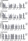
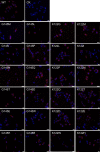
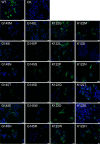
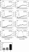
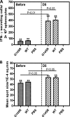
A variety of amino acid substitutions, such as K122I and G145R, have been identified around or within the a determinant of hepatitis B surface antigen (HBsAg), impair HBsAg secretion and antibody binding, and may be responsible for immune escape in patients. In this study, we examined how different substitutions at amino acid positions 122 and 145 of HBsAg influence HBsAg expression, secretion, and recognition by anti-HBs antibodies. The results showed that the hydrophobicity, the presence of the phenyl group, and the charges in the side chain of the amino acid residues at position 145 reduced HBsAg secretion and impaired reactivity with anti-HBs antibodies. Only the substitution K122I at position 122 affected HBsAg secretion and recognition by anti-HBs antibodies. Genetic immunization in mice demonstrated that the priming of anti-HBs antibody response was strongly impaired by the substitutions K122I, G145R, and others, like G145I, G145W, and G145E. Mice preimmunized with wild-type HBsAg (wtHBsAg) or variant HBsAg (vtHBsAg) were challenged by hydrodynamic injection (HI) with a replication-competent hepatitis B virus (HBV) clone. HBsAg persisted in peripheral blood for at least 3 days after HI in mice preimmunized with vtHBsAg but was undetectable in mice preimmunized with wtHBsAg, indicating that vtHBsAgs fail to induce proper immune responses for efficient HBsAg clearance. In conclusion, the biochemical properties of amino acid residues at positions 122 and 145 of HBsAg have a major effect on antigenicity and immunogenicity. In addition, the presence of proper anti-HBs antibodies is indispensable for the neutralization and clearance of HBsAg during HBV infection.
Figures



Similar articles
-
Biological significance of amino acid substitutions in hepatitis B surface antigen (HBsAg) for glycosylation, secretion, antigenicity and immunogenicity of HBsAg and hepatitis B virus replication.J Gen Virol. 2010 Feb;91(Pt 2):483-92. doi: 10.1099/vir.0.012740-0. Epub 2009 Oct 7. J Gen Virol. 2010. PMID: 19812261
-
Amino acid substitutions Q129N and T131N/M133T in hepatitis B surface antigen (HBsAg) interfere with the immunogenicity of the corresponding HBsAg or viral replication ability.Virus Res. 2018 Sep 15;257:33-39. doi: 10.1016/j.virusres.2018.08.019. Epub 2018 Sep 1. Virus Res. 2018. PMID: 30179704
-
The amino Acid residues at positions 120 to 123 are crucial for the antigenicity of hepatitis B surface antigen.J Clin Microbiol. 2007 Sep;45(9):2971-8. doi: 10.1128/JCM.00508-07. Epub 2007 Jul 3. J Clin Microbiol. 2007. PMID: 17609325 Free PMC article.
-
Vaccine- and hepatitis B immune globulin-induced escape mutations of hepatitis B virus surface antigen.J Biomed Sci. 2001 May-Jun;8(3):237-47. doi: 10.1007/BF02256597. J Biomed Sci. 2001. PMID: 11385295 Review.
-
Exchanges in the 'a' determinant of the hepatitis B virus surface antigen revisited.Virology. 2024 Nov;599:110184. doi: 10.1016/j.virol.2024.110184. Epub 2024 Jul 30. Virology. 2024. PMID: 39127000 Review.
Cited by
-
Characterization of Novel Hepatitis B Virus PreS/S-Gene Mutations in a Patient with Occult Hepatitis B Virus Infection.PLoS One. 2016 May 16;11(5):e0155654. doi: 10.1371/journal.pone.0155654. eCollection 2016. PLoS One. 2016. PMID: 27182775 Free PMC article.
-
Detection of S-HBsAg Mutations in Patients with Hematologic Malignancies.Diagnostics (Basel). 2021 May 27;11(6):969. doi: 10.3390/diagnostics11060969. Diagnostics (Basel). 2021. PMID: 34072185 Free PMC article.
-
Characterization of hepatitis B virus in Amerindian children and mothers from Amazonas State, Colombia.PLoS One. 2017 Oct 10;12(10):e0181643. doi: 10.1371/journal.pone.0181643. eCollection 2017. PLoS One. 2017. PMID: 29016603 Free PMC article.
-
Inducible interleukin 32 (IL-32) exerts extensive antiviral function via selective stimulation of interferon λ1 (IFN-λ1).J Biol Chem. 2013 Jul 19;288(29):20927-20941. doi: 10.1074/jbc.M112.440115. Epub 2013 May 31. J Biol Chem. 2013. PMID: 23729669 Free PMC article.
-
Persistence of the recombinant genomes of woodchuck hepatitis virus in the mouse model.PLoS One. 2015 May 5;10(5):e0125658. doi: 10.1371/journal.pone.0125658. eCollection 2015. PLoS One. 2015. PMID: 25942393 Free PMC article.
References
-
- Carman WF. 1997. The clinical significance of surface antigen variants of hepatitis B virus. J. Viral Hepat. 4(Suppl. 1):11–20 - PubMed
-
- Carman WF, et al. 1990. Vaccine-induced escape mutant of hepatitis B virus. Lancet 336:325–329 - PubMed
-
- Chiou HL, Lee TS, Kuo J, Mau YC, Ho MS. 1997. Altered antigenicity of ‘a’ determinant variants of hepatitis B virus. J. Gen. Virol. 78:2639–2645 - PubMed
-
- Cooreman MP, Leroux-Roels G, Paulij WP. 2001. Vaccine- and hepatitis B immune globulin-induced escape mutations of hepatitis B virus surface antigen. J. Biomed. Sci. 8:237–247 - PubMed
Publication types
MeSH terms
Substances
LinkOut - more resources
Full Text Sources

