. 2011 Dec 23;35(6):972-85.
doi: 10.1016/j.immuni.2011.09.019. Epub 2011 Dec 15.

Th17 cells are long lived and retain a stem cell-like molecular signature

Affiliations

Th17 cells are long lived and retain a stem cell-like molecular signature

Pawel Muranski et al. Immunity. .

Abstract

Th17 cells have been described as short lived, but this view is at odds with their capacity to trigger protracted damage to normal and transformed tissues. We report that Th17 cells, despite displaying low expression of CD27 and other phenotypic markers of terminal differentiation, efficiently eradicated tumors and caused autoimmunity, were long lived, and maintained a core molecular signature resembling early memory CD8(+) cells with stem cell-like properties. In addition, we found that Th17 cells had high expression of Tcf7, a direct target of the Wnt and β-catenin signaling axis, and accumulated β-catenin, a feature observed in stem cells. In vivo, Th17 cells gave rise to Th1-like effector cell progeny and also self-renewed and persisted as IL-17A-secreting cells. Multipotency was required for Th17 cell-mediated tumor eradication because effector cells deficient in IFN-γ or IL-17A had impaired activity. Thus, Th17 cells are not always short lived and are a less-differentiated subset capable of superior persistence and functionality.


Figures

Figure 1
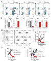
Figure 1. Th17-polarized cells effectively reject large tumors despite phenotypic features suggesting terminal differentiation, but must acquire type 1-like features in vivo.
(A) Representative intracellular staining demonstrating production of IFN-γ IL-17A, IL-17F and Rorγt by Th1 and Th17-polarized TRP-1 cells generated in vitro following 4 hour re-stimulation with phorbol myrystate acetate (PMA) and ionomycin in the presence of brefeldin A. Resting polarized cells were used as negative control. (B) Expression of Tbx21 and Rorc in in vitro polarized Th1 and Th17 cells was measured by quantitative reverse transcription polymerase chain reaction (qRT-PCR) and expressed relative to β-actin (Actb). (C) Secretion of indicated cytokines upon peptide stimulation was measured by ELISA. Error bars for (B) and (C) represent SEM (n=3). (D) Contour plots show expression of CD44 vs. CD62L and CD4 vs. CD45RB and CD27 on the surface of Th1 and Th17-polarized TRP-1 cells at day 7 following stimulation. (E) A total of 1×10⁶ TRP-1 cells polarized into Th1 and Th17 subsets were injected into sublethally (5 Gy) irradiated C57/B6 mice bearing B16.10 tumors. Tumor growth was measured serially and is represented as tumor area. Error bars represent SEM (n=5–7). (F)(G) Relative contribution of IFN-γ and T-bet to the antitumor activity of TCR-engineered Th17-polarized cells. CD4+ cells from WT, Ifng−/− and Tbx21−/− mice were polarized as indicated and transduced with a retroviral vector encoding the TRP-1 TCR. A total of 1.2×10⁶ transduced cells were transferred as in (E). Recipients were treated with exogenous IL-2 and rVV TRP-1 vaccine. Error bars represent SEM (n=4–7). All tumor treatment experiments were reproduced at least twice. See also Figure S1.
Figure 2
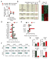
Figure 2. Adoptively transferred Th17 cells evolve in vivo into a distinct Th1-like population
TRP-1 CD4+ T cells (Thy1.2+) were cultured under Th1 and Th17 conditions for 7 days. Serial gene expression profiling was performed on cells preserved on the day of adoptive transfer (day 0) and on highly purified cells recovered on day 5 and 15 from spleens and lymph nodes 5Gy irradiated B6.PL (Thy1.1+) animals (2–4 independent replicates/condition for days 5 and 15) (A) Number of genes 2-fold up- or down-regulated (FDR<0.05 for days 5 and 15) between Th17 and Th1-polarized populations on indicated days is shown in their resting state and upon 2 hour re-stimulation in vitro. (B) Fold differences in expression of indicated genes as measured by microarray on indicated days, red font indicates a fold difference <2 on day 15. (C) Heat map of the genes differentially expressed by Th17 and Th1-derived cells recovered ex vivo on day 15 (FC>2, FDR<0.05). (D) Relative log2 expression of selected differentially expressed genes encoding phenotypic markers of terminal differentiation and end-effector function in Th17 and Th1-derived cells recovered on day 15 after adoptive transfer. (E) Relative log2 expression of selected differentially-expressed genes correlating with in vivo functionality in Th17 and Th1-derived cells recovered at day 15. Error bars for (D) and (E) represent SEM (n=2–4). List of differentially expressed genes at day 0, 5 and 15 is shown in File S1. See also Figure S2. (F)(G) Expression of CD62L, CD27, KLRG1 and CCR7 on surviving Th17 or Th1-derived cells. Persisting Thy1.2+ cells in spleens of B6.PL (Thy1.1+) mice treated with Th17 or Th1 polarized cells were analyzed by flow cytometry on day 18 after adoptive transfer. Representative dot plots for indicated phenotypic markers are shown. Bar graphs depict frequency of positive cells in Th1 and Th17 populations. Error bars represent SEM (n=3), (*=p<0.05, **=p<0.01, ***=p<0.001).
Figure 3
Figure 3. Th17-polarized cells maintain the core molecular signature of a less differentiated T cell subset with stem-cell-like features
Gene sets representing molecular signatures of early memory cells or late memory cells were generated from data set obtained following multiple rounds of in vivo stimulation of CD8+ T cells (Wirth et al., 2010). (A)(B) Gene Set Enrichment Analysis (GSEA) enrichment plot of early memory signature set (NES=1.5557741, p<0.0001, FDR q=0.07) and late memory signature set (NES=1.346455 p<0.0001, FDR q=0.15) of the running enrichment score (ES) and positions of gene set members on the rank ordered list based on fold-change of Th17 versus Th1 gene expression profiles obtained on day 15 after adoptive transfer are shown. Full list of genes contained in the early and late memory signature sets with GSEA core enrichment heat maps for each analysis are shown in Figure S3A and S3B. (C) Relative log2 expression of Tcf7, Lef1 and β-catenin (Ctnnb1) on indicated days following adoptive cell transfer. (D) Presence of stable β-catenin was assessed by Western blot on day 7 in purified CD4+ T cells from WT (C57BL/6) donors (left panel) stimulated with aCD3/aCD28 and in fresh TRP-1 cells (right panel) peptide stimulated under Th1 and Th17-polarizing conditions. (E)(F) WT CD4+ cells were cultured as in (D) and harvested at indicated times, while TRP-1 CD4+ T cells were grown for 7 days. Expression of Tcf7 was measured by qRT-PCR and shown as ratio vs. β-actin (Actb). Error bars represent SEM (n=3); (*=p<0.05, **=p<0.01, ***=p<0.001). See also Figure S3C-S3D.
Figure 4
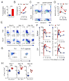
Figure 4. Th17-polarized TRP-1 cells expand upon secondary stimulation in vitro and are long-lived in vivo
(A) 3H-Thymidine incorporation in re-stimulated Th1 and Th17 cells was measured upon overnight incubation using the equal numbers of viable cells. Error bars represent SEM (n=3). (B) Absolute number of viable cells in Th1 and Th17 cultures re-stimulated with anti-CD3/anti-CD28 was calculated serially at indicated days and represented as fold expansion. (C) Annexin V expression was measured in Th1 and Th17 cells following overnight in vitro stimulation with anti-CD3 and anti-CD28 and compared to resting cells. (D) Expression of mRNA encoding Bcl2 was measured serially by qRT-PCR following in vitro re-stimulation of Th1 and Th17-polarized cells. Expression relative to β-actin (Actb) is shown. Error bars represent SEM (n=3). Experiments (A, B and C) are representative and were replicated at least twice. (*=p<0.05, **=p<0.01, ***=p<0.001). (E) A total of 1×10⁶ Th1 and Th17 TRP-1 cells (Thy1.2+) were adoptively transferred into sublethally (5Gy) irradiated B6.PL mice (Thy1.1+). Spleens and inguinal lymph nodes were harvested and analyzed by flow cytometry at indicated timepoints for the presence of Thy1.2+Vβ14+ cells. Representative contour plots illustrate the frequency of Thy1.2+ Vβ14+ cells in spleens of animals injected with Th1 and Th17 polarized TRP-1 cells at the indicated days post transfer. (F) Mean frequency (left panels) and total number (right panels) of persisting Thy1.2+ Vβ14+ cells from animals treated with Th1 or Th17 cells recovered at indicated days from spleens and inguinal lymph nodes are shown. Error bars represent SEM (n=3–6). (G) (H) Purification of in vitro polarized Th17 TRP-1 cells was performed using IL-17A capture reagent and flow cytometric sorting of labeled cells. Dot plots show result of FACS-sorting, as compared with similarly labeled Th1 cells used as a negative control. A total of 4×10⁵ Th1 and purified Th17 cells were adoptively transferred into B6.PL mice as described in (E). Frequency of Thy1.2+CD4+Vβ14 T cells was measured in blood of recipient animals at indicated time point. Error bars represent SEM (n=5–7, *=p<0.05, **=p<0.01, ***=p<0.001). See also Figure S4.
Figure 5
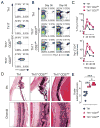
Figure 5. Expression level of CD27 on Th17 cells does not affect their ability to survive and function in vivo
In vitro cultured Th1 and Th17 TRP-1 cells were labeled for the expression of Thy1.2 and CD27. CD27hi and CD27lo Th17 subsets sorted using Th1 population as a positive control. 1×10⁶ cells were adoptively transferred into 5Gy irradiated B6.PL (Thy1.1) recipient animals. (A) Expression of CD27 is shown on Th17 cells before and after sorting into CD27hi and CD27lo subsets. (B) Dot plots demonstrate frequency of surviving CD4+Thy1.2+ TRP-1 cells in spleens of recipient animals treated with indicated cell subset at day 36 and 58 after adoptive cell transfer. (C) Mean frequency (upper panel) and total number (lower panel) of CD4+Thy1.2+ cells recovered at indicated time points from the spleens of recipient animals (n=3–4 for Th1 and Th17 CD27lo cells, n=2–3 for Th17 CD27hi cells). (D) Eyes from animals analyzed on day 58 in (B) and (C) were H&E stained and examined for evidence of autoimmunity in cornea, iris, photoreceptors and choroid. Representative examples for indicated experimental groups are shown. (E) Degree of self-tissue destruction in eyes harvested from indicated experimental groups on day 58 after adoptive cell transfer was evaluated using masked autoimmunity score as described in materials and methods. Error bars represent SEM (n=4–8); (*=p<0.05, **=p<0.01, ***=p<0.001)
Figure 6
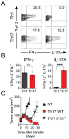
Figure 6. IL-17A-secreting cells maintain the ability to self-renew upon transfer into a highly activating environment and contribute to the effective rejection of tumor
A total of 1×10⁶ in vitro polarized Th17 and Th1 TRP-1 cells (Thy1.2+) were adoptively transferred into sublethally irradiated (5Gy) B6.PL (Thy1.1+) mice. On day 36 following transfer spleens from recipient mice were harvested and stimulated for 6 hours with TRP-1 peptide in presence of brefeldin A. (A) Representative dot plots show intracellular IFN-γ and IL-17A in Thy1.2+ cells in the indicated experimental groups. (B) Bar graphs show mean frequency of IFN-γ- and IL-17A-secreting cells among surviving Thy1.2+ Th1- and Th17-derived cells 36 days after adoptive transfer. (C) Purified CD4+ cells from C57/B6 and IL-17A−/− donors were stimulated under Th17 conditions and transduced with retroviral vector encoding for TRP-1 TCR. A total of 1.2×10⁶ cells were adoptively transferred into 5Gy irradiated C57BL/6 mice. Tumor growth was measured serially. Error bars represent SEM (n=5–6); (*=p<0.05, **=p<0.01, ***=p<0.001).

Comment in

Similar articles

Cited by

References

    1. Ballesteros-Tato A, Randall Troy D. Memory: The Incomplete Unhappening of Differentiation. Immunity. 2011;35:496–498. - PubMed
    1. Bending D, De La Pena H, Veldhoen M, Phillips JM, Uyttenhove C, Stockinger B, Cooke A. Highly purified Th17 cells from BDC2.5NOD mice convert into Th1-like cells in NOD/SCID recipient mice. The Journal of clinical investigation 2009 - PMC - PubMed
    1. Benoist C, Germain RN, Mathis D. A plaidoyer for ‘systems immunology’. Immunological reviews. 2006;210:229–234. - PubMed
    1. Berger C, Jensen MC, Lansdorp PM, Gough M, Elliott C, Riddell SR. Adoptive transfer of effector CD8(+) T cells derived from central memory cells establishes persistent T cell memory in primates. J Clin Invest. 2008;118:294–305. - PMC - PubMed
    1. Beyersdorf N, Ding X, Tietze JK, Hanke T. Characterization of mouse CD4 T cell subsets defined by expression of KLRG1. European journal of immunology. 2007;37:3445–3454. - PubMed

MeSH terms

Substances

Associated data