. 2011 Nov 22;4(200):ra79.
doi: 10.1126/scisignal.2002223.

Ric-8 proteins are molecular chaperones that direct nascent G protein α subunit membrane association

Affiliations

Ric-8 proteins are molecular chaperones that direct nascent G protein α subunit membrane association

Meital Gabay et al. Sci Signal. .

Abstract

Ric-8A (resistance to inhibitors of cholinesterase 8A) and Ric-8B are guanine nucleotide exchange factors that enhance different heterotrimeric guanine nucleotide-binding protein (G protein) signaling pathways by unknown mechanisms. Because transgenic disruption of Ric-8A or Ric-8B in mice caused early embryonic lethality, we derived viable Ric-8A- or Ric-8B-deleted embryonic stem (ES) cell lines from blastocysts of these mice. We observed pleiotropic G protein signaling defects in Ric-8A(-/-) ES cells, which resulted from reduced steady-state amounts of Gα(i), Gα(q), and Gα(13) proteins to <5% of those of wild-type cells. The amounts of Gα(s) and total Gβ protein were partially reduced in Ric-8A(-/-) cells compared to those in wild-type cells, and only the amount of Gα(s) was reduced substantially in Ric-8B(-/-) cells. The abundances of mRNAs encoding the G protein α subunits were largely unchanged by loss of Ric-8A or Ric-8B. The plasma membrane residence of G proteins persisted in the absence of Ric-8 but was markedly reduced compared to that in wild-type cells. Endogenous Gα(i) and Gα(q) were efficiently translated in Ric-8A(-/-) cells but integrated into endomembranes poorly; however, the reduced amounts of G protein α subunits that reached the membrane still bound to nascent Gβγ. Finally, Gα(i), Gα(q), and Gβ(1) proteins exhibited accelerated rates of degradation in Ric-8A(-/-) cells compared to those in wild-type cells. Together, these data suggest that Ric-8 proteins are molecular chaperones required for the initial association of nascent Gα subunits with cellular membranes.


Conflict of interest statement

Competing interests: A.J.M., D.M.V., and G.D.Y. are employees of Regeneron Pharmaceuticals Inc. Use of the Ric-8A– or Ric-8B–null mice requires a materials transfer agreement (MTA) from Regeneron Pharmaceuticals Inc.

Figures

Fig. 1
Fig. 1
Mouse ES cell lines derived from blastocysts deficient in Ric-8A or Ric-8B are viable. (A) Wild-type (C57BL/6J), Ric-8A+/−, Ric-8A−/−, Ric-8B+/−, and Ric-8B−/− mES cell lines were derived from blastocysts and cultured on mitotically inactivated wild-type MEFs (columns 1 and 2) and adapted to grow in feeder-free culture (columns 3 to 6). β-Galactosidase activity from LacZ transgenes under control of the endogenous Ric-8A or Ric-8B promoters was measured in each cell line (column 2). mES colonies were costained with DAPI (column 4) and with antibody against Oct3/4 (column 5) to show homogeneous Oct3/4 abundance (Merge, column 6). Scale bar, 200 μm. (B) Quantitative Western blotting analysis of the amounts of Ric-8A, Ric-8B, actin, Oct3/4, and GAPDH was performed with whole-cell lysates prepared from two independently derived wild-type (WT), Ric-8A−/−, and Ric-8B−/− mES cell lines. The amounts of the indicated proteins relative to that of GAPDH were quantified by pixel densitometry analysis with Adobe Photoshop 4.0 (Adobe Systems Inc.). Error bars represent the means ± SEM of three experiments with averaged results from two independent cell lines of each genotype.
Fig. 2
Fig. 2
Steady-state amounts of heterotrimeric G proteins are selectively abrogated in Ric-8A– or Ric-8B–deleted mES cells. (A) Whole-cell lysates (WCL) or crude membrane fractions (from 150,000g centrifugation, P150) prepared from two independently derived wild-type, Ric-8A−/−, and Ric-8B−/− mES cell lines were subjected to quantitative Western blotting for actin, α-tubulin, and the G protein subunits Gαi1/2, Gαo, Gαq, Gα13, Gαs, and total Gβ1. The amounts of the indicated proteins in membrane fractions compared to that of α-tubulin were quantified by pixel densitometry, as described earlier. Error bars represent the means ± SEM obtained from duplicate cell lines of each genotype. (B) Stable expression of the Ric-8A cDNA as a trans-gene (Tg) in a Ric-8A−/− cell line restored G protein and actin protein amounts as determined by quantitative Western blotting. Error bars represent the means ± SEM. (C) Relative amounts of Gαi1, Gα13, Gαs, Gαq, Gβ1, Ric-8A, and Ric-8B mRNAs in Ric-8A−/− and Ric-8B−/− cell lines were measured by quantitative real-time PCR. Error bars represent the means ± SEM obtained from duplicate, independently derived cell lines of each genotype.
Fig. 3
Fig. 3
G proteins are localized less efficiently at the plasma membrane in Ric-8A– or Ric-8B–deleted mES cells than in wild-type cells. Wild-type mES cells, Ric-8A−/− mES cells stably expressing pcDNA or Ric-8A cDNA transgene (Tg), and Ric-8B−/− mES cells were transiently transfected with plasmids encoding GFP-Gαi1, GFP-Gαq, and YFP-GαsL or the split YFP constructs, YFP–N′-Gβ1 and YFP–C′-Gγ7. Cells were visualized by confocal fluorescence microscopy. The signal gain in these images was uniformly enhanced to observe the weak plasma membrane–localized fluorescence of G proteins in the respective Ric-8−/− cells. The enhancement resulted in some saturated cell images, such that the apparent differences in G protein plasma membrane signal intensity are not linear. Scale bar, 20 μm.
Fig. 4
Fig. 4
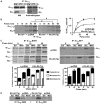
Nascent Gαi1/2 and Gαq are defective in their initial association with the membrane in Ric-8A–deleted mES cells. (A) Nucleotide-dependent coimmunoprecipitation of endogenous Gαi1/2 and Gβ subunits. WCLs from wild-type mES cells that were metabolically labeled with [35S]methionine and [35S]cysteine for 1 hour and were supplemented with GDP or Mg+2–GTP-γ-S were subjected to immunoprecipitation (IP) with antiserum against Gαi1/2. The immunoprecipitated samples were resolved by SDS-PAGE and subjected to autoradiography or were analyzed by Western blotting (WB) for Gαi1/2 and Gβ1. (B) Nascent Gαi1/2 synthesis and association with nascent Gβ subunits over a 1-hour time course of metabolic pulse labeling. Ric-8A−/− mES cells expressing pcDNA or Ric-8A (Tg) were metabolically labeled for the indicated times. WCLs were prepared and subjected to quantitative immunoprecipitation with antiserum against Gαi1/2. Immunoprecipitated Gαi1/2 and associated Gβ subunits were resolved by SDS-PAGE and visualized by autoradiography. Molar ratios of the amount of Gβ to that of Gαi1/2 were quantified by pixel densitometry, as described earlier, after factoring in a 22:18 ratio of methionines and cysteines within the two processed proteins. Error bars represent the means ± SEM of three independent experiments. *P < 0.0085; #P < 0.0091. (C) Subcellular fractionation of newly synthesized Gαq and Gαi1/2 with associated nascent Gβ subunits was measured after metabolic pulse (10 min) and chase (to 30 min) labeling of Ric-8A−/− cells expressing sham cDNA or Ric-8A cDNA. Labeled proteins were immunoprecipitated from cytosolic (Sol) and crude membrane (Mem) fractions and subjected to quantitative autoradiography. Error bars on the accompanying histographs represent the means ± SEM of three independent experiments. (D) Metabolic 2-hour pulse-labeling and subcellular fractionation analysis of Gαq in cytosolic (Sol) and crude membrane (Mem) fractions was measured by immunoprecipitation with antisera against Gαq (Z808 and Z811). The panel shown is representative of three independent experiments.
Fig. 5
Fig. 5
Newly synthesized Gα subunits and Gβ1 are degraded rapidly in Ric-8A−/− mES cells. Ric-8A−/− cells stably expressing Ric-8A (Tg) or pcDNA were pulse-labeled with [35S]methionine and [35S]cysteine for 1 hour and chased in the presence of unlabeled amino acids for 0 to 24 hours. (A to D) Endogenous Gαi1/2 (A), Gαq (B), Gβ1 (C), and γ-tubulin (D) and stably expressed Ric-8A were immunoprecipitated at each time point, resolved by SDS-PAGE, and visualized by autoradiography. The percentages of proteins that remained with time were quantified by pixel densitometry analysis as described earlier. The data were fit to monoexponential turnover functions to calculate half-lives with GraphPad Prism 5.0 software. Error bars represent the means ± SEM of three independent experiments.
Fig. 6
Fig. 6
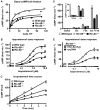
Basal and β-AR–stimulated AC activity is attenuated in Ric-8A– and Ric-8B–deleted mES cells. (A) Wild-type, Ric-8A−/−, and Ric-8B−/− (1 × 10⁴ to 75 × 104) mES cell lines were seeded in duplicate 96-well plates 24 hours before basal cAMP measurements were made. Cell counts were determined from one parallel plate at the time of basal cAMP measurement. Results are the average of two independent experiments from duplicate cell lines of each genotype. (B) Accumulated cAMP was measured after treatment of cells with isoproterenol (0 to 10 μM) for 2 min at 22°C. Error bars represent the means ± SEM for three independent experiments with duplicate cell lines of each genotype. (C) Accumulated cAMP was measured after cell treatment with a saturating concentration of isoproterenol (10 μM) for the indicated times at 22°C. Error bars represent the means ± SEM for three independent experiments with duplicate cell lines of each genotype. (D) Basal and accumulated cAMP amounts were measured after cell treatment with vehicle (basal), isoproterenol (100 nM, EC20), forskolin (50 μM, EC20), or isoproterenol and forskolin (EC20s) for 5 min at 22°C. Error bars represent the means ± SEM for three independent experiments with duplicate cell lines of each genotype. (E) Induction of cAMP accumulation by isoproterenol (0 to 10 μM) was measured in Ric-8A−/− mES cell lines stably transfected with Ric-8A cDNA (Tg) or sham vector. Error bars represent the means ± SEM for three independent experiments.
Fig. 7
Fig. 7
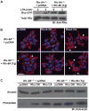
Basal and LPA receptor–stimulated Rho signaling is attenuated in Ric-8A–deleted mES cells. (A) Ric-8A−/− cells stably expressing Ric-8A or sham cDNA were not stimulated or were stimulated with LPA (10 μM) for 3 min. Rho-GTP was isolated from equivalent amounts of cell lysates in GST-Rhotekin pull-down assays. Relative amounts of total Rho and Rho-GTP were visualized by Western blotting with an antibody against Rho (Anti-Rho). Data are representative of two independent experiments. (B) Ric-8A−/− cells stably expressing Ric-8A (Tg) or sham cDNA were transiently transfected with control plasmid (pcDNA), plasmid encoding dominant-negative (DN) RhoA T19N, or plasmid encoding constitutively active (CA) RhoA G14V. After 48 hours, cells were costained with DAPI and rhodamine-phalloidin to visualize nuclei and F-actin structures, respectively. Arrowheads denote actin filopodia. Scale bar, 20 μm. Data are representative of three or more independent experiments. (C) Ric-8A−/− cells stably expressing Ric-8A (Tg) or sham cDNA were transiently transfected with pcDNA, RhoA (DN), or RhoA (CA) and fractionated by centrifugation of postnuclear supernatants at 150,000g. Relative amounts of G-actin and F-actin were visualized by Western blotting analysis with antibody against actin (Anti-actin). Data are representative of two independent experiments.
Fig. 8
Fig. 8
Proposed model for the action of Ric-8 during G protein biosynthesis. Gα subunits are translated on free, cytosolic ribosomes. (i) The emerging Gα chain may be bound by Ric-8 and cellular chaperones during initial protein folding, or (ii) it may be folded as part of a ternary complex withCCTand Ric-8. (iii) The nucleotide-free Ric-8–Gα complex then translocates to a putative, ER membrane–specific docking factor (X) that may also serve as the cellular activator (iv) of Ric-8 nucleotide exchange stimulatory activity, enabling production of (v) dissociated Ric-8 and membrane-bound Gα-GTP. (vi) Gα hydrolyzes GTP, perhaps through the aid of organelle-specific RGS GAPs (GTPase-activating proteins). The resultant Gα-GDP binds to Gβγ before the newly formed G protein heterotrimer traffics to the plasma membrane.

Similar articles

Cited by

References

    1. Chan P, Gabay M, Wright FA, Tall GG. Ric-8B is a GTP-dependent G protein αs guanine nucleotide exchange factor. J Biol Chem. 2011;286:19932–19942. - PMC - PubMed
    1. Tall GG, Krumins AM, Gilman AG. Mammalian Ric-8A (synembryn) is a heterotrimeric Gα protein guanine nucleotide exchange factor. J Biol Chem. 2003;278:8356–8362. - PubMed
    1. Miller KG, Alfonso A, Nguyen M, Crowell JA, Johnson CD, Rand JB. A genetic selection for Caenorhabditis elegans synaptic transmission mutants. Proc Natl Acad Sci U S A. 1996;93:12593–12598. - PMC - PubMed
    1. Miller KG, Emerson MD, McManus JR, Rand JB. RIC-8 (synembryn): A novel conserved protein that is required for Gqα signaling in the C. elegans nervous system. Neuron. 2000;27:289–299. - PMC - PubMed
    1. Miller KG, Rand JB. A role for RIC-8 (synembryn) and GOA-1 (Goα) in regulating a subset of centrosome movements during early embryogenesis in Caenorhabditis elegans. Genetics. 2000;156:1649–1660. - PMC - PubMed

Publication types

MeSH terms

Substances

LinkOut - more resources