. 2011 Nov 11;147(4):773-88.
doi: 10.1016/j.cell.2011.08.054.

ncRNA- and Pc2 methylation-dependent gene relocation between nuclear structures mediates gene activation programs

Affiliations

ncRNA- and Pc2 methylation-dependent gene relocation between nuclear structures mediates gene activation programs

Liuqing Yang et al. Cell. .

Erratum in

  • Cell. 2013 Oct 10;155(2):478

Abstract

Although eukaryotic nuclei contain distinct architectural structures associated with noncoding RNAs (ncRNAs), their potential relationship to regulated transcriptional programs remains poorly understood. Here, we report that methylation/demethylation of Polycomb 2 protein (Pc2) controls relocation of growth-control genes between Polycomb bodies (PcGs) and interchromatin granules (ICGs) in response to growth signals. This movement is the consequence of binding of methylated and unmethylated Pc2 to the ncRNAs TUG1 and MALAT1/NEAT2, located in PcGs and ICGs, respectively. These ncRNAs mediate assembly of multiple corepressors/coactivators and can serve to switch mark recognition by "readers" of the histone code. Additionally, binding of NEAT2 to unmethylated Pc2 promotes E2F1 SUMOylation, leading to activation of the growth-control gene program. These observations delineate a molecular pathway linking the actions of subnuclear structure-specific ncRNAs and nonhistone protein methylation to relocation of transcription units in the three-dimensional space of the nucleus, thus achieving coordinated gene expression programs.


Figures

Figure 1
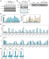
Figure 1. Suv39h1 Methylates Pc2 at K191
(A) Pc2 is a non-histone substrate for Suv39h1. In vitro methylation assay was performed by incubating GST-Suv39h1 with recombinant His-Pc2, His-E2F1, histone H3, E1, E2, and SUMO1 in the presence of [3H]-SAM. (B) The enzymatic activity of Suv39h1 is required for Pc2 methylation. In vitro methylation assay was performed by incubating GST-Suv39h1 (wt, H320R or H324L mutant) with His-Pc2 in the presence of [3H]-SAM. (C) Suv39h1 methylates Pc2 at lysine 191 in vivo. FLAG-Pc2 immunoprecipitated from 293T cells (indicated by red arrow in left panel) was subjected to MOLDI-TOF analysis (right panel) and modified peptides are indicated by the red box. (D) Suv39h1 methylates Pc2 at K191 in vitro. In vitro methylation assay was performed by incubating GST-Suv39h1 with wt Pc2 or K191R mutant in the presence of [3H]-SAM. (E) Validation of Pc2K191me2 and Pc2K191 antibodies. Methylated and unmethylated His-Pc2 was subjected to autoradiography (top panel) or immunoblotting with antibodies indicated (middle panels). (F and G) Suv39h1 methylates Pc2 at K191 in vivo. Cell lysates from HeLa cells transfected with siRNAs (F) or expression vectors (G) as indicated were immunoblotted with antibodies indicated. For A, B and D, the reaction products were separated by SDS-PAGE followed by Coomassie blue staining (CBB) (left panel) or autoradiography (right panel). See also Figure S1.
Figure 2
Figure 2. Growth Control Gene Activation Requires KDM4C-Mediated Pc2K191me2 Demethylation
(A) KDM4C demethylates Pc2 in vitro. Histone Demethylase assay was performed by incubating purified FLAG-histone demethylases with in vitro methylated His-Pc2. Changes in substrate methylation levels were analyzed by autoradiography (upper panel) and Pc2K191me2 antibodies (middle panel). Immunoblotting with antibodies against His tag was used to demonstrate equal loading (bottom panel). (B) Enzymatic activity of KDM4C is required for Pc2K191me2 demethylation. His-KDM4C wt or H190G/E192A mutant was incubated with in vitro methylated His-Pc2 in the presence or absence of Fe (II). The reactions were separated by SDS-PAGE followed by autoradiography (upper panel), immunoblotting with Pc2K191me2 antibodies (middle panel) or Coomassie blue staining (lower panel). (C) Serum-induced demethylation of Pc2K191me2 in vivo. HeLa cells were serum-starved followed by restimulation and the cell lysates were immunoprecipitated with Pc2K191 or Pc2K191me2 antibodies followed by immunoblotting with pan Pc2 antibodies. (D) Serum-induced demethylation of Pc2 on growth control gene promoters. HeLa cells were serum-starved followed by restimulation and ChIP analyses were performed using Pc2K191, Pc2K191me2 and pan Pc2 antibodies on indicated regions. For each primer pair, Pc2K191me2 (left panel) and Pc2K191 ChIP values (right panel) were normalized to the corresponding total pan Pc2 ChIP values. (E and F) Serum-induced KDM4C recruitment is responsible for Pc2K191me2 demethylation on growth control gene promoters. HeLa cells transfected with blank vector, FLAG-KDM4C wt or H190G/E192A mutant were serum-starved followed by restimulation and ChIP analyses using FLAG antibody (E) or Pc2K191me2 antibody (F) were performed on indicated regions. (G) KDM4C is required for serum-induced growth control gene expression. HeLa cells transfected with control or validated KDM4C siRNAs were serum-starved followed by restimulation and relative mRNA levels of indicated genes were determined by qRT-PCR. Mean±SEM, *p<0.05. See also Figure S2.
Figure 3
Figure 3. Pc2 Methylation/Demethylation Controls Mitogenic Signal-Induced Cell Proliferation
(A and B) Pc2 is essential for serum-induced growth control gene expression and cell proliferation. HeLa cells transfected with Pc2 siRNAs were serum-starved followed by restimulation and mRNA level of indicated genes (A) and cell proliferation (B) were analyzed, respectively. (C) Pc2K191 methylation is a key regulator for cell proliferation. HeLa cells transfected with Pc2 plasmids (wt or K191R mutant) were serum-starved followed by restimulation and cell proliferation were analyzed. (D and E) IMR-90 cells were serum-starved followed by restimulation and Pc2K191 methylation level (D) and cell proliferation (E) was analyzed by immunoblotting and flow cytometry, respectively. For B, C and E, cell proliferation was analyzed by monitoring the EDU incorporation into DNA synthesis in S-phase cells. Mean±SEM, *p<0.05 and **p<0.01. See also Figure S3.
Figure 4
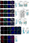
Figure 4. Transition of E2F1-regulated Genes from PcG Bodies to Interchromatin Granules following Serum Induction
(A–C) Methylated and unmethylated Pc2 exhibit distinct immunostaining patterns with respect to Bmi1 (A) or Ring1A (B) as a marker of PcG bodies, and SC35 (C) of interchromatin granules. (D and E) Serum-induced relocation of growth control gene locus between PcG bodies and interchromatin granules. HeLa cells were serum-starved followed by restimulation and Immuno-FISH analyses were performed using antibodies against Pc2K191me2 (D) or Ring1A (E) and FISH probes targeting RBL1 (D) or MCM3 (E). (F) Effect of knockdown of Pc2, Bmi1 and PHC1 on relocation of MCM3 locus between PcG bodies and interchromatin granules. HeLa cells microinjected with indicated siRNAs were serum-starved followed by restimulation and Immuno-FISH analyses were performed using antibodies and FISH probes as indicated. (G) Interaction between endogenous Pc2 and other core components of PRC1 complex demonstrated by co-immunoprecipitation of Pc2K191me2 with Bmi1, Ring1A and PHC1 in HeLa cell extracts. The input represents 10% of the protein amount used for immunoprecipitation. (H) KDM4C is required for serum-induced dissociation of MCM3 locus from PcG bodies. HeLa cells microinjected with control siRNA or validated siRNA against KDM4C were serum-starved followed by restimulation and Immuno-FISH analyses were performed using Ring1A antibodies and probes targeting MCM3 locus. (I) Demethylated Pc2 is required for relocation of MCM3 locus. HeLa cells microinjected with blank vector, Pc2 wt or K191R mutant were serum-starved followed by restimulation and Immuno-FISH analyses were performed using Ring1A, SC35 antibodies and probes targeting MCM3 locus. Statistical analyses on colocalization of MCM3 locus with Ring1A staining (left panel) and SC35 staining (right panel) were shown. For A, B, C, D and E, left panel shows representative images and right panel shows statistical analyses. Mean±SEM, *p<0.05 and **p<0.01. See also Figure S4.
Figure 5
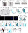
Figure 5. NcRNAs-Pc2 Interactions Underlie Relocation and Transcriptional Activation of Growth Control Genes
(A) Methylated and unmethylated Pc2 bind to RNAs. CLIP assay using Pc2K191me2 (left panel) or Pc2K191 (right panel) antibodies were performed and specific protein-RNA complexes (indicated by red box) were visualized by autoradiography. (B) Validation of interactions of TUG1 and NEAT2 with Pc2. In vitro transcribed biotinylated TUG1 RNA (left panel) or NEAT2 RNA (right panel) were incubated with HeLa nuclear extract. The elution of Monoavadin beads was subjected to immunoblotting with indicated antibodies. (C) The selective binding of Pc2 to TUG1 or NEAT2 depends on K191 methylation state. RNA gel shift assay was performed using synthesized TUG1, NEAT2 RNA and their corresponding DNA oligos and recombinant His-Pc2 with or without in vitro methylation. (D–F) TUG1 and NEAT2 function as modulators of Pc2 chromodomain for “reading” the histone code. MODified Histone Peptide ArrayR was incubated with Recombinant His-Pc2 chromodomain in the presence of yeast tRNA (D), in vitro transcribed TUG1 (117-3390) (E), or NEAT2 (2281-5611) (F) RNA fragments and subjected to immunoblotting with indicated antibodies. The Myc tag antibody was added to detect Myc tagged peptides on position P21 as positive control. The binding specificity calculated by Array Analyses Software based on two arrays (images shown in Figures S5F–S5H) was shown. (G) TUG1 is localized in PcG bodies. HeLa cells microinjected with molecular beacon probes targeting TUG1 were subjected to immunostaining with Pc2K191me2 antibodies. (H) TUG1 and NEAT2 are required for serum-induced relocation of the MCM3 locus. HeLa cells microinjected with validated siRNA targeting TUG1 or NEAT2 were serum-starved followed by restimulation and Immuno-FISH analyses were performed using Ring1A, SC35 antibodies and probes targeting MCM3 locus. Statistical analyses on colocalization of MCM3 loci with Ring1A staining (left panel) and SC35 staining (right panel) are shown. (I and J) Effect of TUG1 or NEAT2 knockdown on serum-induced growth control gene expression and cell proliferation. HeLa cells transfected with TUG1 or NEAT2 siRNA were serum-starved followed by restimulation and relative mRNA levels of indicated genes and cell proliferation were determined by qRT-PCR (I) and flow cytometry (J). (K) A list of TUG1 and NEAT2 ncRNA- associated proteins identified from two independent MS experiments. Mean±SEM, *p<0.05, **p<0.01 and ***p<0.001. See also Figure S5 and Table S1–S3.
Figure 6
Figure 6. Pc2-mediated E2F1 SUMOylation Is Required for Growth Control Gene Activation
(A) Serum-induced E2F1 SUMOylation. HeLa cells were serum-starved followed by restimulation and the cell lysates were immunoprecipitated with E2F1 antibodies under denaturing conditions followed by immunoblotting with antibodies indicated. (B) Pc2 promotes E2F1 SUMOylation in vivo. HeLa cells were transfected with indicated siRNAs/plasmids and the cell lysates were immunoprecipitated with E2F1 antibodies under denaturing conditions followed by immunoblotting with E2F1 (left panel) or Myc tag (right panel) antibodies. (C) Pc2 promotes E2F1 SUMOylation in vitro. Bacterially-expressed His-Pc2, His-E2F1 were incubated with recombinant E1, E2 and SUMO1 or SUMO1 mutant. The reactions were separated by SDS-PAGE followed by Coomassie blue staining (left panel), immunoblotting with E2F1 (middle panel) or SUMO1 (right panel) antibodies. (D and E) E2F1 is SUMOylated at lysine 266. HeLa cells were transfected with wt or single K mutated E2F1 (D) or K266R mutant (E) as indicated and the cell lysates were immunoprecipitated with FLAG antibodies under denaturing conditions followed by immunoblotting with E2F1 (upper panel) or Myc tag (bottom panel) antibodies (D) and E2F1 (left panel) or SUMO1 (right panel) antibodies (E). (F) E2F1 SUMOylation on growth control gene promoters. HeLa cells were transfected with indicated plasmids followed by two-step ChIP analyses using FLAG and SUMO1 antibodies on indicated regions. (G) K191 methylation inhibits Pc2-mediated E2F1 SUMOylation in vivo. HeLa cells were transfected with indicated plasmids and the cell lysates were immunoprecipitated with E2F1 antibodies under denaturing conditions followed by immunoblotting with E2F1 (left panel), Myc tag (middle panel) or SUMO1 (right panel) antibodies. (H) Pc2-mediated E2F1 SUMOylation is RNA dependent. Immunoprecipitates of Pc2K191me2 or Pc2K191 from HeLa cells (right panel) were incubated with His-tagged E2F1, recombinant E1, E2 and SUMO1 or SUMO1 mutant with or without RNase I treatment. The reaction products were subjected to immunoblotting using antibodies targeting E2F1 (left panel). (I) TUG1 and NEAT2 regulate Pc2-mediated E2F1 SUMOylation. Bacterially-expressed His-E2F1 were incubated with recombinant E1, E2, SUMO1 (wt or mutant), and His- Pc2 (unmethylated or methylated) in the presence of synthesized sense or antisense TUG1 or NEAT2 RNA oligos. The reactions were subjected to immunoblotting using antibodies against E2F1. See also Figure S6.
Figure 7
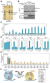
Figure 7. CDCA7L Interacts with SUMOylated E2F1 and Functions as a H2B Monoubiquitinase to Promote Growth Control Gene Activation
(A) CDCA7L interacts with K266 SUMOylated E2F1. FLAG-E2F1 immunoprecipitates from HeLa cells were separated by SDS-PAGE followed by silver staining. The band indicated was cut and subjected to MS analysis. (B) CDCA7L interacts with SUMOylated E2F1 via its SUMO-interacting motif. HeLa cells transfected with indicated plasmids were serum-starved followed by restimulation and the cells lysates were immunoprecipitated with E2F1 antibodies followed by immunoblotting with antibodies indicated. (C) Serum-induced recruitment of CDCA7L on growth control gene promoters. ChIP analyses were performed in serum-starved and restimulated HeLa cells using CDCA7L antibodies on indicated regions. (D) CDCA7L is required for growth control gene activation. HeLa cells transfected with shRNA against CDCA7L were serum-starved followed by restimulation and relative mRNA levels of indicated genes were determined by qRT-PCR. Mean±SEM, *p<0.05 and **p<0.01. (E) CDCA7L is required for serum-induced histone H2B ubiquitination on growth control gene promoters. HeLa cells transfected with shRNA against CDCA7L were serum-starved followed by restimulation and ChIP analyses were performed using antibodies targeting UbH2B and H2B on indicated regions. For each primer pair, UbH2B values were normalized to the corresponding total H2B ChIP. (F) Model: Interplay between ncRNAs and methylated vs. unmethylated Pc2 determines intranuclear localization of growth control genes in PcG bodies or interchromatin granules, which dictates gene repression vs. activation events. See also Figure S7.

Comment in

Similar articles

Cited by

References

    1. Aagaard L, Laible G, Selenko P, Schmid M, Dorn R, Schotta G, Kuhfittig S, Wolf A, Lebersorger A, Singh PB, et al. Functional mammalian homologues of the Drosophila PEV-modifier Su(var)3-9 encode centromere-associated proteins which complex with the heterochromatin component M31. EMBO J. 1999;18:1923–1938. - PMC - PubMed
    1. Ait-Si-Ali S, Guasconi V, Fritsch L, Yahi H, Sekhri R, Naguibneva I, Robin P, Cabon F, Polesskaya A, Harel-Bellan A. A Suv39h-dependent mechanism for silencing S-phase genes in differentiating but not in cycling cells. EMBO J. 2004;23:605–615. - PMC - PubMed
    1. Akhtar A, Zink D, Becker PB. Chromodomains are protein-RNA interaction modules. Nature. 2000;407:405–409. - PubMed
    1. Bagchi S, Weinmann R, Raychaudhuri P. The retinoblastoma protein copurifies with E2F-I, an E1A-regulated inhibitor of the transcription factor E2F. Cell. 1991;65:1063–1072. - PubMed
    1. Bernstein E, Duncan EM, Masui O, Gil J, Heard E, Allis CD. Mouse polycomb proteins bind differentially to methylated histone H3 and RNA and are enriched in facultative heterochromatin. Mol Cell Biol. 2006;26:2560–2569. - PMC - PubMed

Publication types

MeSH terms