

Top 10 plant viruses in molecular plant pathology
- PMID: 22017770
- PMCID: PMC6640423
- DOI: 10.1111/j.1364-3703.2011.00752.x
Top 10 plant viruses in molecular plant pathology
Abstract
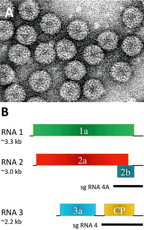
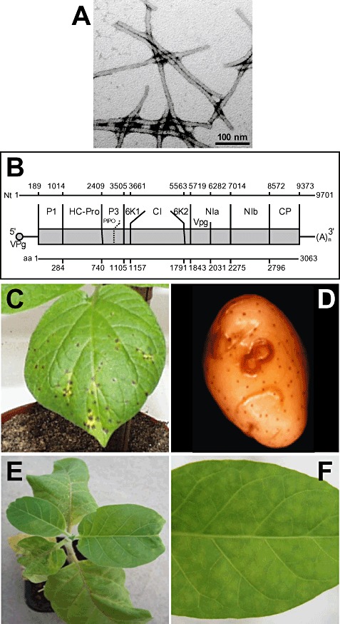
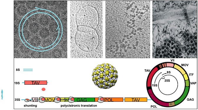
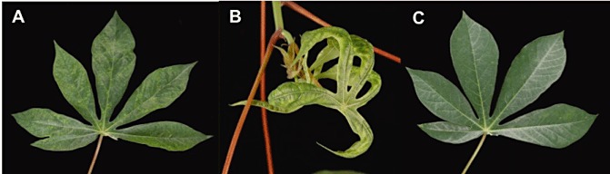
Many scientists, if not all, feel that their particular plant virus should appear in any list of the most important plant viruses. However, to our knowledge, no such list exists. The aim of this review was to survey all plant virologists with an association with Molecular Plant Pathology and ask them to nominate which plant viruses they would place in a 'Top 10' based on scientific/economic importance. The survey generated more than 250 votes from the international community, and allowed the generation of a Top 10 plant virus list for Molecular Plant Pathology. The Top 10 list includes, in rank order, (1) Tobacco mosaic virus, (2) Tomato spotted wilt virus, (3) Tomato yellow leaf curl virus, (4) Cucumber mosaic virus, (5) Potato virus Y, (6) Cauliflower mosaic virus, (7) African cassava mosaic virus, (8) Plum pox virus, (9) Brome mosaic virus and (10) Potato virus X, with honourable mentions for viruses just missing out on the Top 10, including Citrus tristeza virus, Barley yellow dwarf virus, Potato leafroll virus and Tomato bushy stunt virus. This review article presents a short review on each virus of the Top 10 list and its importance, with the intent of initiating discussion and debate amongst the plant virology community, as well as laying down a benchmark, as it will be interesting to see in future years how perceptions change and which viruses enter and leave the Top 10.
© 2011 The Authors. Molecular Plant Pathology © 2011 BSPP and Blackwell Publishing Ltd.
Figures


Similar articles
-
Top 10 plant pathogenic bacteria in molecular plant pathology.Mol Plant Pathol. 2012 Aug;13(6):614-29. doi: 10.1111/j.1364-3703.2012.00804.x. Epub 2012 Jun 5. Mol Plant Pathol. 2012. PMID: 22672649 Free PMC article. Review.
-
The Top 10 fungal pathogens in molecular plant pathology.Mol Plant Pathol. 2012 May;13(4):414-30. doi: 10.1111/j.1364-3703.2011.00783.x. Mol Plant Pathol. 2012. PMID: 22471698 Free PMC article. Review.
-
Oligonucleotide microarray-based detection and identification of 10 major tomato viruses.J Virol Methods. 2010 Sep;168(1-2):133-40. doi: 10.1016/j.jviromet.2010.05.003. Epub 2010 May 12. J Virol Methods. 2010. PMID: 20470828
-
Status of tobacco viruses in Serbia and molecular characterization of tomato spotted wilt virus isolates.Acta Virol. 2011;55(4):337-47. doi: 10.4149/av_2011_04_337. Acta Virol. 2011. PMID: 22149499
-
Evidence for plant viruses in the region of Argentina Islands, Antarctica.FEMS Microbiol Ecol. 2007 Feb;59(2):409-17. doi: 10.1111/j.1574-6941.2006.00242.x. FEMS Microbiol Ecol. 2007. PMID: 17328120
Cited by
-
Geminivirus protein structure and function.Mol Plant Pathol. 2013 Aug;14(6):635-49. doi: 10.1111/mpp.12032. Epub 2013 Apr 25. Mol Plant Pathol. 2013. PMID: 23615043 Free PMC article. Review.
-
The Tomato yellow leaf curl virus V2 protein forms aggregates depending on the cytoskeleton integrity and binds viral genomic DNA.Sci Rep. 2015 May 5;5:9967. doi: 10.1038/srep09967. Sci Rep. 2015. PMID: 25940862 Free PMC article.
-
Tomato yellow leaf curl virus: No evidence for replication in the insect vector Bemisia tabaci.Sci Rep. 2016 Aug 1;6:30942. doi: 10.1038/srep30942. Sci Rep. 2016. PMID: 27476582 Free PMC article.
-
Identification of new sources of resistance to resistance-breaking isolates of tomato spotted wilt virus.Saudi J Biol Sci. 2021 May;28(5):3094-3099. doi: 10.1016/j.sjbs.2021.02.053. Epub 2021 Feb 21. Saudi J Biol Sci. 2021. PMID: 34025184 Free PMC article.
-
Rescue of tomato spotted wilt virus entirely from complementary DNA clones.Proc Natl Acad Sci U S A. 2020 Jan 14;117(2):1181-1190. doi: 10.1073/pnas.1910787117. Epub 2019 Dec 26. Proc Natl Acad Sci U S A. 2020. PMID: 31879355 Free PMC article.
References
-
- Abel, P.P. , Nelson, R.S. , De, B. , Hoffmann, N. , Rogers, S.G. , Fraley, R.T. and Beachy, R.N. (1986) Delay of disease development in transgenic plants that express the tobacco mosaic virus coat protein gene. Science, 232, 738–743. - PubMed
-
- Adams, M.J. , Antoniw, J.F. , Bar‐Joseph M., Brunt, A.A. , Candresse, T. , Foster, G.D. , Martelli, G.P. , Milne, R.G. , Zavriev, S.K. and Fauquet, C.M. (2004) The new plant virus family Flexiviridae and assessment of molecular criteria for species demarcation. Arch. Virol. 149, 1045–1060. - PubMed
-
- Adkins, S. (2000) Tomato spotted wilt virus—positive steps to negative success. Mol. Plant Pathol. 1, 151–157. - PubMed
Publication types
MeSH terms
LinkOut - more resources
Full Text Sources
Other Literature Sources
