. 2011 Oct 19:10:131.
doi: 10.1186/1476-4598-10-131.

Aurora-A inactivation causes mitotic spindle pole fragmentation by unbalancing microtubule-generated forces

Affiliations

Aurora-A inactivation causes mitotic spindle pole fragmentation by unbalancing microtubule-generated forces

Italia A Asteriti et al. Mol Cancer. .

Abstract

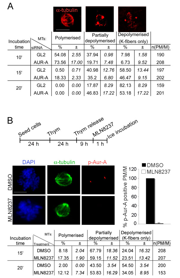
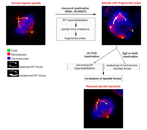
Background: Aurora-A is an oncogenic kinase playing well-documented roles in mitotic spindle organisation. We previously found that Aurora-A inactivation yields the formation of spindles with fragmented poles that can drive chromosome mis-segregation. Here we have addressed the mechanism through which Aurora-A activity regulates the structure and cohesion of spindle poles.

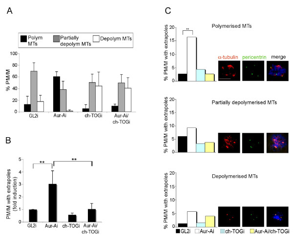
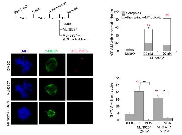
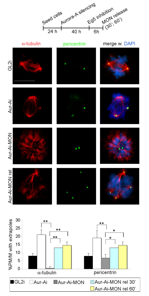
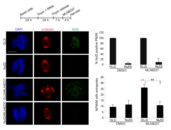
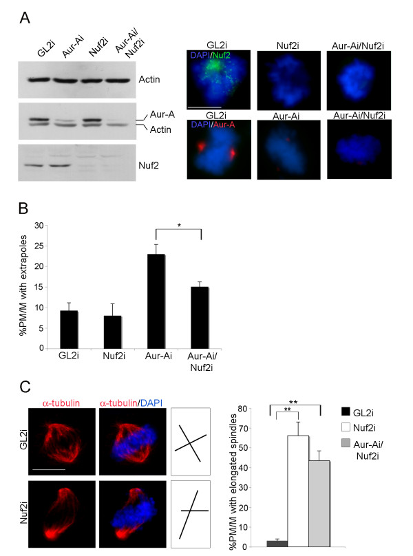
Results: We inactivated Aurora-A in human U2OS osteosarcoma cells either by RNA-interference-mediated silencing or treating cultures with the specific inhibitor MLN8237. We show that mitotic spindle pole fragmentation induced by Aurora-A inactivation is associated with microtubule hyperstabilisation. Silencing of the microtubule-stabilising factor ch-TOG prevents spindle pole fragmentation caused by inactivation of Aurora-A alone and concomitantly reduces the hyperstabilisation of microtubules. Furthermore, decreasing pole-directed spindle forces by inhibition of the Eg5 kinesin, or by destabilisation of microtubule-kinetochore attachments, also prevents pole fragmentation in Aurora-A-inactivated mitoses.

Conclusions: Our findings indicate that microtubule-generated forces are imbalanced in Aurora-A-defective cells and exert abnormal pressure at the level of spindle poles, ultimately causing their fragmentation. This study therefore highlights a novel role of the Aurora-A kinase in regulating the balance between microtubule forces during bipolar spindle assembly.


Figures

Figure 1
Figure 1
Aurora-A inactivation induces hyperstabilisation of spindle MTs. A. PM and M cells from control (GL2) and Aurora-Ai cultures incubated on ice for the indicated times were classified according to the status of MTs, examplified in the IF panels. The rightmost column indicates the number (n) of scored PM/M cells in 2 independent experiments; mean values (%) and s.d. (italics) are indicated. B. The protocol of MLN8237 treatment in synchronous cultures is shown (time intervals not represented to scale). Histograms show the percentage of PM and M cells displaying active pThr288-Aurora-A (p-Aur-A-positive) in control (DMSO) or MLN8237-treated cultures (200 counted cells per condition, 2 experiments; error bars represent s.d.); representative images are shown in the IF panels. In the table below, DMSO- or MLN8237-treated cells subjected to the ice-induced depolymerisation assay were classified as in A (2 independent experiments). Scale bars: 10 μm.
Figure 2
Figure 2
Aurora-A and ch-TOG modulate the stability of spindle MTs in opposite manners. A. Cells interfered (i) with the indicated siRNAs were incubated on ice for 15 minutes. Histograms represent the distribution of PM/M in the three MT categories identified in Figure 1A (at least 350 counted cells per condition, 2 experiments). B. PM/M with extrapoles were quantified (fold induction) in cultures interfered as indicated and incubated for 15 minutes of ice (at least 600 counted cells per condition, 2 experiments). C. Histograms represent the distribution of mitoses with fragmented spindle poles among the MT stability categories shown in Figure 1A (examples are shown in the IF panels). Around 700 PM/M were counted for each interference (2 experiments). Error bars represent s.d. **: p < 0.001, χ2 test. Scale bar: 10 μm.
Figure 3
Figure 3
Eg5 inhibition counteracts the induction of spindle pole fragmentation by Aurora-A inactivation. The protocol to inhibit Aurora-A by MLN8237 in cells progressing towards mitosis is depicted (time intervals not represented to scale). Control cultures were treated with solvent (DMSO) in the same time window. When indicated, MON was added 1 hour before harvesting. Note the absence of active phosphorylated (pThr288) Aurora-A (in red in IF panels) in cells treated with MLN8237. Upper histograms represent the percentage of all spindle and MT abnormalities in control and MLN8237-treated cultures (200 counted PM/M per condition in 2 experiments); the grey fraction of the histograms represents mitoses with spindle extrapoles, while other defects (monopolar or disorganised spindles, few and short MTs) are in white. Lower histograms and IF panels show that concomitant Eg5 inhibition by MON prevents MLN8237-induced spindle pole fragmentation (note the failure of centrosome migration reflecting Eg5 inactivation in lower IF panels). 200 PM/M per condition were counted in 2 experiments. Error bars represent s.d. **: p < 0.001, χ2 test. Red asterisks indicate significant differences with respect to DMSO controls, and black asterisks significant differences between Aurora-Ai mitoses with active or inactive Eg5. Scale bar: 10 μm.
Figure 4
Figure 4
Spindle pole fragmentation in Aurora-Ai mitoses depends on Eg5 activity. A schematisation of the protocol is shown (time intervals not represented to scale). IF panels show spindles displaying normal or fragmented poles in control and Aurora-Ai cells, respectively (first and second row); monopolar spindles in Aurora-Ai cells treated with MON (third row); spindles displaying pole fragmentation in Aurora-Ai cells after MON release (MON-rel; lower row). Histograms represent the percentage of PM/M displaying fragmented poles, as assessed by alpha-tubulin (left) and pericentrin (right) staining (200 to 400 counted cells per condition in 2-4 experiments; s.d are shown). *: p < 0.01, **: p < 0.001, χ2 test. Scale bar: 10 μm.
Figure 5
Figure 5
Nuf2 silencing prevents MLN8237-induced spindle pole fragmentation. A schematisation of the protocol for concomitant Nuf2 RNAi and Aurora-A inactivation (MLN8237) in cultures synchronously progressing from S-phase to mitosis is shown (time intervals not represented to scale). The efficiency of Nuf2 depletion following the RNAi protocol is quantified in the upper histograms (Nuf2 staining; 100-200 counted cells per condition in 2 experiments; see representative images in IF panels). Lower histograms (PM/M cells with fragmented poles by alpha-tubulin staining; 150-250 counted cells per condition, 2 experiments) and IF panels show that generation of extrapoles is reduced by Nuf2/Aurora-A co-inactivation compared to Aurora-A inactivation alone. **: p < 0.001, χ2 test. Red asterisks indicate significant differences with respect to DMSO controls, and black asterisks significant differences between MLN8237-treated mitoses with (GL2i) or without (Nuf2i) the Nuf2 protein. s.d are shown. Scale bar: 10 μm.
Figure 6
Figure 6
Spindle pole fragmentation in Aurora-Ai mitoses depends on active Nuf2. A. The efficiency of Aurora-A and Nuf2 depletion after RNAi was assessed by WB (left panels) and IF (right panels) analyses. B. Histograms represent the percentage of PM/M displaying fragmented spindle poles (alpha-tubulin staining) after transfection with the indicated siRNAs (at least 200 cells per condition, 2 experiments). C. Elongated spindles induced by Nuf2 RNAi are not rescued in Aur-Ai/Nuf2i co-inactivated cells (about 200 counted PM/M per condition in 2 experiments). Spindle axes are schematised on the right: the pole-to-pole axis is longer in Nuf2i cells compared to controls. Error bars denote s.d. *: p < 0.01, **: p < 0.001, χ2 test. Scale bar: 10μm.
Figure 7
Figure 7
Aurora-A modulates the balance of forces required for spindle pole integrity: a model. Left upper panel: in a normal mitosis balanced MT forces determine the formation of a symmetrical bipolar spindle. Arrows represent opposite-directed MT forces. Right upper panel: in Aurora-A-defective mitoses, the spindle displays fragmented poles. Lower panel: spindle pole disruption is prevented by either inactivating a MT stabiliser (ch-TOG), or weakening KT-generated (Nuf2 silencing), or inhibiting motor-associated (Eg5 inhibition) centrosome-directed MT forces in the absence of Aurora-A activity. The model suggests therefore that spindle poles fragment consequently to the imbalance in MT-generated forces in Aurora-A-defective mitoses.

Similar articles

Cited by

References

    1. Tanenbaum ME, Medema RH. Mechanisms of centrosome separation and bipolar spindle assembly. Dev Cell. 2010;19:797–806. doi: 10.1016/j.devcel.2010.11.011. - DOI - PubMed
    1. Walczak CE, Cai S, Khodjakov A. Mechanisms of chromosome behaviour during mitosis. Nat Rev Mol Cell Biol. 2010;11:91–102. doi: 10.1038/nrg2737. - DOI - PMC - PubMed
    1. Dumont S, Mitchison TJ. Force and length in the mitotic spindle. Curr Biol. 2009;19:R749–761. doi: 10.1016/j.cub.2009.07.028. - DOI - PMC - PubMed
    1. Gatlin JC, Bloom K. Microtubule motors in eukaryotic spindle assembly and maintenance. Semin Cell Dev Biol. 2010;21:248–254. doi: 10.1016/j.semcdb.2010.01.015. - DOI - PMC - PubMed
    1. Kapoor TM, Mayer TU, Coughlin ML, Mitchison TJ. Probing spindle assembly mechanisms with monastrol, a small molecule inhibitor of the mitotic kinesin Eg5. J Cell Biol. 2000;150:975–988. doi: 10.1083/jcb.150.5.975. - DOI - PMC - PubMed

Publication types

MeSH terms

LinkOut - more resources