. 2011 Sep;13(9):874-86.
doi: 10.1593/neo.11324.

Lymph node stromal cells enhance drug-resistant colon cancer cell tumor formation through SDF-1α/CXCR4 paracrine signaling

Affiliations

Lymph node stromal cells enhance drug-resistant colon cancer cell tumor formation through SDF-1α/CXCR4 paracrine signaling

David A Margolin et al. Neoplasia. 2011 Sep.

Abstract

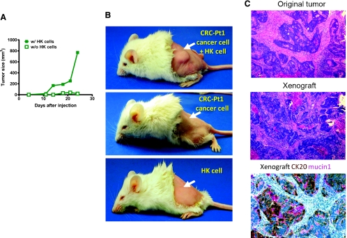
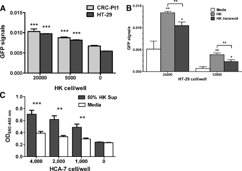
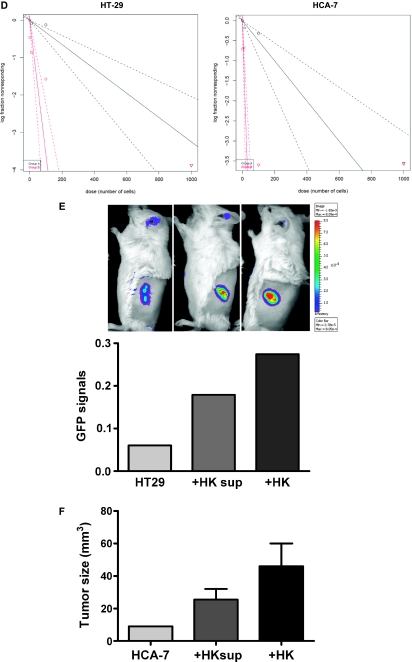
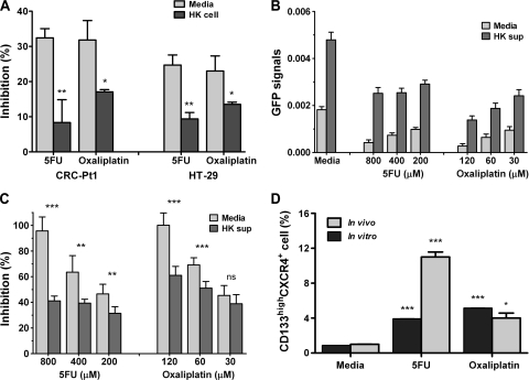
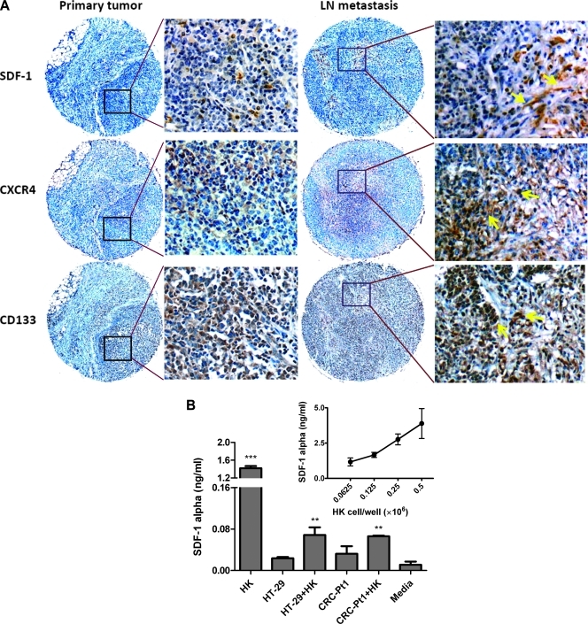
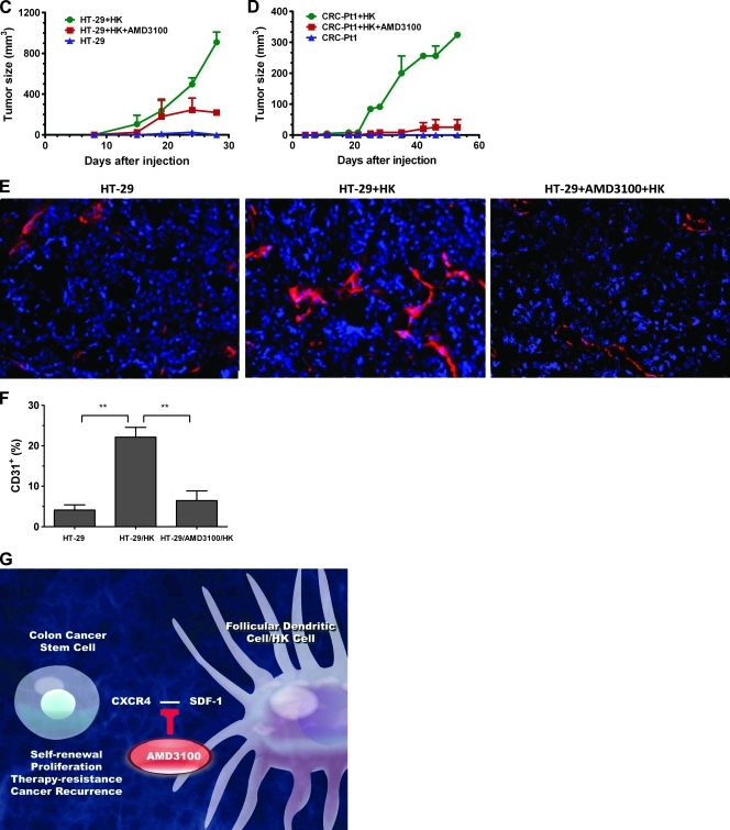
Colorectal cancer (CRC) is the third most common malignancy and the second leading cause of cancer-related deaths in America. Nearly two thirds of newly diagnosed CRC cases include lymph node (LN) involvement, and LN metastasis is one of the strongest negative prognostic factors for CRC. It is thought that CRC tumors contain a small population of drug-resistant CRC tumor-initiating cells (Co-TICs) that may be responsible for cancer recurrence. To evaluate the effects of the LN stromal cells on Co-TICs, we established a unique xenoplant model using CRC cells isolated by enzymatic digestion from consented patient specimens, HT-29 cells, HCA-7 cells, and LN stromal cell line HK cells. We found that HK cells and HK cell-conditioned media enhanced CRC tumor formation and tumor angiogenesis. Cells expressing CD133(+) and the stromal cell-derived factor 1α (SDF-1α) receptor CXCR4 were enriched in chemotherapeutic-resistant CRC cells. CD133(+)CXCR4(+) Co-TICs isolated from patient specimens are more tumorigenic than unsorted tumor cells. Furthermore, the inhibitors specific to HK cell-derived SDF-1α reduced tumor formation and tumor angiogenesis. Our results have demonstrated a role for Co-TICs in tumor growth and defined the influence of LN stromal cells on Co-TICs. We have identified a major Co-TIC/LN microenvironment-specific mechanism for CRC resistance to chemotherapeutic agents and established experimental platforms for both in vitro and in vivo testing, indicating that SDF-1α and its receptor, CXCR4, may be targets for clinical therapy.


Figures

Figure 1
Figure 1
FDC/HK cells support HT-29 cell and CRC patient cancer cell tumor formation in immunodeficient mice. (A) HT-29 cell tumor formation in nude mice. HT-29 cells (2 x 10⁵) were subcutaneously injected with or without HK cells (1 x 10⁶) into the posterior flank of nude mice. Graph shows tumor size measured with a caliper in three dimensions at the indicated days after cell injection. (B) Single-suspension cancer cells from patient 1 (2 x 10⁵) were subcutaneously injected with or without HK cells (1 x 10⁶) into the left posterior flank of NOD/SCID mice (arrows). Images show patient's colon cancer cells form tumor in the presence of HK cells (top), whereas no tumor was formed without HK cells (middle) or with HK cells alone (bottom). (C) Hematoxylin and eosin staining of paraffin-embedded original tumor and tumor from xenograft showing similarity in cancer morphology, and cytokeratin 20 (CK20) and Mucin 1 staining positive in the xenograft tumor. Images were captured by a deconvoluting microscope using SlideBook software. Original magnification, x400.
Figure 2
Figure 2
FDC/HK cells support CRC cell proliferation, sphere formation, and tumor formation. (A–C) CRC cell proliferation. GFP-tagged HT-29 (3000 cells/well) and CRC-Pt1 (1000 cells/well) cells were cocultured with indicated numbers of HK cells in 96 well plates for 72 hours (A). GFP-tagged HT-29 cells were cultured alone, with HK cells, or with HK cells separated by a transwell in 24-well plates for 48 hours (B). GFP signals were evaluated by IVIS Lumina Imaging System (A and B), and WST-1 assay for HCA-7 cells was measured by OD650−450 nm after 72 hours of incubation with or without 50% HK cell-conditioned medium (C). (D) A log-fraction plot of the limiting dilution model fitted to the data in Table W1. The slope of the line is the sphere formation cell fraction for HT-29 and HCA-7 cells. The dotted lines give the 95% confidence interval. Black line for SCM and red lines for 50% HK cell-conditioned medium. (E and F) HK cells and HK cell-conditioned medium support HT-29 and HCA-7 cell tumor formation in NOD/SCID mice. CRC tumor formation was indicated by GFP signal imaging (for GFP-HT-29 cells), and tumor size was measured by a caliper (for HCA-7 cells) in the absence or the presence of HK cell or HK cell-conditioned media.
Figure 2
Figure 2
FDC/HK cells support CRC cell proliferation, sphere formation, and tumor formation. (A–C) CRC cell proliferation. GFP-tagged HT-29 (3000 cells/well) and CRC-Pt1 (1000 cells/well) cells were cocultured with indicated numbers of HK cells in 96 well plates for 72 hours (A). GFP-tagged HT-29 cells were cultured alone, with HK cells, or with HK cells separated by a transwell in 24-well plates for 48 hours (B). GFP signals were evaluated by IVIS Lumina Imaging System (A and B), and WST-1 assay for HCA-7 cells was measured by OD650−450 nm after 72 hours of incubation with or without 50% HK cell-conditioned medium (C). (D) A log-fraction plot of the limiting dilution model fitted to the data in Table W1. The slope of the line is the sphere formation cell fraction for HT-29 and HCA-7 cells. The dotted lines give the 95% confidence interval. Black line for SCM and red lines for 50% HK cell-conditioned medium. (E and F) HK cells and HK cell-conditioned medium support HT-29 and HCA-7 cell tumor formation in NOD/SCID mice. CRC tumor formation was indicated by GFP signal imaging (for GFP-HT-29 cells), and tumor size was measured by a caliper (for HCA-7 cells) in the absence or the presence of HK cell or HK cell-conditioned media.
Figure 3
Figure 3
Detecting Co-TICs in CRC. (A) Paraffin-embedded tumor tissue sections from CRC patients 1 and 4 were double stained for CD133 (brown) and CXCR4 (pink) and control Ab staining. Arrows show double-positive cells. Hematoxylin was used as a counterstain. Original magnification, x400. (B) Frozen tumor tissue sections from CRC patients 5 and 8 were double stained for CD133 (red) and CXCR4 (green, arrows). DAPI was used as a nuclear counterstain. Original magnification, x400. (C) Double staining of CD133 and CXCR4 on patient's normal colon cells or CRC patient cells (left panels) and HT-29 cells or HCA-7 cells (right panels). Numbers indicate percentage of cells in quadrants. Isotype control mouse IgGs (mIgG) were used for background-level staining.
Figure 4
Figure 4
FDC/HK cells support chemoresistant colon cancer cells in vitro and in vivo. (A) Suboptimal doses of 5-FU (400 µM) and oxaliplatin (60 µM) were added to the coculture of GFP-tagged CRC cells with HK cells in 96-well plates for 72 hours. Percentage inhibition of cell growth by GFP signal levels was calculated against that of untreated cells. (B–C) GFP-tagged HT-29 cells were cultured with or without 50% HK cell-conditioned media and then treated with 5-FU and oxaliplatin for 72 hours. GFP signals (B) and percentage inhibition of viable cells by GFP signal levels (C) are shown. (D) CD133+CXCR4+ cells are enriched in chemoresistant HT-29 cells. HT-29 cells after 72 hours of chemotherapy in A were collected. Tumor-bearing mice were treated with control and chemotherapeutic drugs for 2 weeks as described in Results. Enzyme-digested single-cell suspension tumor cells were collected. Both cells were stained with CD133 and CXCR4 and analysis by FACS similar to Figure 3C. Data are presented as mean ± SD. Data show percentage of double-positive cells.
Figure 5
Figure 5
FDC/HK cells support Co-TIC by providing SDF-1α and enhance host angiogenesis. (A) Immunohistochemistry staining of colon cancer original tumor, LN metastasis paraffin slides for SDF-1α, CXCR4, and CD133 expression. Arrows show Ab staining-positive cells. (B) SDF-1α protein levels in 72 hours of culture supernatants of HK cells and/or colon cancer cells were measured by specific ELISA (R&D Systems). Insert shows SDF-1α levels in HK cell dose titration. (C and D) HK cells promote HT-29 (C) and CRC-Pt1 (D) cells to form tumors in nude and NOD/SCID mice, respectively. HT-29 or CRC-Pt1 cells (1 x 10⁵) with or without HK cells (1 x 10⁶) were subcutaneously injected into the posterior flank of immunodeficient mice. In some experiments, cancer cells were pretreated with AMD3100. Tumor sizes (mm3) were measured at the indicated time points. (E) Immunohistochemistry staining of frozen slides from tumors formed by HT-29 cells with or without HK cells using antimouse CD31 Abs (red). DAPI is used for nuclear staining (blue). Images were captured by a deconvoluting microscope using SlideBook software. Original magnification, x400. (F) Quantitative analysis of CD31-positive staining (% area) in tumors formed with HT-29 cells. The analysis was done with PhotoShop 7.0 software. (G) Cartoon showing in vitro and in vivo Co-TIC/stromal cell coculture models.
Figure 5
Figure 5
FDC/HK cells support Co-TIC by providing SDF-1α and enhance host angiogenesis. (A) Immunohistochemistry staining of colon cancer original tumor, LN metastasis paraffin slides for SDF-1α, CXCR4, and CD133 expression. Arrows show Ab staining-positive cells. (B) SDF-1α protein levels in 72 hours of culture supernatants of HK cells and/or colon cancer cells were measured by specific ELISA (R&D Systems). Insert shows SDF-1α levels in HK cell dose titration. (C and D) HK cells promote HT-29 (C) and CRC-Pt1 (D) cells to form tumors in nude and NOD/SCID mice, respectively. HT-29 or CRC-Pt1 cells (1 x 10⁵) with or without HK cells (1 x 10⁶) were subcutaneously injected into the posterior flank of immunodeficient mice. In some experiments, cancer cells were pretreated with AMD3100. Tumor sizes (mm3) were measured at the indicated time points. (E) Immunohistochemistry staining of frozen slides from tumors formed by HT-29 cells with or without HK cells using antimouse CD31 Abs (red). DAPI is used for nuclear staining (blue). Images were captured by a deconvoluting microscope using SlideBook software. Original magnification, x400. (F) Quantitative analysis of CD31-positive staining (% area) in tumors formed with HT-29 cells. The analysis was done with PhotoShop 7.0 software. (G) Cartoon showing in vitro and in vivo Co-TIC/stromal cell coculture models.

Similar articles

Cited by

References

    1. Jemal A, Siegel R, Xu J, Ward E. Cancer statistics, 2010. CA Cancer J Clin. 2010;60:277–300. - PubMed
    1. Reya T, Morrison SJ, Clarke MF, Weissman IL. Stem cells, cancer, and cancer stem cells. Nature. 2001;414:105–111. - PubMed
    1. Todaro M, Francipane MG, Medema JP, Stassi G. Colon cancer stem cells: promise of targeted therapy. Gastroenterology. 2010;138:2151–2162. - PubMed
    1. SEER Cancer Statistics Review. [April 18, 2011]. Available at: http://seer.cancer.gov/statfacts/html/colorect.html.
    1. Nakagawa H, Liyanarachchi S, Davuluri RV, Auer H, Martin EW, Jr, de la Chapelle A, Frankel WL. Role of cancer-associated stromal fibroblasts in metastatic colon cancer to the liver and their expression profiles. Oncogene. 2004;23:7366–7377. - PubMed

MeSH terms

LinkOut - more resources