Review
. 2011 Aug 23;30(18):3667-83.
doi: 10.1038/emboj.2011.307.

Deciphering the rules of programmed cell death to improve therapy of cancer and other diseases

Affiliations
Review

Deciphering the rules of programmed cell death to improve therapy of cancer and other diseases

Andreas Strasser et al. EMBO J. .

Abstract

Apoptosis, the major form of programmed cell death in metazoan organisms, plays critical roles in normal development, tissue homeostasis and immunity, and its disturbed regulation contributes to many pathological states, including cancer, autoimmunity, infection and degenerative disorders. In vertebrates, it can be triggered either by engagement of 'death receptors' of the tumour necrosis factor receptor family on the cell surface or by diverse intracellular signals that act upon the Bcl-2 protein family, which controls the integrity of the mitochondrial outer membrane through the complex interactions of family members. Both pathways lead to cellular demolition by dedicated proteases termed caspases. This review discusses the groundbreaking experiments from many laboratories that have clarified cell death regulation and galvanised efforts to translate this knowledge into novel therapeutic strategies for the treatment of malignant and perhaps certain autoimmune and infectious diseases.


Conflict of interest statement

The authors declare that their research at the Walter and Eliza Hall Institute includes a joint programme with Genentech Inc. and Abbott Labs to develop novel anti-cancer therapeutics.

Figures
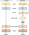
Figure 1
Figure 1
Comparison of the pathway of programmed cell death in C. elegans with the major one in vertebrates. The worm has a homologue of Bcl-2 (CED-9), of its BH3-only apoptosis inducers (EGL-1), of the caspase-activator Apaf-1 (CED-4) and of the proteolytic caspases (CED-3). However, a striking difference is that CED-9 directly binds and inhibits CED-4, until CED-4 is displaced by EGL-1, whereas vertebrate Bcl-2 does not bind to Apaf-1 but instead prevents the activation of its pro-apoptotic siblings Bax and Bak, thereby preventing their permeabilisation of the MOM and release of cytochrome c (cyt c), an essential cofactor for Apaf-1.
Figure 2
Figure 2
The two major pathways to caspase activation in vertebrates: the death receptor or extrinsic pathway, engaged by the indicated members of the TNF receptor family on the cell surface, and the Bcl-2-regulated mitochondrial or intrinsic pathway. The death receptors lead, via the adaptor FADD (with help by TRADD in certain death receptors), to activation of caspase-8, which then activates the effector caspases-3, -6 and -7. Caspase-8 also processes the BH3-only protein Bid, and the truncated Bid (tBid) can then activate the Bcl-2-regulated pathway. Upon MOMP, that pathway leads to effector caspase activation via Apaf-1 and caspase-9. The cytosolic E3 ubiquitin ligase XIAP can inhibit caspases-3 and -7 (and perhaps caspase-9), but that inhibition is blocked by SMAC/DIABLO when it is released from mitochondria. E3 ubiquitin ligases cIAP1 and cIAP2 work instead in part by preventing formation of the pro-apoptotic signalling complex from TNF-R1 and by regulating pro-survival NF-κB survival pathways.
Figure 3
Figure 3
(A) The three functional subgroups of the Bcl-2 family. Sequences most homologous to Bcl-2 (BH domains) and α-helical regions are indicated. The BH3-only proteins share sequence homology only within the BH3 domain, which mediates association between family members. Bid has a defined 3D structure but the others are relatively unstructured. The pro-survival group, which shares four regions of sequence homology, includes Bcl-B in humans but its mouse homologue (Boo) appears to be inactive due to a mutation of essential residues in BH1. The pro-apoptotic Bax/Bak group, which includes the little studied Bok, is remarkably similar in sequence and structure to the pro-survival group, including an α-helix resembling BH4 near the N-terminus (Kvansakul et al, 2008). Most family members contain a C-terminal hydrophobic trans-membrane (TM) region, which mediates their targeting and anchoring to the MOM and/or the ER, either constitutively (e.g. Bcl-2 or Bak) or after an apoptotic stimulus (e.g. Bcl-xL or Bax). (B,C) Predominant interactions within the Bcl-2 family, including those of BH3-only proteins with their pro-survival relatives (B) and the major interactions of the latter with Bax and Bak (C).
Figure 4
Figure 4
Models for how the BH3-only proteins activate Bax and Bak. (A) In the direct activation model, the activator BH3-only proteins (Bim, tBid and probably Puma), via their BH3 domain (red triangle), can directly engage and activate Bax (or Bak), but in healthy cells the pro-survival family members (‘Bcl-2 et al’) prevent this by sequestering the BH3-only activators. After an apoptotic signal, the sensitiser BH3-only proteins (e.g. Bad, Noxa, Bik) free the activators to target Bax or Bak. Inactive cytosolic Bax has its BH3 domain buried and its C-terminal hydrophobic helix (α9) lies in its surface groove, but during activation that helix is freed and can target Bax to the membrane. (B) In the indirect activation model, the BH3-only proteins need only target their pro-survival relatives, which primarily prevent activation of Bax and Bak by sequestering any Bax or Bak that becomes active (‘primed’) by exposure of its BH3 domain (red triangle). (C) The priming–capture–displacement model proposed here incorporates features of both direct and indirect activation. In it, any Bax or Bak that becomes primed, either spontaneously or by BH3-only proteins or other potential signals (e.g. phosphorylation), is immediately captured by a pro-survival relative, until the primed Bax or Bak is displaced by further activation of BH3-only proteins (e.g. Bad or Bim). The displaced Bax or Bak can then form dimers and higher oligomers (see Figure 5).
Figure 5
Figure 5
Models for the oligomerisation of Bax and Bak. (A) In this dimer multimerisation model, upon activation by an apoptotic signal, Bak (or Bax) first extrudes its BH3 domain, which allows it to engage another ‘primed’ Bak (or Bax) molecule to form a ‘symmetric’ (face-to-face) dimer (Dewson et al, 2008). These dimers then multimerise by a different interface, such as one between α6 helices, to form the large oligomers that provoke MOMP, allowing cytochrome c (cyt c) to escape to the cytosol (Dewson et al, 2009). (B) In an alternative model (Shamas-Din et al, 2011), prompted by the proposal that certain BH3 domains can engage a novel ‘rear’ site on Bax, ‘primed’ Bak (or Bax) can engage the proposed rear site of another activated molecule to form an ‘asymmetric (face-to-back) dimer’, which could be extended by monomer recruitment. In this model, insertion of the α5–α6 hairpin as well as α9 is depicted. That insertion might also feature in the model shown in (A).

Similar articles

Cited by

References

    1. Adams JM, Cory S (2007) The Bcl-2 apoptotic switch in cancer development and therapy. Oncogene 26: 1324–1337 - PMC - PubMed
    1. Akhtar RS, Klocke BJ, Strasser A, Roth KA (2008) Loss of BH3-only protein Bim inhibits apoptosis of hemopoietic cells in the fetal liver and male germ cells but not neuronal cells in bcl-x-deficient mice. J Histochem Cytochem 56: 921–927 - PMC - PubMed
    1. Allan JM, Travis LB (2005) Mechanisms of therapy-related carcinogenesis. Nat Rev Cancer 5: 943–955 - PubMed
    1. Arbour N, Vanderluit JL, Le Grand JN, Jahani-Asl A, Ruzhynsky VA, Cheung EC, Kelly MA, MacKenzie AE, Park DS, Opferman JT, Slack RS (2008) Mcl-1 is a key regulator of apoptosis during CNS development and after DNA damage. J Neurosci 28: 6068–6078 - PMC - PubMed
    1. Bardwell PD, Gu J, McCarthy D, Wallace C, Bryant S, Goess C, Mathieu S, Grinnell C, Erickson J, Rosenberg SH, Schwartz AJ, Hugunin M, Tarcsa E, Elmore SW, McRae B, Murtaza A, Wang LC, Ghayur T (2009) The Bcl-2 family antagonist ABT-737 significantly inhibits multiple animal models of autoimmunity. J Immunol 182: 7482–7489 - PubMed

Publication types