

Critical role for NLRP3 in necrotic death triggered by Mycobacterium tuberculosis
- PMID: 21740493
- PMCID: PMC3257557
- DOI: 10.1111/j.1462-5822.2011.01625.x
Critical role for NLRP3 in necrotic death triggered by Mycobacterium tuberculosis
Abstract
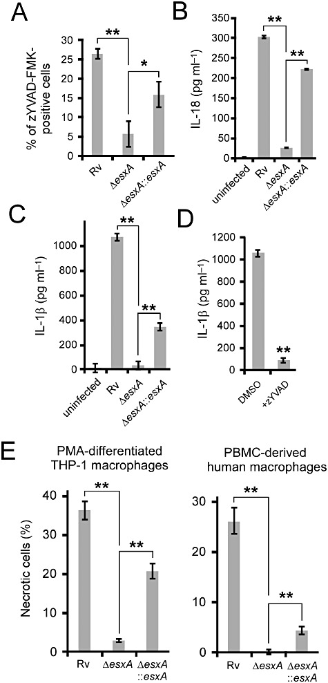
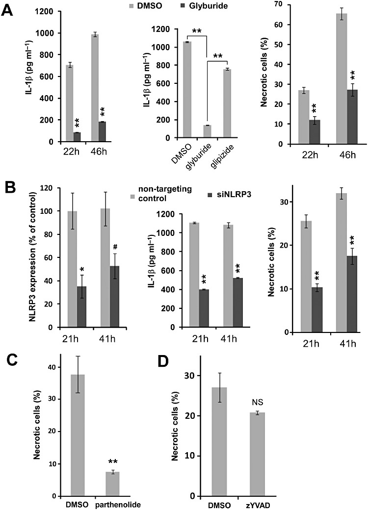
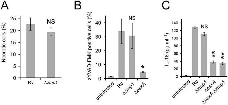
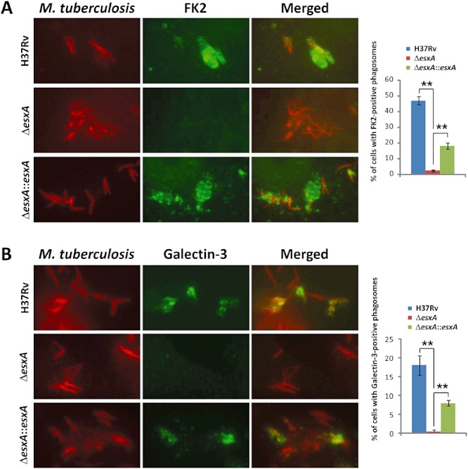
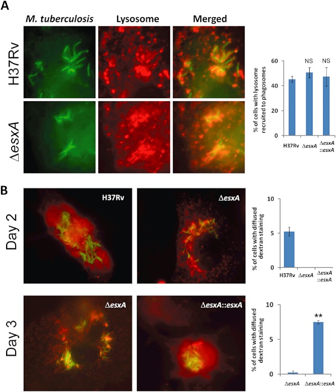
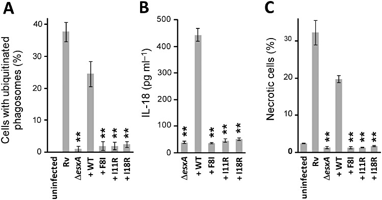
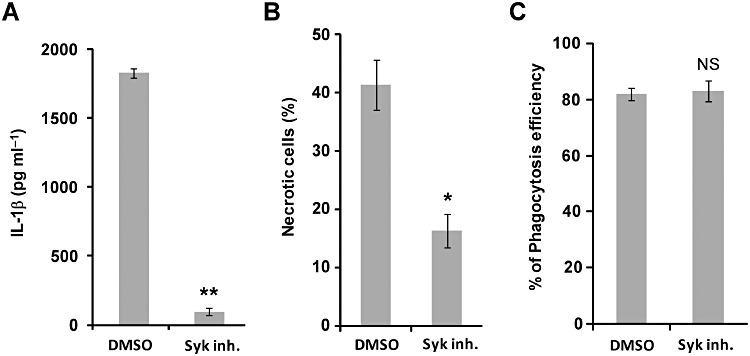
Induction of necrotic death in macrophages is a primary virulence determinant of Mycobacterium tuberculosis. The ESX-1 secretion system and its substrate ESAT-6 are required for M. tuberculosis to induce necrosis, but host factors that mediate the ESAT-6-promoted necrosis remain unknown. Here we report that ESAT-6-promoted necrotic death in THP-1 human macrophages is dependent on the NLRP3 inflammasome, as shown by RNA interference and pharmacological inhibitions. Phagosomes containing ESAT-6-expressing M. tuberculosis recruit markers previously associated with damaged phagosomal membrane, such as galectin-3 and ubiquitinated protein aggregates. In addition, ESAT-6 promoted lysosomal permeabilization by M. tuberculosis. ESAT-6 mutants defective for ubiquitination were unable to trigger NLRP3 activation and necrotic death. Furthermore, Syk tyrosine kinase, recently implicated in NLRP3 activation during fungal and malarial infections, was necessary for mediating the ESAT-6-promoted necrosis and NLRP3 activation. Our results thus link phagosomal damage and Syk activity to NLRP3-mediated necrotic death triggered by M. tuberculosis ESAT-6 during infection.
© 2011 Blackwell Publishing Ltd.
Figures
Similar articles
-
Mycobacterium tuberculosis protein ESAT-6 is a potent activator of the NLRP3/ASC inflammasome.Cell Microbiol. 2010 Aug;12(8):1046-63. doi: 10.1111/j.1462-5822.2010.01450.x. Epub 2010 Feb 9. Cell Microbiol. 2010. PMID: 20148899
-
Human macrophages infected with a high burden of ESAT-6-expressing M. tuberculosis undergo caspase-1- and cathepsin B-independent necrosis.PLoS One. 2011;6(5):e20302. doi: 10.1371/journal.pone.0020302. Epub 2011 May 26. PLoS One. 2011. PMID: 21637850 Free PMC article.
-
The C terminus of the mycobacterium ESX-1 secretion system substrate ESAT-6 is required for phagosomal membrane damage and virulence.Proc Natl Acad Sci U S A. 2022 Mar 15;119(11):e2122161119. doi: 10.1073/pnas.2122161119. Epub 2022 Mar 10. Proc Natl Acad Sci U S A. 2022. PMID: 35271388 Free PMC article.
-
ESAT-6 proteins: protective antigens and virulence factors?Trends Microbiol. 2004 Nov;12(11):500-8. doi: 10.1016/j.tim.2004.09.007. Trends Microbiol. 2004. PMID: 15488391 Review.
-
ESAT-6 a Major Virulence Factor of Mycobacterium tuberculosis.Biomolecules. 2023 Jun 9;13(6):968. doi: 10.3390/biom13060968. Biomolecules. 2023. PMID: 37371548 Free PMC article. Review.
Cited by
-
COVID-19: captures iron and generates reactive oxygen species to damage the human immune system.Autoimmunity. 2021 Jun;54(4):213-224. doi: 10.1080/08916934.2021.1913581. Epub 2021 Apr 26. Autoimmunity. 2021. PMID: 33899609 Free PMC article.
-
Vitamin D modulates human macrophage response to Mycobacterium tuberculosis DNA.Tuberculosis (Edinb). 2019 May;116S:S131-S137. doi: 10.1016/j.tube.2019.04.021. Epub 2019 May 3. Tuberculosis (Edinb). 2019. PMID: 31085128 Free PMC article.
-
Expression of NLRP3 inflammasome in leprosy indicates immune evasion of Mycobacterium leprae.Mem Inst Oswaldo Cruz. 2020 Feb 27;115:e190324. doi: 10.1590/0074-02760190324. eCollection 2020. Mem Inst Oswaldo Cruz. 2020. PMID: 32130367 Free PMC article.
-
MyD88 in Mycobacterium tuberculosis infection.Med Microbiol Immunol. 2017 Jun;206(3):187-193. doi: 10.1007/s00430-017-0495-0. Epub 2017 Feb 20. Med Microbiol Immunol. 2017. PMID: 28220253 Review.
-
Vaccines Against Tuberculosis: Problems and Prospects (Review).Appl Biochem Microbiol. 2020;56(5):497-504. doi: 10.1134/S0003683820050129. Epub 2020 Sep 22. Appl Biochem Microbiol. 2020. PMID: 32981943 Free PMC article.
References
-
- Bardarov S, Bardarov S, Jr, Pavelka MS, Jr, Sambandamurthy V, Larsen M, Tufariello J, et al. Specialized transduction: an efficient method for generating marked and unmarked targeted gene disruptions in Mycobacterium tuberculosis, M. bovis BCG and M. smegmatis. Microbiology. 2002;148:3007–3017. - PubMed
-
- Brodin P, de Jonge MI, Majlessi L, Leclerc C, Nilges M, Cole ST, Brosch R. Functional analysis of early secreted antigenic target-6, the dominant T-cell antigen of Mycobacterium tuberculosis, reveals key residues involved in secretion, complex formation, virulence, and immunogenicity. J Biol Chem. 2005;280:33953–33959. - PubMed
-
- van Bruggen R, Koker M, Jansen M, van Houdt M, Roos D, Kuijpers T, van den Berg T. Human NLRP3 inflammasome activation is Nox1-4 independent. Blood. 2010;115:5398–5400. - PubMed
Publication types
MeSH terms
Substances
Grants and funding
LinkOut - more resources
Full Text Sources
Other Literature Sources
Miscellaneous
