. 2011 May;7(5):e1002054.
doi: 10.1371/journal.ppat.1002054. Epub 2011 May 19.

Reactive oxygen species hydrogen peroxide mediates Kaposi's sarcoma-associated herpesvirus reactivation from latency

Affiliations

Reactive oxygen species hydrogen peroxide mediates Kaposi's sarcoma-associated herpesvirus reactivation from latency

Fengchun Ye et al. PLoS Pathog. 2011 May.

Abstract

Kaposi's sarcoma-associated herpesvirus (KSHV) establishes a latent infection in the host following an acute infection. Reactivation from latency contributes to the development of KSHV-induced malignancies, which include Kaposi's sarcoma (KS), the most common cancer in untreated AIDS patients, primary effusion lymphoma and multicentric Castleman's disease. However, the physiological cues that trigger KSHV reactivation remain unclear. Here, we show that the reactive oxygen species (ROS) hydrogen peroxide (H₂O₂) induces KSHV reactivation from latency through both autocrine and paracrine signaling. Furthermore, KSHV spontaneous lytic replication, and KSHV reactivation from latency induced by oxidative stress, hypoxia, and proinflammatory and proangiogenic cytokines are mediated by H₂O₂. Mechanistically, H₂O₂ induction of KSHV reactivation depends on the activation of mitogen-activated protein kinase ERK1/2, JNK, and p38 pathways. Significantly, H₂O₂ scavengers N-acetyl-L-cysteine (NAC), catalase and glutathione inhibit KSHV lytic replication in culture. In a mouse model of KSHV-induced lymphoma, NAC effectively inhibits KSHV lytic replication and significantly prolongs the lifespan of the mice. These results directly relate KSHV reactivation to oxidative stress and inflammation, which are physiological hallmarks of KS patients. The discovery of this novel mechanism of KSHV reactivation indicates that antioxidants and anti-inflammation drugs could be promising preventive and therapeutic agents for effectively targeting KSHV replication and KSHV-related malignancies.


Conflict of interest statement

The authors have declared that no competing interests exist.

Figures
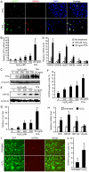
Figure 1
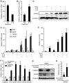
Figure 1. Exogenous H2O2 induces KSHV reactivation in PEL and endothelial cells.
(A) Cells undergoing lytic replication in both uninduced BCBL1 cells and BCBL1 cells induced for lytic replication with phorbol ester TPA for 48 h also had high level of intracellular H2O2. Lytic cells were identified by staining for ORF65 protein (red), a late KSHV lytic protein while intracellular H2O2 level was monitored with a H2O2 sensor protein cpYFP. DAPI was used to label the nuclei. (B–C) Exogenous H2O2 induced the expression of KSHV RTA transcript and protein in BCBL1 cells in a dose-dependent manner. Cells were induced for 24 h. RTA transcript was detected by RT-qPCR (B). RTA protein was detected by Western-blotting usingβ-tubulin for calibration of sample loading (C). (D) Exogenous H2O2 induced the expression of KSHV lytic transcripts of ORF57, ORF59, kbZIP and ORF65 in BCBL1 cells detected by RT-qPCR while latent transcripts of LANA and vFLIP had minimal changes. Cells were induced for 24 h. (E–G) Exogenous H2O2 induced the expression of KSHV lytic proteins ORF65 and ORF59, and production of infectious virions in BCBL1 cells in a dose-dependent manner. ORF65 was detected by Western-blotting following 96 h of induction using β-tubulin for calibration of sample loading (E). ORF59 protein was detected by immunofluorescence staining following 48 h of induction (F). Relative virus titers were determined by using supernatants collected at 5 days of treatment to infect endothelial cells and calculating the numbers of GFP-positive cells at 48 hpi (G). (H) Exogenous H2O2 (150 µM) induced the expression of KSHV lytic transcripts of RTA, ORF57, ORF59, and ORF-K2 but not latent vCyclin transcript detected by RT-qPCR in latent KSHV-infected primary human umbilical vein endothelial cells (HUVEC). Cells were induced for 24 h. (I–J) Exogenous H2O2 (150 µM) induced the expression of KSHV lytic protein ORF65 in latent KSHV-infected HUVEC. ORF65 was detected by immunofluorescence staining following 96 h of induction (I). Quantification of ORF65-positive cells (J).
Figure 2
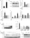
Figure 2. Intracellular ROS H2O2 induces KSHV reactivation.
(A–B) Treatment of BCBL1 cells with catalase inhibitor ATZ reduced catalase activity (A) and increased intracellular H2O2 level (B). Cells were treated with 1 mM of ATZ for 12 h. (C–E) Induction of KSHV lytic replication in BCBL1 cells by treatment with ATZ, H2O2 and TPA alone or in combination. ATZ was used at 1 mM, H2O2 at 300 µM, and TPA at 20 ng/ml. KSHV RTA, kbZIP, ORF65 and vFLIP transcripts were detected by RT-qPCR following 24 h of treatment (C). Relative lytic protein ORF65 protein levels shown in numbers were detected by Western-blotting following 96 h of treatments using β-tubulin for calibration of sample loading (D). Relative virus titers were measured by using the supernatants to infect endothelial cells and calculating the numbers of GFP-positive cells at 48 hpi (E). (F–H) Silencing of catalase induced KSHV lytic replication. BCBL1 cells harboring BAC36 were stably transfected with siRNA specific to catalase or scrambled control. Transcripts of catalase, and KSHV RTA, kbZIP, ORF57, ORF59 and ORF65 were measured by RT-qPCR (F). Catalase, KSHV lytic protein ORF65, and β-tubulin were detected by Western-blotting (G). Relative virus titers were measured as described in “E” (H).
Figure 3
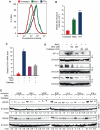
Figure 3. TPA-induced KSHV reactivation is mediated by H2O2.
(A) Intracellular H2O2 levels in untreated BCBL1 cells and BCBL1 cells treated with TPA for 6 h. (B) Catalase protein levels in untreated BCBL1 cells and BCBL1 cells treated with TPA for 6 and 12 h. Catalase protein was detected by Western-blotting and calibrated with β-tubulin for sample loading. (C) H2O2 scavengers reduced TPA-induction of intracellular H2O2 levels measured by flow cytometry analysis of fluorescence intensity in BCBL1 cells stably expressing a H2O2 sensor protein cpYFP. Cells were treated with 20 ng/ml of TPA without any scavengers, or with scavenger catalase at 400 U/ml, reduced glutathione at 400 µM or NAC at 400 µM for 6 h. (D–E) H2O2 scavengers reduced TPA-induction of RTA transcript and protein in BCBL1 cells. Treatments of the cells were the same as in (C). RTA transcript was detected by RT-qPCR (D). RTA protein was detected by Western-blotting and calibrated with β-tubulin for sample loading (E). (F–G) H2O2 mediated the activation of lytic RTA but not latent LANA promoter. RTA promoter was induced by H2O2 and TPA, and this induction effect was inhibited by NAC (F). Latent LANA promoter was marginally activated by TPA but not by H2O2 (G). Luciferase activities were measured for 293 cells transfected with promoter reporter plasmids for 24 h, and treated with H2O2 (150 µM) or TPA (20 ng/ml) with or without NAC (400 µM) for 12 h. (I–H) H2O2 scavengers reduced TPA-induction of ORF65 protein and production of infectious virions in BCBL1 cells in a dose-dependent manner. TPA was used at 20 ng/ml. Relative ORF65 protein levels shown in numbers were detected following 72 h of treatment by Western-blotting and calibrated with β-tubulin for sample loading (H). Relative virus titers were determined by using supernatants collected at 5 days of treatment to infect endothelial cells and calculating the numbers of GFP-positive cells at 48 hpi (I). (J) H2O2 scavengers inhibited KSHV spontaneous lytic replication. ORF65 protein in BCBL1 cells treated with catalase (400 U/ml) and NAC (400 µM) for 1 and 6 day was determined by Western-blotting.
Figure 4
Figure 4. H2O2 mediates KSHV reactivation induced by hypoxia, and proinflammatory and proangiogenic cytokines.
(A–B) Sodium azide (NaN3) and TPA increased intracellular H2O2 levels measured by flow cytometry analysis of fluorescence intensity in BCBL1 cells stably expressing a H2O2 sensor protein cpYFP. Cells were treated with NaN3 (10 mM) for 90 min or 20 ng/ml of TPA for 12 h, and their fluorescence levels measured by flowcytometry shown in histogram (A) and median fluorescence intensity (B). (C) H2O2 scavengers inhibited NaN3 induction of RTA transcript. BCBL1 cells were treated with NaN3 (10 mM) for 90 min with and without NAC (400 µM) or catalase (400 U/ml). RTA transcript was detected by RT-qPCR following elimination of NaN3, and continuous culture with NAC or catalase for another 24 h. (D) H2O2 scavengers inhibited NaN3 induction of HIF-1α, RTA and ORF65 proteins. BCBL1 cells were treated with NaN3 (10 mM) for 90 min with and without NAC (400 µM) or catalase (400 U/ml). Following elimination of NaN3, cells were cultured with NAC or catalase, and Western-blotting was performed to measure the expression of HIF-1α protein at 6 h, RTA protein at 24 h, and ORF65 protein at 72 h. (E) H2O2 scavengers inhibited the expression of ORF65 protein in BCBL1 cells induced by proinflammatory and angiogenic cytokines. ORF65 protein was determined by Western-blotting in BCBL1 cells with or without co-culture with U973 cells treated with cytokines with or without NAC (400 µM) or catalase (400 U/ml) for 72 h. The concentrations of the cytokines were stated in the Materials and Methods. Relative ORF65 protein level was calculated using untreated BCBL1 cells as a reference (1-fold) and after calibration for protein loading with β-tubulin.
Figure 5
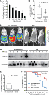
Figure 5. Antioxidant NAC inhibits spontaneous KSHV lytic replication in vitro and in vivo.
(A) NAC inhibited the luciferase activities of BCBL1 cells harboring Δ65Luc in a dose-dependent fashion. The luciferase activities were measured following 72 h of treatment. (B-E) NAC inhibited KSHV lytic replication in a mouse PEL model. NOD/SCID mice intraperitoneally inoculated with BCBL1 cells harboring Δ65Luc at 5×10⁶ cells per mouse were examined for evidence of KSHV lytic replication. Representative images of the untreated control and NAC-treated groups of mice examined for luciferase activities five weeks after inoculation using a Xenogen IVIS 200 small animal imaging system (B). Luciferase activities of the untreated control (N = 44) and NAC-treated mice (N = 36) (C). Detection of KSHV ORF65 protein by Western-blotting in lymphoma cells from representative mice (D). Cells from 100 µl ascitic fluid from each mouse were examined. β-tubulin was used to calibrate the number of total cells. Relative viral loads in blood samples of control and NAC-treated mice (E). Kaplan-Meier analysis of the survival of the two groups of mice showing that NAC treatment significantly extended the lifespan of the mice compared to untreated mice (F).
Figure 6
Figure 6. H2O2 induction of KSHV reactivation is mediated by ERK1/2, JNK, and p38 MAPK pathways.
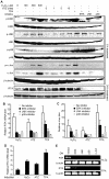
(A) Treatment with H2O2, ATZ or TPA activated ERK1/2, JNK and p38 pathways and their downstream transcriptional factor c-Jun, and induced the expression of RTA protein in BCBL1 cells harboring BAC36. Cells were treated with different concentrations of H2O2 and ATZ, or TPA at 20 ng/ml with DMSO control or inhibitors of MAPK pathways including 10 µM U0126 (ERK inhibitor), 50 µM JNK inhibitor II, and 50 µM SB203580 (p38 inhibitor) for 12 h. Total ERK1/2, JNK, p38 andc-Jun, and their phosphorylated forms, RTA protein, and β-tubulin were detected by Western-blotting. (B–C) Inhibitors of MAPK pathways inhibited the induction of RTA and production of infectious virions by H2O2, ATZ and TPA in BCBL1 cells harboring BAC36. H2O2, ATZ and TPA were used at concentrations of 300 µM, 5 mM and 20 ng/ml, respectively. Inhibitors of MAPK pathways were used at concentrations described in (A). Relative RTA transcript level at 24 h of treatment was detected by RT-qPCR with untreated cells set as “1” (B). Relative virus titers were determined by using supernatants collected at 5 days of treatment to infect endothelial cells and calculating the numbers of GFP-positive cells at 48 hpi (C). Virus titers from untreated cells were set as “1”. (D) H2O2 and ATZ, and TPA at concentrations of 300 µM, 5 mM and 20 ng/ml, respectively, induced the expression of RTA transcript in 293T cells harboring BAC36. RTA transcript was detected by RT-qPCR following 24 h of treatment. (E) Dominant negative (DN) constructs of MAPK pathways inhibited the induction of RTA transcript by H2O2 and TPA in 293T cells. 293T cells harboring BAC36 were transiently transfected with the control plasmid and DN constructs of ERK, JNK, p38, and c-Jun for 24 h, and treated with H2O2 at 300 µM and TPA at 20 ng/ml for an additional 12 h.

Similar articles

Cited by

References

    1. Speck SH, Ganem D. Viral latency and its regulation: lessons from the gamma-herpesviruses. Cell Host Microbe. 2010;8:100–115. - PMC - PubMed
    1. Chang Y, Cesarman E, Pessin MS, Lee F, Culpepper J, et al. Identification of herpesvirus-like DNA sequences in AIDS-associated Kaposi's sarcoma. Science. 1994;266:1865–1869. - PubMed
    1. Cesarman E, Chang Y, Moore PS, Said JW, Knowles DM. Kaposi's sarcoma-associated herpesvirus-like DNA sequences in AIDS-related body-cavity-based lymphomas. N Engl J Med. 1995;332:1186–1191. - PubMed
    1. Soulier J, Grollet L, Oksenhendler E, Cacoub P, Cazals-Hatem D, et al. Kaposi's sarcoma-associated herpesvirus-like DNA sequences in multicentric Castleman's disease. Blood. 1995;86:1276–1280. - PubMed
    1. Staskus KA, Zhong W, Gebhard K, Herndier B, Wang H, et al. Kaposi's sarcoma-associated herpesvirus gene expression in endothelial (spindle) tumor cells. J Virol. 1997;71:715–719. - PMC - PubMed

Publication types

MeSH terms