. 2011 Aug;39(15):6489-99.
doi: 10.1093/nar/gkr230. Epub 2011 Apr 21.

DNA double-strand breaks in heterochromatin elicit fast repair protein recruitment, histone H2AX phosphorylation and relocation to euchromatin

Affiliations

DNA double-strand breaks in heterochromatin elicit fast repair protein recruitment, histone H2AX phosphorylation and relocation to euchromatin

Burkhard Jakob et al. Nucleic Acids Res. 2011 Aug.

Abstract

DNA double-strand breaks (DSBs) can induce chromosomal aberrations and carcinogenesis and their correct repair is crucial for genetic stability. The cellular response to DSBs depends on damage signaling including the phosphorylation of the histone H2AX (γH2AX). However, a lack of γH2AX formation in heterochromatin (HC) is generally observed after DNA damage induction. Here, we examine γH2AX and repair protein foci along linear ion tracks traversing heterochromatic regions in human or murine cells and find the DSBs and damage signal streaks bending around highly compacted DNA. Given the linear particle path, such bending indicates a relocation of damage from the initial induction site to the periphery of HC. Real-time imaging of the repair protein GFP-XRCC1 confirms fast recruitment to heterochromatic lesions inside murine chromocenters. Using single-ion microirradiation to induce localized DSBs directly within chromocenters, we demonstrate that H2AX is early phosphorylated within HC, but the damage site is subsequently expelled from the center to the periphery of chromocenters within ∼ 20 min. While this process can occur in the absence of ATM kinase, the repair of DSBs bordering HC requires the protein. Finally, we describe a local decondensation of HC at the sites of ion hits, potentially allowing for DSB movement via physical forces.


Figures
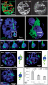
Figure 1.
Figure 1.
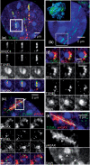
Bending of linear ion-induced γH2AX streaks indicates chromatin density-dependent damage relocation. (a) HeLa cells expressing GFP-tagged histone H2B were nickel ion-irradiated at low angle and immunostained for γH2AX after 15 min. Left image (merged): Chromatin is visualized by H2B-GFP (green), brighter staining indicating higher density as observed around nucleoli (red arrowheads). The depicted linear γH2AX streaks (red) show a slight but consistent bending following the course of bright perinucleolar chromatin staining. Separate channels are shown on the right. (b) Single slice image of a MEF nucleus irradiated with xenon ions to induce linear damage streaks visualized by γH2AX (green) 1 h post-irradiation. Intense DNA-stained regions (DAPI; blue) represent heterochromatic compartments (chromocenters), as confirmed by histone H3-K9me3 staining (Supplementary Figure S1A). The original ion track (arrow) was derived from 3D analysis of the confocal image stack allowing interpolation of the γH2AX streak. Chromocenters traversed by the interpolated trajectory were defined as ion-hit. 3D analysis is exemplarily shown for an ion-hit chromocenter as a rendered 3D-image (right panel) and (c) as a montage of different z-planes (Δ = 0.2 µm). Three types of γH2AX patterns, each shown in a MEF nucleus and as a schematic drawing, were observed at ion-hit chromocenters: bent streaks (d), interrupted streaks (e) and internal signals (f). Bent and interrupted streaks represent external signals, only distinguished by the direction of damage translocation indicated by arrows in the schemes (d and e). (g) Distribution of internal signals over time post-irradiation. Internal γH2AX signals within ion-hit chromocenters were significantly reduced from 15 min to 1 h post-irradiation (P < 0.05 using t-test; asterisk). Error bars represent the SEM of four independent experiments, n is the number of analyzed ion-hit chromocenters.
Figure 2.
Figure 2.
Damage markers remain co-localized with break sites bending around HC. Shown are streaks of directly visualized DSBs (green, labeled by TUNEL) along uranium ion tracks in MEF nuclei. Chromocenters are indicated by intense DAPI staining (blue) (a) Merged projection image (top) showing DSBs (green) co-localizing with the damage marker XRCC1 (red) 5 min post-irradiation. Bottom: selected confocal slices indicating the internal location of DSBs within the traversed chromocenter (box). Shown are the separate color channels and the merged image. (b) DSBs (green) located within HC 5 min post-irradiation (box). The insert shows the corresponding 3D surface image. (c) DSBs co-stained with γH2AX (red) are depicted in the merged projection image (top) and selected confocal slices (bottom, separate channels and merged) demonstrating the bending of the DSB streak around heterochromatic regions and the co-localization with γH2AX at 30 min post-irradiation. The arrow in the upper image indicates the reconstructed original ion track crossing the chromocenter. (d) DSBs co-localizing with RPA (red) at the periphery of a chromocenter are depicted in single confocal slices 30 min after ion exposure as merged image (top) or separate channels below. (e) Ion track passing nearby chromocenters (arrow) with γH2AX staining (red) 30 min post-irradiation showing that chromocenters are precluded from γH2AX spreading. Separate channels of γH2AX and DAPI are depicted below. White arrowheads indicate the border of three bypassed chromocenters that form a barrier for the propagation of the γH2AX signal.
Figure 3.
Figure 3.
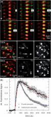
Fast recruitment of the repair protein XRCC1 to heterochromatic DNA damage. (a) Live cell imaging at the high energy beamline showing the recruitment of GFP-XRCC1 (green) to linear 1 GeV/u uranium ion-induced tracks traversing hetero (HC)- and euchromatic (EC) regions in MEF nuclei. Chromocenters are marked by co-expression of HC-associated Cherry-tagged HP1α (red). Top: depicted are selected image stacks (times in seconds as indicated in the inset) of one centrally hit chromocenter and three EC-foci along the ion track. Each time frame is displayed as six central consecutive z-slices of the image stacks acquired. The shift of the brightest HC-associated XRCC1 signals at 160 s to upper z-slices at 440 s indicates movement to the chromocenter top. Bottom: projection image of the whole nucleus 100 s (upper row) and 550 s after irradiation showing the preferential loss of the euchromatic XRCC1 signal. The box indicates the area from which the z-slices are taken. On the right, the separate channels (XRCC1 and HP1α) are shown. The total observation time of ∼10 min is shown in the Supplementary Movies for this (Movie S1, nucleus turned by 90°) and another nucleus with a hit chromocenter (Supplementary Movies S2 and S3). (b) Kinetics of GFP-XRCC1 recruitment in HC (18 foci) and EC (78 foci) measured as described in (a) and fitted by exponential functions. Error bars indicate SEM.
Figure 4.
Figure 4.
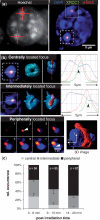
Relocation dynamics of damage sites centrally induced within chromocenters. The MEF nucleus was irradiated with single sulfur ions and immunostained 5 min post-irradiation. For details of microbeam targeting see Figure S3 (a) H2AX is phosphorylated and XRCC1 accumulates at heterochromatic DSBs directly after single-ion irradiation. The left-hand image shows the aimed targeting of chromocenters (red crosses) for single-ion irradiation using Hoechst 33342 (grey scale) as a marker in nuclei of living MEF cells. The right-hand image shows the same nucleus after fixation at 5 min post-irradiation. DNA damage-induced foci of the repair factor XRCC1 (green) and γH2AX (red) are clearly visualized at the sites of ion traversal. Both proteins co-localize within each of the targeted chromocenters (blue: DAPI DNA staining). (b) Definition of different radiation-induced foci positions relative to the ion-hit chromocenter. The depicted hit MEF chromocenters [marked boxes in (a), right panel] stained as detailed above upper panel: co-localizing XRCC1 and γH2AX IRIF are centrally located within a chromocenter as illustrated in the projection (left) and corresponding rendered surface images for γH2AX and DAPI (mid images). The intensity profile measured along the respective dotted line in the single slice image indicates that DNA staining (blue) is depleted (black arrows) at the damage site marked by XRCC1 (green) and γH2AX (red). The depleted DNA staining intensity co-localizes with the damage markers. Mid panel: co-localizing XRCC1 and γH2AX IRIF are located off-centered within the chromocenter (rendered surface images, mid images) defining the IRIF as intermediately located. The intensity profile measured as above shows the depleted DNA staining (blue) co-localizing with the damage markers (green and red) also at the intermediate position. Lower panels: gallery of consecutive light-optical sections (Δ = 0.2 µm) showing the xy position of the damage sites marked by XRCC1 and γH2AX foci. Arrowheads (Frames 2 and 8) mark the initial positions of the damage induced at the ion traversal. The displacement between the damage (foci) and its original induction site (arrowhead) shown in Frame 8 demonstrates the relocation of the damage from the center to the periphery of the chromocenter. The resulting bending of the γH2AX signal surrounding the chromocenter is additionally shown in a rendered 3D image. (c) Analysis of the time-dependent localization of XRCC1 and γH2AX IRIF (positions defined in b). Relative frequencies of each position are given for the indicated post-irradiation intervals and (n), the total number of ion-hit chromocenters from three independent experiments, is indicated. Error bars represent the SEM.
Figure 5.
Figure 5.
Chromatin-density and absence of ATM influence the repair kinetics of carbon ion-induced DSBs. (a) Normalized repair kinetics for γH2AX foci following low angle carbon ion irradiation. The number of γH2AX foci was enumerated in G1 phase wild-type (green) and ATM−/− MEF cells (blue) at the indicated time points after ion exposure. γH2AX signals exhibiting distinct maxima were counted as individual foci. Foci were discriminated to be either associated with the intensively DAPI stained heterochromatic chromocenters (HC) or with euchromatic regions (EC) using software-aided visual inspection. Only foci directly adjacent to HC are classified as being HC-associated, and signals spatially separated are classified EC-associated. At 30 min post-irradiation about 1/3 of the foci were found associated with HC. Repair of HC-associated damages in wt-MEFs (solid green line) is delayed compared to EC-associated DSBs in either wt MEFs (dashed green line) or ATM−/− MEFs (dashed blue line), whereas in ATM−/− MEFs the repair of HC-associated DSBs is abolished (solid blue line). For each time point ∼30 nuclei were analyzed. Error bars represent the SEM. (b) ATM is not required for the damage response within HC. ATM−/− MEF nucleus (left) targeted with a single samarium ion at the microprobe. The hit chromocenter shows XRCC1 (green) recruitment and γH2AX (red) formation within highly compacted DNA (blue: ToPro3). The intensity profile (right panel) measured along the dotted line in the single slice image of the chromocenter shows depleted DNA staining at the damage site (black arrows). (c) Depicted ATM−/− MEF nuclei were carbon ion irradiated at low angle. Staining for γH2AX (green) shows bending patterns around ion-hit chromocenters 30 min post-irradiation. DNA was stained using DAPI (blue).
Figure 6.
Figure 6.
Decondensation of heterochromatic DNA in MEF chromocenters traversed by low energy ions. (a) Chromocenter of a MEF nucleus immunostained 7 min after targeted irradiation with single sulfur ions. Image of the ion-hit chromocenter (left) and intensity profile (right) along the dotted line showing depleted DNA (DAPI) staining (blue) at the damage site marked by XRCC1 (green) and p-ATM (red) marked by black arrow. (b) 3D analysis of a gold ion-hit chromocenter (box) showing depleted DNA staining along the whole ion traversal (red arrows within the binary threshold 3D-slice images; red dotted lines indicate the positions of the depicted xz- and yz-plane). Note that in (a) and (b), independent of the DNA stain and the ion species, the depleted DNA staining intensity clearly co-localizes with the damage markers. (c) Real-time decondensation of uranium-hit chromocenters was measured in a living MEF by local depletion of DNA-bound Hoechst 33342 fluorescence. Sites of ion traversal are indicated by GFP-XRCC1 accumulation (green). (d) Two centrally hit chromocenters (white arrows in c) were individually analyzed (blue symbols and line) in a central projection of a 3D-data set. For comparison, the mean values for three non-hit chromocenter are shown (red symbols; red line). Fluorescence intensity was corrected for bleaching and normalized to the initial intensity. The continuous fluorescence decline at the hit HC site indicates a biological decondensation process, ruling out an instantaneous DNA staining depletion related to disruption of the DNA or Hoechst dye due to particle traversal. (e and f) Images of the two chromocenters analyzed in (d) before (upper panels) and 4 min after ion traversal (bottom panels). Depicted are the Hoechst 33342 stained DNA (left panels) and the XRCC1 fluorescence (right panels) for each chromocenter in (e) and (f), respectively. Besides depletion of the DNA staining, major structural alterations of the chromocenters are not apparent.

Similar articles

Cited by

References

    1. Celeste A, Petersen S, Romanienko PJ, Fernandez-Capetillo O, Chen HT, Sedelnikova OA, Reina-San-Martin B, Coppola V, Meffre E, Difilippantonio MJ, et al. Genomic instability in mice lacking histone H2AX. Science. 2002;296:922–927. - PMC - PubMed
    1. Bassing CH, Chua KF, Sekiguchi J, Suh H, Whitlow SR, Fleming JC, Monroe BC, Ciccone DN, Yan C, Vlasakova K, et al. Increased ionizing radiation sensitivity and genomic instability in the absence of histone H2AX. Proc. Natl. Acad. Sci. USA. 2002;99:8173–8178. - PMC - PubMed
    1. Burma S, Chen BP, Murphy M, Kurimasa A, Chen DJ. ATM phosphorylates histone H2AX in response to DNA double-strand breaks. J. Biol. Chem. 2001;276:42462–42467. - PubMed
    1. Stiff T, O'Driscoll M, Rief N, Iwabuchi K, Löbrich M, Jeggo PA. ATM and DNA-PK function redundantly to phosphorylate H2AX after exposure to ionizing radiation. Cancer Res. 2004;64:2390–2396. - PubMed
    1. Rogakou EP, Boon C, Redon C, Bonner WM. Megabase chromatin domains involved in DNA double-strand breaks in vivo. J. Cell Biol. 1999;146:905–916. - PMC - PubMed

Publication types

MeSH terms