

Histone deacetylases (HDACs) in XPC gene silencing and bladder cancer
- PMID: 21507255
- PMCID: PMC3108377
- DOI: 10.1186/1756-8722-4-17
Histone deacetylases (HDACs) in XPC gene silencing and bladder cancer
Abstract
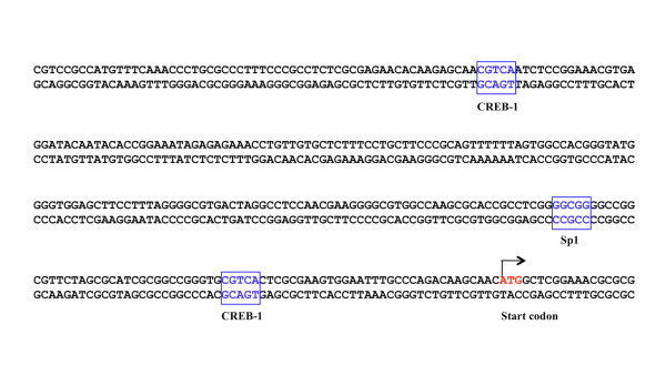
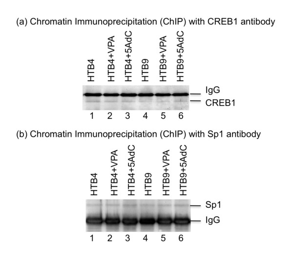
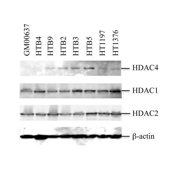
Bladder cancer is one of the most common malignancies and causes hundreds of thousands of deaths worldwide each year. Bladder cancer is strongly associated with exposure to environmental carcinogens. It is believed that DNA damage generated by environmental carcinogens and their metabolites causes development of bladder cancer. Nucleotide excision repair (NER) is the major DNA repair pathway for repairing bulk DNA damage generated by most environmental carcinogens, and XPC is a DNA damage recognition protein required for initiation of the NER process. Recent studies demonstrate reduced levels of XPC protein in tumors for a majority of bladder cancer patients. In this work we investigated the role of histone deacetylases (HDACs) in XPC gene silencing and bladder cancer development. The results of our HDAC inhibition study revealed that the treatment of HTB4 and HTB9 bladder cancer cells with the HDAC inhibitor valproic acid (VPA) caused an increase in transcription of the XPC gene in these cells. The results of our chromatin immunoprecipitation (ChIP) studies indicated that the VPA treatment caused increased binding of both CREB1 and Sp1 transcription factors at the promoter region of the XPC gene for both HTB4 and HTB9 cells. The results of our immunohistochemistry (IHC) staining studies further revealed a strong correlation between the over-expression of HDAC4 and increased bladder cancer occurrence (p < 0.001) as well as a marginal significance of increasing incidence of HDAC4 positivity seen with an increase in severity of bladder cancer (p = 0.08). In addition, the results of our caspase 3 activation studies demonstrated that prior treatment with VPA increased the anticancer drug cisplatin-induced activation of caspase 3 in both HTB4 and HTB9 cells. All of these results suggest that the HDACs negatively regulate transcription of the XPC gene in bladder cancer cells and contribute to the severity of bladder tumors.
Figures


Similar articles
-
Attenuated expression of xeroderma pigmentosum group C is associated with critical events in human bladder cancer carcinogenesis and progression.Cancer Res. 2007 May 15;67(10):4578-85. doi: 10.1158/0008-5472.CAN-06-0877. Cancer Res. 2007. PMID: 17510383
-
Attenuated XPC expression is not associated with impaired DNA repair in bladder cancer.PLoS One. 2015 Apr 30;10(4):e0126029. doi: 10.1371/journal.pone.0126029. eCollection 2015. PLoS One. 2015. PMID: 25927440 Free PMC article.
-
XPC deficiency leads to centrosome amplification by inhibiting BRCA1 expression upon cisplatin-mediated DNA damage in human bladder cancer.Cancer Lett. 2019 Mar 1;444:136-146. doi: 10.1016/j.canlet.2018.12.004. Epub 2018 Dec 21. Cancer Lett. 2019. PMID: 30579971
-
Valproic acid: an old drug newly discovered as inhibitor of histone deacetylases.Ann Hematol. 2004;83 Suppl 1:S91-2. doi: 10.1007/s00277-004-0850-2. Ann Hematol. 2004. PMID: 15124690 Review.
-
Revisiting Histone Deacetylases in Human Tumorigenesis: The Paradigm of Urothelial Bladder Cancer.Int J Mol Sci. 2019 Mar 14;20(6):1291. doi: 10.3390/ijms20061291. Int J Mol Sci. 2019. PMID: 30875794 Free PMC article. Review.
Cited by
-
Effects of novel HDAC inhibitors on urothelial carcinoma cells.Clin Epigenetics. 2018 Jul 31;10(1):100. doi: 10.1186/s13148-018-0531-y. Clin Epigenetics. 2018. PMID: 30064501 Free PMC article.
-
HDAC inhibition as a treatment concept to combat temsirolimus-resistant bladder cancer cells.Oncotarget. 2017 Nov 6;8(66):110016-110028. doi: 10.18632/oncotarget.22454. eCollection 2017 Dec 15. Oncotarget. 2017. PMID: 29299126 Free PMC article.
-
Histone deacetylases and mechanisms of regulation of gene expression.Crit Rev Oncog. 2015;20(1-2):35-47. doi: 10.1615/critrevoncog.2015012997. Crit Rev Oncog. 2015. PMID: 25746103 Free PMC article. Review.
-
Overexpression of deubiquitinating enzyme USP28 promoted non-small cell lung cancer growth.J Cell Mol Med. 2015 Apr;19(4):799-805. doi: 10.1111/jcmm.12426. Epub 2015 Feb 5. J Cell Mol Med. 2015. PMID: 25656529 Free PMC article.
-
CREPT Disarms the Inhibitory Activity of HDAC1 on Oncogene Expression to Promote Tumorigenesis.Cancers (Basel). 2022 Sep 30;14(19):4797. doi: 10.3390/cancers14194797. Cancers (Basel). 2022. PMID: 36230720 Free PMC article.
References
-
- Kufe DW, Bast RBJ, Hait WH, Hong WH, Pollock RE, Weichselbaum RR, Holland JF, Frei EI. Cancer Medicine 7. BC Decker Inc. 2006.
-
- Friedberg EC, Walker GC, Siede W. DNA repair and mutagenesis. First. ASM published, Washington, D.C; 1995.
-
- Friedberg EC, Walker GC, Siede W, Wood RD, Schultz RA, Ellenberger T. DNA repair and mutagenesis. Second. ASM Press, Washington D.C; 2006.
Publication types
MeSH terms
Substances
Grants and funding
LinkOut - more resources
Full Text Sources
Medical
Research Materials
