. 2011 May;138(9):1759-69.
doi: 10.1242/dev.058420.

The Drosophila STUbL protein Degringolade limits HES functions during embryogenesis

Affiliations

The Drosophila STUbL protein Degringolade limits HES functions during embryogenesis

Kevin C Barry et al. Development. 2011 May.

Abstract

Degringolade (Dgrn) encodes a Drosophila SUMO-targeted ubiquitin ligase (STUbL) protein similar to that of mammalian RNF4. Dgrn facilitates the ubiquitylation of the HES protein Hairy, which disrupts the repressive activity of Hairy by inhibiting the recruitment of its cofactor Groucho. We show that Hey and all HES family members, except Her, interact with Dgrn and are substrates for its E3 ubiquitin ligase activity. Dgrn displays dynamic subcellular localization, accumulates in the nucleus at times when HES family members are active and limits Hey and HES family activity during sex determination, segmentation and neurogenesis. We show that Dgrn interacts with the Notch signaling pathway by it antagonizing the activity of E(spl)-C proteins. dgrn null mutants are female sterile, producing embryos that arrest development after two or three nuclear divisions. These mutant embryos exhibit fragmented or decondensed nuclei and accumulate higher levels of SUMO-conjugated proteins, suggesting a role for Dgrn in genome stability.


Figures
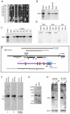
Fig. 1.
Fig. 1.
Dgrn interacts physically with Hey and all members of the HES family of bHLH transcription factors, except Her. (A) Alignment of the basic regions of Hey and all fly HES family members showing the conserved nature of this domain. (B-D) Dgrn binds to Hey and all HES family members except Her. GST pulldown assays with 35S-labeled in vitro translated proteins on the left and GST fusion proteins or GST alone across the top. (E) Map of the dgrn locus indicating the size of the deletion in the dgrnDK mutation as well as the Dgrn protein structure and pieces of Dgrn used for northern blot analysis. (F) Northern blots using probes made to regions A and B of the Dgrn protein demonstrating that dgrn mRNA is found in the fly ovary and in 0- to 4-hour-old embryos. (G) Western blots using the polyclonal antibody generated to GST-Dgrn showing that this antibody recognizes Dgrn in both nuclear extracts (NE) and whole-cell embryo extracts (WCE). (H) Western blots of wild-type and dgrnDK mutant embryos and wild-type and dgrn RNAi cell lysates (S2 cells) showing the specificity for the Dgrn antibody for Dgrn.
Fig. 2.
Fig. 2.
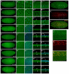
Dgrn exhibits dynamic cellular and subcellular localization. (A-I″″) Surface projections of progressively older wild-type embryos stained with anti-Dgrn (green). Nuclear division cycle (nc) is indicated. Higher magnification sections (A′-I″) and tangential sections (A‴-I″″) of the corresponding embryos A-I are shown. DAPI staining (blue; visualizing nuclei) is included in panels A″-I″ and A″″-I″″. Note the alternating nuclear accumulation (arrows) and nuclear exclusion (arrowheads) of Dgrn. (J-L) Surface projections of wild-type stage 9 embryos stained with anti-Dgrn (green) and anti-Hb (red; to indicate neuroblasts). Note nuclear Dgrn in developing neuroblasts. Dashed boxes indicate region of higher magnification projections (J-L′) of corresponding embryos J-L.
Fig. 3.
Fig. 3.
Nuclei fall from the surface of dgrn null embryos and subcortical actin forms abnormal figures. (A-C′) dgrnDK null mutant embryos stained with DAPI showing arrest of mutants at nuclear cycle 2-3. A′-C′ show higher magnification projections of corresponding embryos A-C. (D,D′) dgrnDK null embryo stained with anti-Lamin (green) to visualize nuclear envelope and DAPI (blue) to visualize nuclei. Boxed region in D is shown at higher magnification in D′. (E-N) DAPI staining of progressively older wild-type and dgrnDK null embryos that make it past the early arrest phenotype. (M,N) Projections of cross-sections of wild-type (M) and dgrnDK (N) null embryos. Note the accumulation of nuclei in the center and just below the surface of the dgrn mutant embryo. (O-P′) Surface projections of wild-type (O,O′) and late arrest dgrnDK null (P,P′) embryos stained with anti-Lamin (green) and DAPI (blue). Boxed regions in O and P are shown at higher magnification in O′ and P′, respectively. Note the abnormal mitotic figures and mis-shapen nuclear envelopes in dgrn mutant embryos (P′). (Q-R‴) Still images from a time lapse of wild-type (Q-Q‴) and dgrnDK mutant (R-R‴) embryos expressing a histone-GFP fusion reporter where nuclei fall from the surface and aggregate in the center of the dgrn mutant embryo (R‴). (S,S′) Surface projection of a dgrnDK null embryo stained with the centrosome marker anti-γ-tubulin (green) and DAPI (blue). Boxed region in S is shown at higher magnification in S′ and shows regions where no nuclei or centrosomes are at the surface (arrows) and where centrosomes are on the surface but nuclei have fallen inwards (arrowheads). (T,T′) Surface projection of a dgrnDK null embryo stained with anti-α-tubulin (red) to visualize microtubules, anti-γ-tubulin (green) to visualize centrosomes and DAPI (blue). Boxed region in T is shown at higher magnification in T′ and shows the proper association of free centrosomes with microtubules, as well as abnormal mitotic figures. (U-V‴) Still images from a time lapse of wild-type (U-U‴) and dgrnDK null (V-V‴) embryos expressing the sGCMA reporter to visualize actin. (W,X) Cross-sections of wild-type (W) and dgrnDK null (X) embryos expressing the sGCMA reporter (green) and stained with DAPI (blue). Note the actin accumulation at the periphery of the embryo and the accumulation of nuclei in the center of the embryo. (Y) Western blot analysis of SUMOylated proteins in embryo extracts from wild-type (wt) and dgrnDK null embryos. Note that dgrn mutants show a 2-fold increase in SUMO expression compared with wild type.
Fig. 4.
Fig. 4.
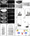
Dgrn antagonizes Dpn and Gro in controlling the transcriptional regulation of the master sex determinant protein Sex Lethal. (A,B) Female wild-type embryos express Sxl (A), whereas males do not (B). (C,C′) A representative dgrnDK null mutant embryo showing no Sxl expression (C) and stained with DAPI (C′). (D-K) Sxl staining of embryos ectopically expressing Dgrn constructs. (D,E) Ectopic expression of wild-type Dgrn leads to an overexpression of Sxl protein in females (D) and a misexpression of Sxl in males (E); however, ectopic expression of mutant forms of Dgrn does not affect Sxl expression (F-K). (L) Western blot analysis of Sxl expression in embryo extracts from wild-type and dgrnDK null embryos. Note that dgrnDK null mutants show an 93% decrease in Sxl expression compared with wild type. (M) Western blot analysis of Sxl expression on embryo extracts from wild type and the four Dgrn overexpression lines: UAS-Dgrn, UAS-DgrnHC/AA, UAS-DgrnI268A and UAS-DgrnΔSIMs. Note that UAS-Dgrn embryos show a 4.6-fold increase in Sxl expression compared with wild type. (N-P) A transgenic Sxl-PE-lacZ reporter was incorporated into a wild-type (N,O) and dgrnDK null (P) background. This in vivo Sxl-lacZ reporter is not activated in dgrnDK null embryos. (Q-S) Transcription assays using the Sxl-PE promoter-luciferase fusion that is activated by Da and Sc. Dpn (Q), Gro (R) and Ubc9 (S) repress transcription of Sxl, which is alleviated by the co-expression of wild-type Dgrn. By contrast, DgrnHC/AA, and to a lesser extent DgrnΔSIMs, are unable to antagonize this repression of Sxl. Error bars represent s.e.m. *P<0.01, **P<0.001, ***P<0.0001. (T) In vitro ubiquitylation assay using in vitro translated 35S-Methionine-Dpn protein and bacterially expressed then purified recombinant ubiquitin activating enzyme (E1), UbcD2 (E2) and His6-Dgrn. Conj., Ub-protein conjugates. (U) Model for Dgrn function in sex determination. Dgrn binds to Dpn via its RING domain and simultaneously associates with SUMOylated-Gro via its SIM domains. Ubiquitylation of Dpn by Dgrn prevents association of Dpn with Gro, allowing Da-Sc to activate Sxl expression. SUMO-Gro and its associated Gro-oligomers are then inactivated by sequestration.
Fig. 5.
Fig. 5.
Ectopic Dgrn expression suppresses E(spl) activity during Drosophila neurogenesis. (A-D) Overexpression of UAS-GFP (A), UAS-Dgrn (B), UAS-DgrnHC/AA (C) or UAS-DgrnΔSIMs (D) does not affect adult thoracic bristle development. (E,F) Ectopic expression of UAS-E(spl)m8 leads to a loss of thoracic bristles in the adult fly (E); however, this ‘bald’ phenotype is suppressed by co-expression of UAS-Dgrn (F). Note the increase of thoracic bristles. (G,H) Co-expression of UAS-E(spl)m8 along with either UAS-DgrnHC/AA (G) or UAS-DgrnΔSIMs (H) does not suppress the E(spl)m8 bald phenotype.
Fig. 6.
Fig. 6.
Dgrn interacts genetically with the Notch signaling pathway. (A,B) Tip of adult wing in wild type (A) and in an N1/+ female (B) displaying a notched adult wing phenotype. (C,D) Reducing the dose of dgrn in the N1 background results in a rescue of the male lethality with the adult males displaying wild-type wings (C), whereas females are not affected and continue to display a notched wing phenotype (D). (E-G) Adult wing showing longitudinal vein 4 (L4) and 5 (L5) from wild type (E) and heterozygous NAX1682 (F). Note that these veins are truncated in the NAX1682 mutant (arrow in F indicates L4 truncation). Reducing the dose of dgrn in trans to the heterozygous NAX1682 allele partially rescues the vein phenotype caused by this N gain-of-function allele (arrow in G).

Similar articles

Cited by

References

    1. Abed M., Barry K. C., Kenyagin D., Koltun B., Phippen T. M., Delrow J. J., Parkhurst S. M., Orian A. (2011). Degringolade, a SUMO-targeted ubiquitin ligase, inhibits Hairy/Groucho-mediated repression. EMBO J. (in press). - PMC - PubMed
    1. Abrams E. W., Vining M. S., Andrew D. J. (2003). Constructing an organ: the Drosophila salivary gland as a model for tube formation. Trends Cell Biol. 13, 247-254 - PubMed
    1. Barbash D. A., Cline T. W. (1995). Genetic and molecular analysis of the autosomal component of the primary sex determination signal of Drosophila melanogaster. Genetics 141, 1451-1471 - PMC - PubMed
    1. Ben-Saadon R., Zaaroor D., Ziv T., Ciechanover A. (2006). The polycomb protein Ring1B generates self atypical mixed ubiquitin chains required for its in vitro Histone H2A ligase activity. Mol. Cell 24, 701-711 - PubMed
    1. Bianchi-Frias D., Orian A., Delrow J. J., Vazquez J., Rosales-Nieves A. E., Parkhurst S. M. (2004). Hairy transcriptional repression targets and cofactor recruitment in Drosophila. PLoS Biol. 2, E178 - PMC - PubMed

Publication types

MeSH terms

LinkOut - more resources