

Cutaneous denervation of psoriasiform mouse skin improves acanthosis and inflammation in a sensory neuropeptide-dependent manner
- PMID: 21471984
- PMCID: PMC3116081
- DOI: 10.1038/jid.2011.60
Cutaneous denervation of psoriasiform mouse skin improves acanthosis and inflammation in a sensory neuropeptide-dependent manner
Abstract
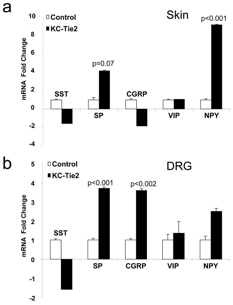
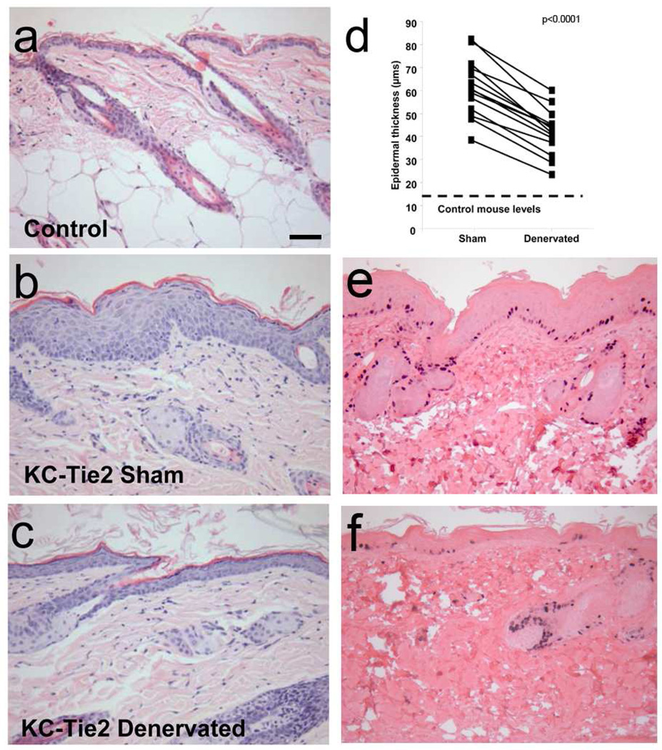
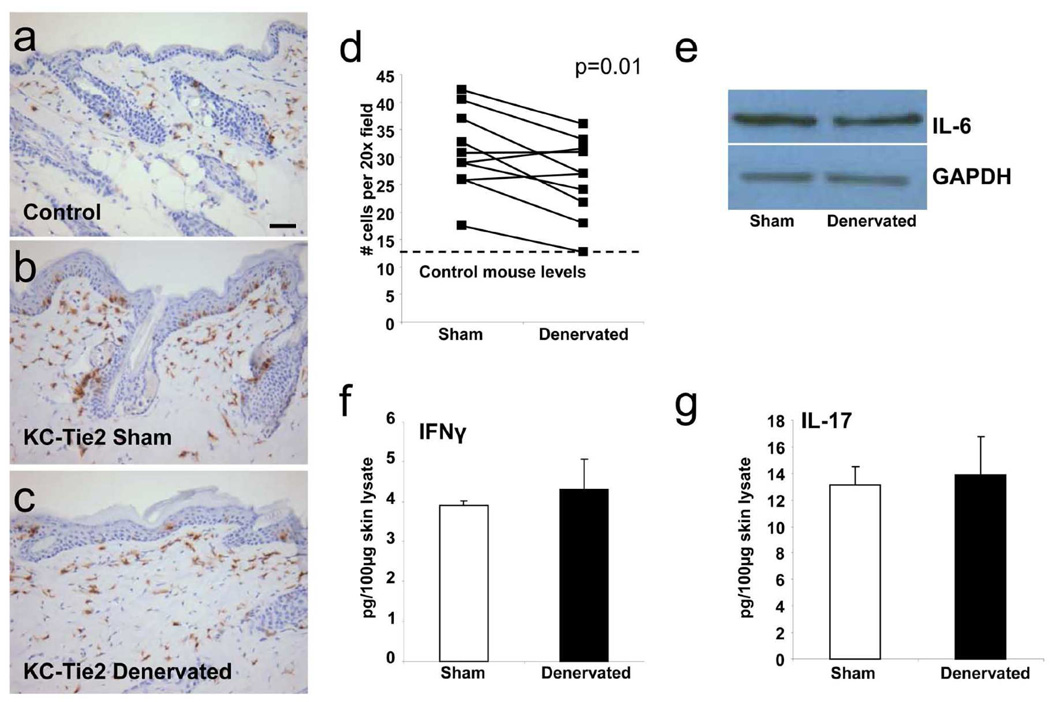
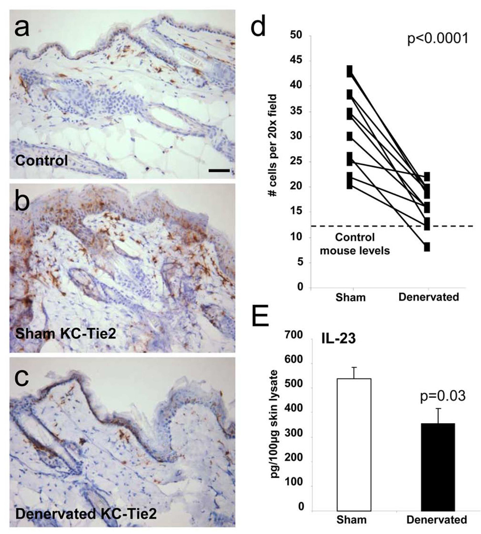
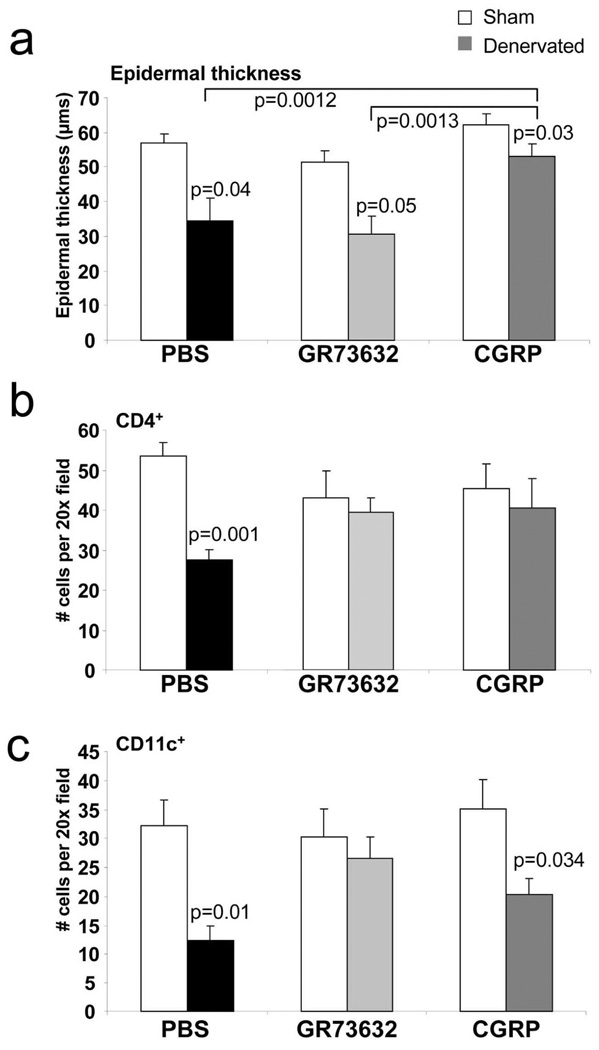
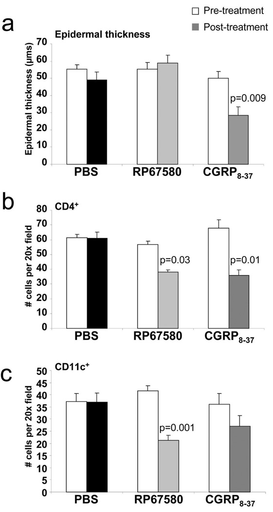
Nervous system involvement in psoriasis pathogenesis is supported by increases in nerve fiber numbers and neuropeptides in psoriatic skin and by reports detailing spontaneous plaque remission following nerve injury. Using the KC-Tie2 psoriasiform mouse model, we investigated the mechanisms by which nerve injury leads to inflammatory skin disease remission. Cutaneous nerves innervating dorsal skin of KC-Tie2 animals were surgically axotomized and beginning 1 day after denervation, CD11c(+) cell numbers decreased by 40% followed by a 30% improvement in acanthosis at 7 days and a 30% decrease in CD4(+) T-cell numbers by 10 days. Restoration of substance P (SP) signaling in denervated KC-Tie2 skin prevented decreases in CD11c(+) and CD4(+) cells, but had no effect on acanthosis; restoration of calcitonin gene-related peptide (CGRP) signaling reversed the improvement in acanthosis and prevented denervated-mediated decreases in CD4(+) cells. Under innervated conditions, small-molecule inhibition of SP in KC-Tie2 animals resulted in similar decreases to those observed following surgical denervation for cutaneous CD11c(+) and CD4(+) cell numbers; whereas small-molecule inhibition of CGRP resulted in significant reductions in CD4(+) cell numbers and acanthosis. These data demonstrate that sensory nerve-derived peptides mediate psoriasiform dendritic cell and T-cell infiltration and acanthosis and introduce targeting nerve-immunocyte/KC interactions as potential psoriasis therapeutic treatment strategies.
Conflict of interest statement
The authors state no conflict of interest.
Figures
Similar articles
-
Downregulation of lysine 2-hydroxyisobutyrylation of ErbB3 binding protein 1 at amino acid 210 promotes keratinocyte proliferation via induction of transcription initiation factor IA-mediated rRNA synthesis.Br J Dermatol. 2024 Dec 23;192(1):92-103. doi: 10.1093/bjd/ljae194. Br J Dermatol. 2024. PMID: 38752336
-
Defining the optimum strategy for identifying adults and children with coeliac disease: systematic review and economic modelling.Health Technol Assess. 2022 Oct;26(44):1-310. doi: 10.3310/ZUCE8371. Health Technol Assess. 2022. PMID: 36321689 Free PMC article.
-
A multicomponent psychosocial intervention to reduce substance use by adolescents involved in the criminal justice system: the RISKIT-CJS RCT.Public Health Res (Southampt). 2023 Mar;11(3):1-77. doi: 10.3310/FKPY6814. Public Health Res (Southampt). 2023. PMID: 37254608
-
Etrolizumab for induction of remission in ulcerative colitis.Cochrane Database Syst Rev. 2015 Dec 2;2015(12):CD011661. doi: 10.1002/14651858.CD011661.pub2. Cochrane Database Syst Rev. 2015. PMID: 26630451 Free PMC article. Review.
-
Impact of residual disease as a prognostic factor for survival in women with advanced epithelial ovarian cancer after primary surgery.Cochrane Database Syst Rev. 2022 Sep 26;9(9):CD015048. doi: 10.1002/14651858.CD015048.pub2. Cochrane Database Syst Rev. 2022. PMID: 36161421 Free PMC article. Review.
Cited by
-
Linear psoriasis following the typical distribution of the sciatic nerve.J Dermatol Case Rep. 2015 Mar 31;9(1):6-11. doi: 10.3315/jdcr.2015.1189. eCollection 2015 Mar 31. J Dermatol Case Rep. 2015. PMID: 25932056 Free PMC article.
-
Understanding and modeling nerve-cancer interactions.Dis Model Mech. 2023 Jan 1;16(1):dmm049729. doi: 10.1242/dmm.049729. Epub 2023 Jan 9. Dis Model Mech. 2023. PMID: 36621886 Free PMC article. Review.
-
Cutaneous nerve fibers participate in the progression of psoriasis by linking epidermal keratinocytes and immunocytes.Cell Mol Life Sci. 2022 Apr 30;79(5):267. doi: 10.1007/s00018-022-04299-x. Cell Mol Life Sci. 2022. PMID: 35488965 Free PMC article.
-
Nociceptive Sensory Fibers Drive Interleukin-23 Production in a Murine Model of Psoriasis via Calcitonin Gene-Related Peptide.Front Immunol. 2021 Oct 22;12:743675. doi: 10.3389/fimmu.2021.743675. eCollection 2021. Front Immunol. 2021. PMID: 34745116 Free PMC article.
-
Using optical coherence tomography for the longitudinal non-invasive evaluation of epidermal thickness in a murine model of chronic skin inflammation.Skin Res Technol. 2012 May;18(2):225-31. doi: 10.1111/j.1600-0846.2011.00558.x. Epub 2011 Sep 14. Skin Res Technol. 2012. PMID: 22092854 Free PMC article.
References
-
- Casasco A, Marchetti C, Calligaro A, Casasco M, Poggi P, Beolchini M. Immunocytochemical labelling of Merkel cells of human oral mucosa by means of antibodies to protein gene product 9.5. Bull Group Int Rech Sci Stomatol Odontol. 1990;33:61–64. - PubMed
-
- Chiang HY, Huang IT, Chen WP, Chien HF, Shun CT, Chang YC, et al. Regional difference in epidermal thinning after skin denervation. Exp Neurol. 1998;154:137–145. - PubMed
Publication types
MeSH terms
Substances
Grants and funding
LinkOut - more resources
Full Text Sources
Other Literature Sources
Medical
Molecular Biology Databases
Research Materials
Miscellaneous
