A viral microRNA cluster strongly potentiates the transforming properties of a human herpesvirus
- PMID: 21379335
- PMCID: PMC3040666
- DOI: 10.1371/journal.ppat.1001294
A viral microRNA cluster strongly potentiates the transforming properties of a human herpesvirus
Abstract
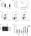
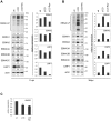
Epstein-Barr virus (EBV), an oncogenic human herpesvirus, induces cell proliferation after infection of resting B lymphocytes, its reservoir in vivo. The viral latent proteins are necessary for permanent B cell growth, but it is unknown whether they are sufficient. EBV was recently found to encode microRNAs (miRNAs) that are expressed in infected B cells and in some EBV-associated lymphomas. EBV miRNAs are grouped into two clusters located either adjacent to the BHRF1 gene or in introns contained within the viral BART transcripts. To understand the role of the BHRF1 miRNA cluster, we have constructed a virus mutant that lacks all its three members (Δ123) and a revertant virus. Here we show that the B cell transforming capacity of the Δ123 EBV mutant is reduced by more than 20-fold, relative to wild type or revertant viruses. B cells exposed to the knock-out virus displayed slower growth, and exhibited a two-fold reduction in the percentage of cells entering the cell cycle S phase. Furthermore, they displayed higher latent gene expression levels and latent protein production than their wild type counterparts. Therefore, the BHRF1 miRNAs accelerate B cell expansion at lower latent gene expression levels. Thus, this miRNA cluster simultaneously enhances expansion of the virus reservoir and reduces the viral antigenic load, two features that have the potential to facilitate persistence of the virus in the infected host. Thus, the EBV BHRF1 miRNAs may represent new therapeutic targets for the treatment of some EBV-associated lymphomas.
Conflict of interest statement
The authors have declared that no competing interests exist.
Figures


Similar articles
-
A Viral microRNA Cluster Regulates the Expression of PTEN, p27 and of a bcl-2 Homolog.PLoS Pathog. 2016 Jan 22;12(1):e1005405. doi: 10.1371/journal.ppat.1005405. eCollection 2016 Jan. PLoS Pathog. 2016. PMID: 26800049 Free PMC article.
-
A cluster of virus-encoded microRNAs accelerates acute systemic Epstein-Barr virus infection but does not significantly enhance virus-induced oncogenesis in vivo.J Virol. 2013 May;87(10):5437-46. doi: 10.1128/JVI.00281-13. Epub 2013 Mar 6. J Virol. 2013. PMID: 23468485 Free PMC article.
-
The members of an Epstein-Barr virus microRNA cluster cooperate to transform B lymphocytes.J Virol. 2011 Oct;85(19):9801-10. doi: 10.1128/JVI.05100-11. Epub 2011 Jul 13. J Virol. 2011. PMID: 21752900 Free PMC article.
-
Role of Viral and Host microRNAs in Immune Regulation of Epstein-Barr Virus-Associated Diseases.Front Immunol. 2020 Mar 3;11:367. doi: 10.3389/fimmu.2020.00367. eCollection 2020. Front Immunol. 2020. PMID: 32194570 Free PMC article. Review.
-
Genetics of Epstein-Barr virus microRNAs.Semin Cancer Biol. 2014 Jun;26:52-9. doi: 10.1016/j.semcancer.2014.02.002. Epub 2014 Mar 3. Semin Cancer Biol. 2014. PMID: 24602823 Review.
Cited by
-
Spontaneous lymphoblastoid cell lines from patients with Epstein-Barr virus infection show highly variable proliferation characteristics that correlate with the expression levels of viral microRNAs.PLoS One. 2019 Sep 30;14(9):e0222847. doi: 10.1371/journal.pone.0222847. eCollection 2019. PLoS One. 2019. PMID: 31568538 Free PMC article.
-
Comprehensive profiling of Epstein-Barr virus-encoded miRNA species associated with specific latency types in tumor cells.Virol J. 2013 Oct 26;10:314. doi: 10.1186/1743-422X-10-314. Virol J. 2013. PMID: 24161012 Free PMC article.
-
Epstein-Barr virus and Burkitt lymphoma.Chin J Cancer. 2014 Dec;33(12):609-19. doi: 10.5732/cjc.014.10190. Epub 2014 Nov 21. Chin J Cancer. 2014. PMID: 25418195 Free PMC article. Review.
-
Virus-encoded microRNAs: an overview and a look to the future.PLoS Pathog. 2012 Dec;8(12):e1003018. doi: 10.1371/journal.ppat.1003018. Epub 2012 Dec 20. PLoS Pathog. 2012. PMID: 23308061 Free PMC article. Review.
-
Epstein-Barr Virus Limits the Accumulation of IPO7, an Essential Gene Product.Front Microbiol. 2021 Feb 16;12:643327. doi: 10.3389/fmicb.2021.643327. eCollection 2021. Front Microbiol. 2021. PMID: 33664726 Free PMC article.
References
-
- Rickinson AB, Kieff E. Epstein-Barr virus, In: Knipe DM, Howley PM, Griffin DE, Lamb RA, Martin MA, Roizman B, Straus SE, editors. Fields Virology. Philadelphia: Lippincott Williams & Wilkins; 2007. pp. 2655–2700.
-
- Hammerschmidt W, Sudgen B. Genetic analysis of immortalizing functions of Epstein-Barr virus in human B-lymphocytes. Nature. 1989;340:393–397. - PubMed
Publication types
MeSH terms
Substances
Grants and funding
LinkOut - more resources
Full Text Sources

