. 2011 Apr 29;286(17):15022-31.
doi: 10.1074/jbc.M110.211326. Epub 2011 Feb 22.

Membrane-associated ubiquitin ligase complex containing gp78 mediates sterol-accelerated degradation of 3-hydroxy-3-methylglutaryl-coenzyme A reductase

Affiliations

Membrane-associated ubiquitin ligase complex containing gp78 mediates sterol-accelerated degradation of 3-hydroxy-3-methylglutaryl-coenzyme A reductase

Youngah Jo et al. J Biol Chem. .

Abstract

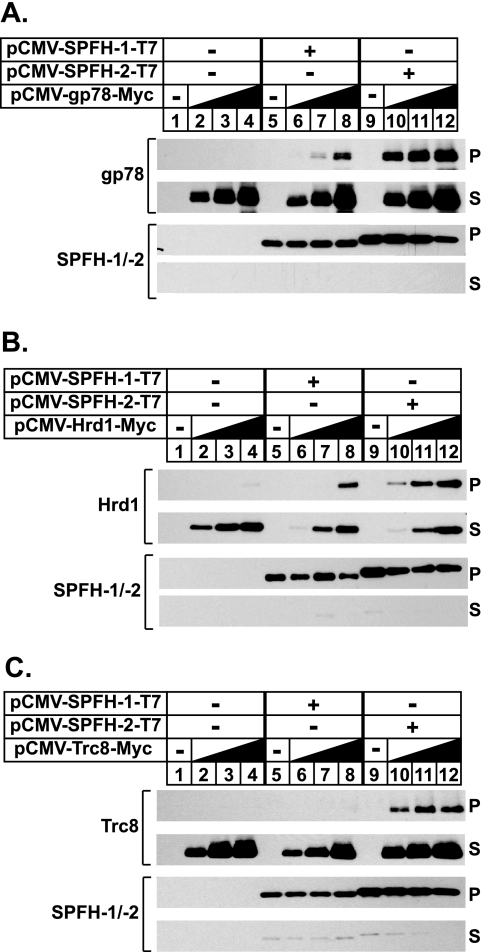
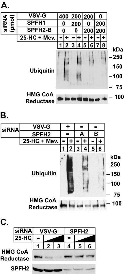
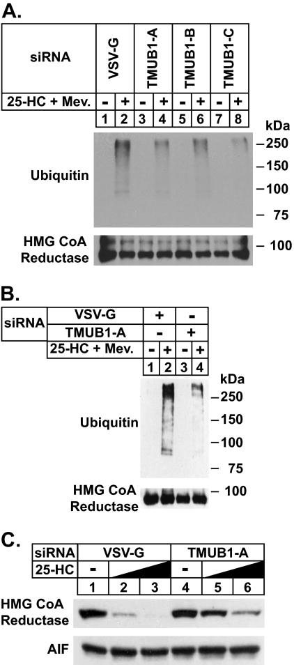
The endoplasmic reticulum (ER)-associated degradation (ERAD) pathway in the yeast Saccharomyces cerevisiae is mediated by two membrane-bound ubiquitin ligases, Doa10 and Hrd1. These enzymes are found in distinct multiprotein complexes that allow them to recognize and target a variety of substrates for proteasomal degradation. Although multiprotein complexes containing mammalian ERAD ubiquitin ligases likely exist, they have yet to be identified and characterized in detail. Here, we identify two ER membrane proteins, SPFH2 and TMUB1, as associated proteins of mammalian gp78, a membrane-bound ubiquitin ligase that bears significant sequence homology with mammalian Hrd1 and mediates sterol-accelerated ERAD of the cholesterol biosynthetic enzyme HMG-CoA reductase. Co-immunoprecipitation studies indicate that TMUB1 bridges SPFH2 to gp78 in ER membranes. The functional significance of these interactions is revealed by the observation that RNA interference (RNAi)-mediated knockdown of SPFH2 and TMUB1 blunts both the sterol-induced ubiquitination and degradation of endogenous reductase in HEK-293 cells. These studies mark the initial steps in the characterization of the mammalian gp78 ubiquitin ligase complex, the further elucidation of which may yield important insights into mechanisms underlying gp78-mediated ERAD.


Figures

FIGURE 1.
FIGURE 1.
Identification of SPFH2 as a gp78-associated protein. A, CHO/gp78-TAP cells were set up on day 0 at 4 × 10⁵ cells/60-mm dish in medium A containing 5% LPDS. On day 1 the cells were switched to medium A supplemented with 5% LPDS, 10 μm sodium compactin, and 50 μm sodium mevalonate. After incubation for 16 h at 37 °C, the cells were re-fed the identical medium supplemented with 5% LPDS and 10 μm compactin in the absence or presence of 10 mm mevalonate plus 0.1–1.0 μg/ml 25-HC. After incubation for 5 h at 37 °C, the cells were harvested and subjected to subcellular fractionation as described under “Experimental Procedures.” Aliquots of the resulting membrane fractions were subjected to SDS-PAGE, the proteins were transferred to nitrocellulose membranes, and immunoblot analysis was carried out with monoclonal IgG-A9 (against reductase), monoclonal anti-T7 IgG (against gp78-TAP), and polyclonal anti-gp78 IgG. B, HEK-293 cells were set up on day 0 at 7 × 10⁵ cells/100-mm dish in medium B supplemented with 10% FCS. On day 2 the cells were depleted of sterols through incubation in medium B containing 10% LPDS, 10 μm compactin, and 50 μm mevalonate. After incubation for 16 h at 37 °C, the cells were re-fed the identical medium in the absence or presence of 10 μm MG-132 for 2 h, after which they received 1 μg/ml 25-HC plus 10 mm mevalonate (Mev.) for additional 2 h as indicated. The cells were then harvested, lysed, and subjected to immunoprecipitation (Immunoppt.) with either control preimmune (Preim.) IgG or anti-gp78 IgG. Aliquots of the resulting pellet and supernatant fractions of the immunoprecipitation were subjected to SDS-PAGE followed by immunoblot analysis with polyclonal anti-gp78 IgG, monoclonal IgG-A9 (against reductase), monoclonal anti-VCP/p97, and polyclonal anti-SPFH2 IgG. Ab, antibody.
FIGURE 2.
FIGURE 2.
Association of SPFH2 with gp78 and other membrane-bound ubiquitin ligases. A–C, CHO-7 cells were set up on day 0 at 5 × 10⁵ cells/60-mm dish in medium A containing 5% LPDS. On day 1 the cells were transfected with 2 μg/dish of empty pcDNA3.1 vector (lanes 1–4), pCMV-SPFH1-T7 (lanes 5–8), or pCMV-SPFH2-T7 (lanes 9–12) together with increasing amounts (0.1, 0.3, and 1.0 μg) of pCMV-gp78-Myc (A), pCMV-Hrd1-Myc (B), or pCMV-Trc8-Myc (C) in medium A containing 5% LPDS as described under “Experimental Procedures.” The total amount of DNA/dish was adjusted to 2 μg by the addition of pcDNA3.1 mock vector. After incubation for 3–6 h at 37 °C, the cells were depleted of sterols by direct addition of medium A containing 5% LPDS, 10 μm compactin, and 50 μm mevalonate (final concentration). After 16 h at 37 °C, the cells were harvested, lysed, and immunoprecipitated with anti-T7 coupled beads. Aliquots of the pellet (P) and supernatant (S) fractions of the immunoprecipitation were subjected to SDS-PAGE, and immunoblot analysis was carried out with polyclonal anti-T7 IgG (against SPFH1 and SPFH2) and monoclonal IgG-9E10 (against gp78, Hrd1, and Trc8).
FIGURE 3.
FIGURE 3.
RNAi-mediated knockdown of SPFH2 blunts ER-associated degradation of HMG-CoA reductase and Insig-1. HEK-293 cells were set up on day 0 in medium B containing 10% FCS at 2 × 10⁵cells/60-mm dish. On days 1 and 3 the cells were transfected in medium B containing 10% FCS with the indicated amount of siRNA duplexes targeted SPFH1, SPFH2, or the control mRNA, vesicular stomatitis virus glycoprotein (VSV-G), as described under “Experimental Procedures.” A and B, after sterol depletion for 16 h at 37 °C, the cells were incubated for 1 h in medium B supplemented with 10% LPDS, 10 μm compactin, and 10 μm MG-132 in the absence or presence of 1 μg/ml 25-HC plus 10 mm mevalonate (Mev.). The cells were subsequently harvested and subjected to immunoprecipitation with polyclonal antibodies against reductase. Aliquots of the immunoprecipitates were subjected sequentially to SDS-PAGE and immunoblot analysis with monoclonal IgG-A9 (against reductase) and monoclonal IgG-P4D1 (against ubiquitin). C, after sterol depletion for 16 h at 37 °C, the cells were incubated for 2.5 h in medium B supplemented with 10% LPDS, 10 μm compactin, and 0, 0.3, or 1.0 μg/ml 25-HC as indicated. The cells were subsequently harvested for subcellular fractionation. Aliquots of the membrane fractions (normalized for equal protein loaded/lane) were subjected to SDS-PAGE followed by immunoblot analysis with IgG-A9 (against reductase) and anti-SPFH2 IgG.
FIGURE 4.
FIGURE 4.
Organization of complex between HMG-CoA reductase, Insig-1, gp78, and SPFH2 as determined by co-immunoprecipitation. A, HEK-293 cells were set for experiments on day 0, depleted of sterols on day 2, and treated in the absence or presence of 10 μm MG-132 and 1 μg/ml 25-HC plus 10 mm mevalonate (Mev.) on day 3 as described in the legend to Fig. 1B. After treatments the cells were harvested, lysed, and subjected to immunoprecipitation (Immunoppt.) with polyclonal antibodies against reductase (Red.). Aliquots of the resulting pellet and supernatant fractions of the immunoprecipitation were subjected to SDS-PAGE, and immunoblot analysis was carried out with monoclonal IgG-A9 (against reductase), polyclonal anti-gp78, anti-SPFH2, and monoclonal anti-VCP/p97 IgGs. Ab, antibody; Preim., Preimmune. B, CHO-7 cells were set up for experiments on day 0, transfected with 1 μg of pCMV-HMG-Red-T7, 0.1 μg of pCMV-Insig-1-Myc, and 0.1 μg of pCMV-SPFH2-Myc, and depleted of sterols on day 1 as described in the legend to Fig. 2. The sterol-depleted cells were then incubated for 2 h at 37 °C in medium A containing 5% LPDS, 10 μm compactin, and 10 μm MG-132 in the absence or presence of 1 μg/ml 25-HC plus 10 mm mevalonate as indicated. The cells were subsequently harvested, lysed, and subjected to anti-T7 immunoprecipitation. Aliquots of the resulting supernatant and pellet fractions were subjected to SDS-PAGE followed by immunoblot analysis with polyclonal anti-T7 IgG (against reductase) and monoclonal IgG-9E10 (against Insig-1 and SPFH2). C, CHO-7 cells were set up on day 0, transfected with 0.1 μg of pCMV-Insig-1-T7, 0.01 μg of pCMV-gp78-Myc, and 0.1 μg of pCMV-SPFH2-Myc and depleted of sterols on day 1 as described in B. After sterol depletion, the cells were incubated for 2 h at 37 °C in medium A supplemented with 5% LPDS, 10 μm compactin, and 10 μm MG-132. The cells were subsequently harvested and lysed, and immunoprecipitation was carried out with anti-T7-coupled beads. Aliquots of the pellet and supernatant fractions of the immunoprecipitation were subjected to SDS-PAGE and immunoblot analysis with polyclonal anti-T7 IgG (against Insig-1) and monoclonal IgG-9E10 (against gp78 and SPFH2). D, CHO-7 cells were set up on day 0, transfected with 0.1 μg/dish pCMV-SPFH2-T7 together with 0.1 μg/dish pCMV-gp78-Myc (lanes 1 and 2, WT), (lanes 3 and 4, TM), or (lanes 5 and 6, Cyto.) as indicated and depleted of sterols on day 1 as described in A. After sterol depletion, the cells were subjected to incubation for 2 h in medium A containing 5% LPDS and 10 μm compactin with or without 1 μg/ml 25-HC plus 10 mm mevalonate as indicated. The cells were then harvested, lysed, and immunoprecipitated with anti-T7 beads. Aliquots of the pellet and supernatant fractions were subjected to SDS-PAGE followed by immunoblot analysis with polyclonal anti-T7 (against SPFH2) and monoclonal IgG-9E10 (against gp78).
FIGURE 5.
FIGURE 5.
Identification of TMUB1 as an associated protein of SPFH2. A, shown is the domain structure of TMUB1. B–E, CHO-7 cells were set up for experiments on day 0, transfected with 0.1 μg of pCMV-SPFH2-T7, 1 μg of pCMV-TMUB1-Myc, 1 μg of pCMV-UbxD2-Myc, and 1 μg of pCMV-UbxD8-Myc (B), 0.1 μg of pCMV-SPFH2-Myc, 1 μg of pCMV-TMUB1 (WT), (ΔHD), (ΔTM1&2), and (ΔTM2)-Myc (C), 0.1 μg of pCMV-gp78-Myc, 0.5 μg of pCMV-TMUB1 (WT), 0.02 μg (ΔHD), 0.2 μg of (ΔTM1&2), 0.05 μg of (ΔTM2), and 1.2 μg of (ΔHD, TM1&2)-T7 (D), and 0.1 μg of pCMV-SPFH2-Myc, 1 μg of pCMV-TMUB1-T7, and 0.01 μg of pCMV-gp78-T7 (E) as indicated and depleted of sterols as described in Fig. 2. After sterol depletion, the cells were re-fed medium A containing 5% LPDS, 10 μm compactin, and 10 μm MG-132 in the absence or presence of 1 μg/ml 25-HC plus 10 mm mevalonate (Mev.) as indicated. The cells were subsequently harvested and subjected to immunoprecipitation with anti-Myc (B and E)- or anti-T7 (C and D)-coupled beads. Aliquots of the resulting pellet and supernatant fractions of the immunoprecipitations were subjected to SDS-PAGE followed by immunoblot analysis with polyclonal or monoclonal anti-T7 (against SPFH2 in B. TMUB1 in C, D, and E. gp78 in E) and monoclonal IgG-9E10 (against TMUB1, UbxD2, and UbxD8 in B, SPFH2 in C and E, and gp78 in D).
FIGURE 6.
FIGURE 6.
RNAi-mediated knockdown of TMUB1 blunts degradation of HMG-CoA reductase and Insig-1. SV-589 cells were set up for experiments on day 0 at 2 × 10⁵ cells/60-mm dish in medium B containing 10% FCS. On days 1 and 2 the cells were transfected with 600 pmol/dish of the indicated siRNA duplex in medium B containing 10% FCS as described under “Experimental Procedures.” A and B, after sterol depletion the cells were incubated for 30 min at 37 °C in medium B containing 10% LPDS, 10 μm compactin, and 10 μm MG-132 in the absence or presence of 1 μg/ml 25-HC plus 10 mm mevalonate (Mev.) as indicated. The cells were then harvested, lysed, and subjected to immunoprecipitation with polyclonal anti-reductase. Aliquots of the immunoprecipitates were subjected to SDS-PAGE followed by immunoblot analysis with monoclonal IgG-A9 (against reductase) and IgG-P4D1 (against ubiquitin). VSV-G, vesicular stomatitis virus glycoprotein. C, sterol-depleted cells were incubated for 2.5 h at 37 °C in medium B containing 10% LPDS and 10 μm compactin in the absence or presence of 25-HC (0.3 or 1.0 μg/ml). The cells were then harvested for subcellular fractionation. Aliquots of the membrane fractions were subjected to SDS-PAGE followed by immunoblot analysis with monoclonal IgG-A9 (against reductase) and polyclonal anti-apoptosis inducing factor (AIF) as a loading control.

Similar articles

Cited by

References

    1. Vembar S. S., Brodsky J. L. (2008) Nat. Rev. Mol. Cell Biol. 9, 944–957 - PMC - PubMed
    1. Hampton R. Y., Garza R. M. (2009) Chem. Rev. 109, 1561–1574 - PMC - PubMed
    1. Meusser B., Hirsch C., Jarosch E., Sommer T. (2005) Nat. Cell Biol. 7, 766–772 - PubMed
    1. Jarosch E., Lenk U., Sommer T. (2003) Int. Rev. Cytol. 223, 39–81 - PubMed
    1. Kostova Z., Tsai Y. C., Weissman A. M. (2007) Semin. Cell Dev. Biol. 18, 770–779 - PMC - PubMed

Publication types

MeSH terms