. 2010 Dec 23;6(12):e1001238.
doi: 10.1371/journal.ppat.1001238.

Interaction of c-Cbl with myosin IIA regulates Bleb associated macropinocytosis of Kaposi's sarcoma-associated herpesvirus

Affiliations

Interaction of c-Cbl with myosin IIA regulates Bleb associated macropinocytosis of Kaposi's sarcoma-associated herpesvirus

Mohanan Valiya Veettil et al. PLoS Pathog. .

Erratum in

  • PLoS Pathog. 2011;7(2). doi:10.1371/annotation/79109603-41dd-40b8-a9ec-8df2c7fa42eb

Abstract

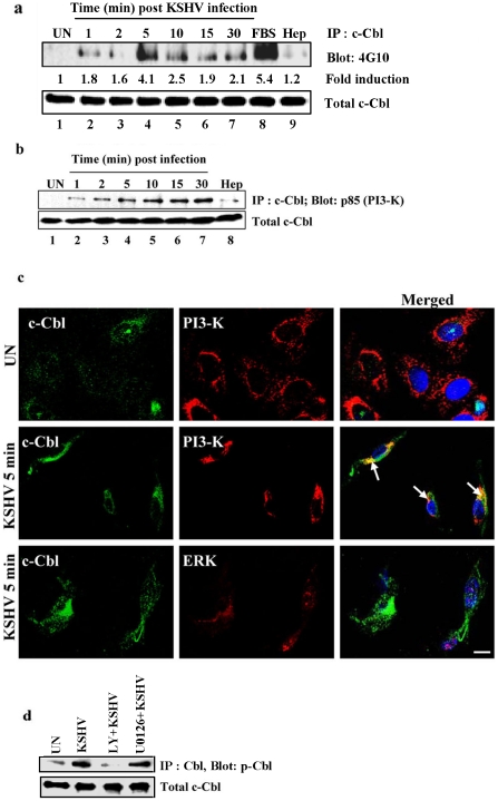
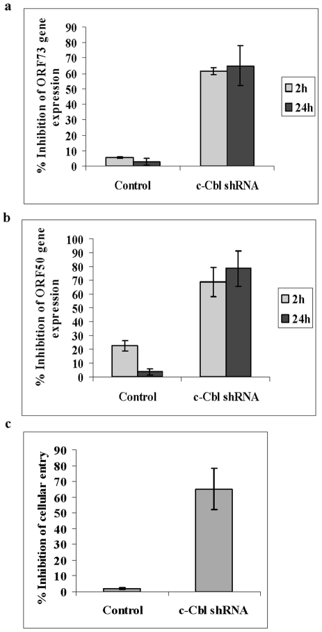
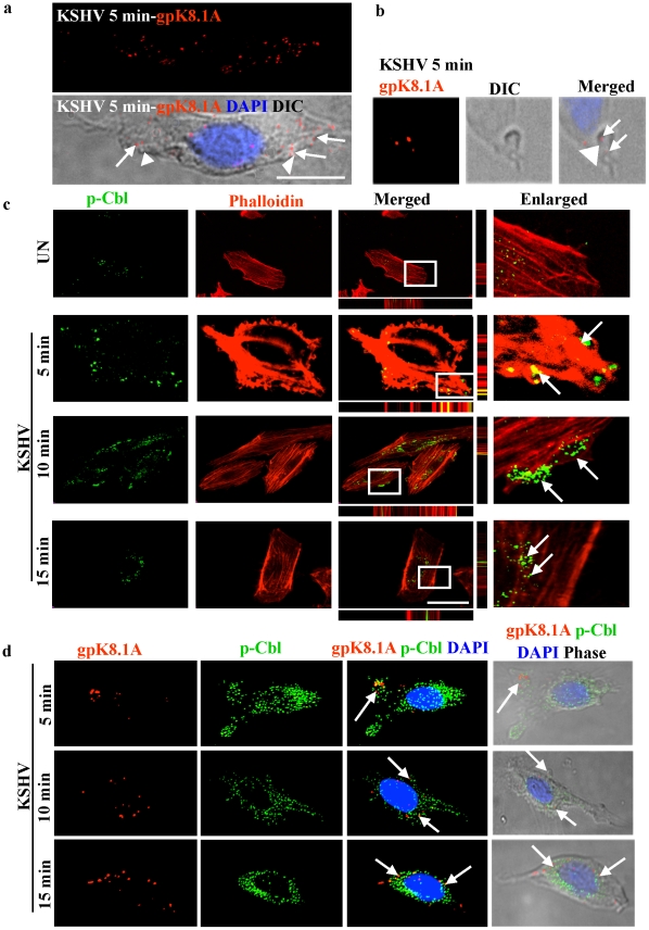
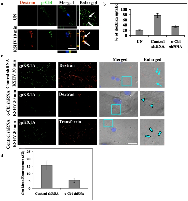
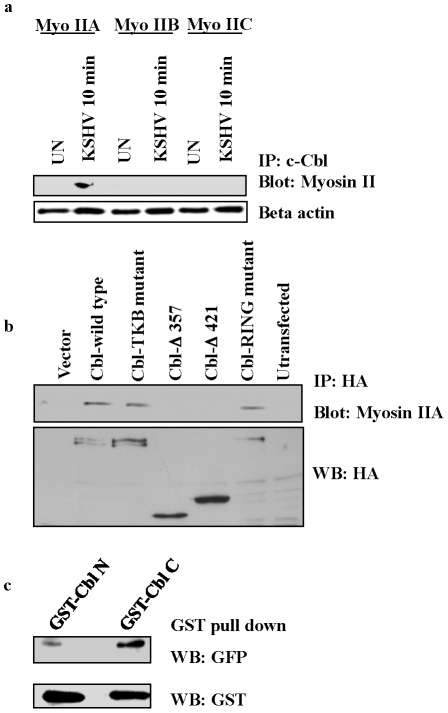
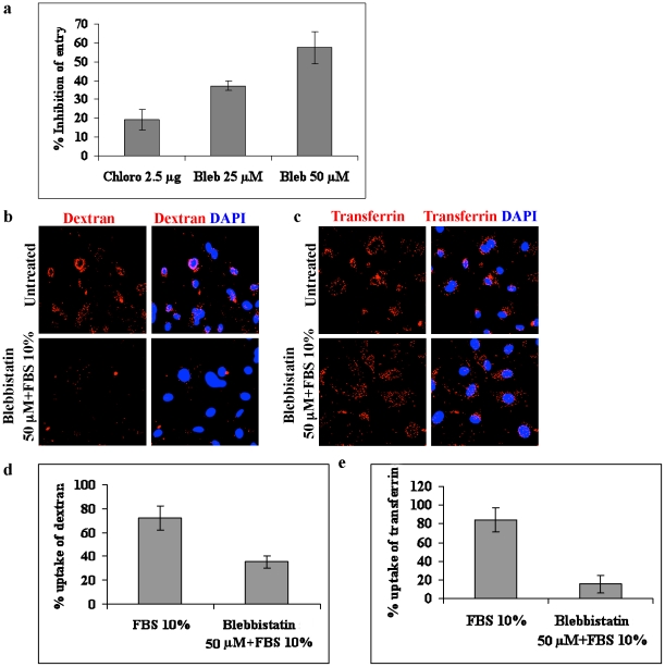
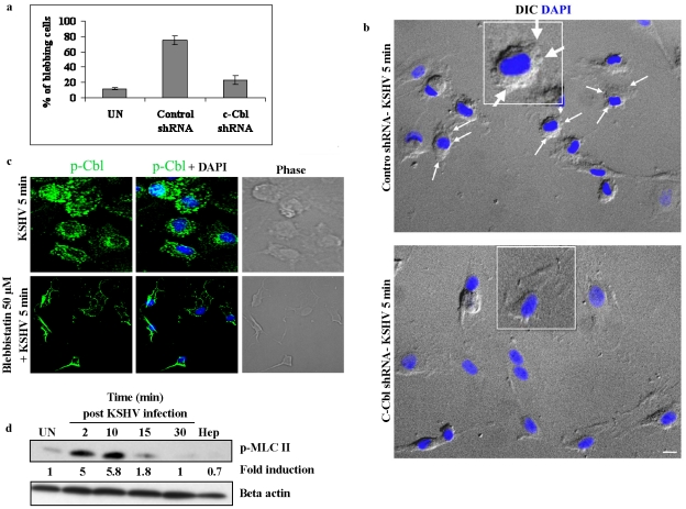
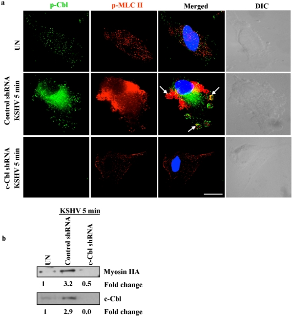
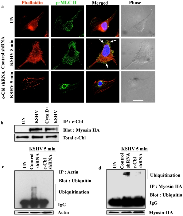
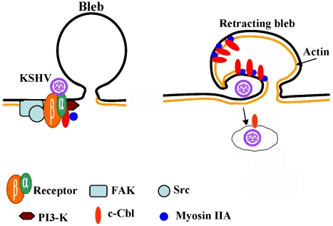
KSHV is etiologically associated with Kaposi's sarcoma (KS), an angioproliferative endothelial cell malignancy. Macropinocytosis is the predominant mode of in vitro entry of KSHV into its natural target cells, human dermal microvascular endothelial (HMVEC-d) cells. Although macropinocytosis is known to be a major route of entry for many viruses, the molecule(s) involved in the recruitment and integration of signaling early during macropinosome formation is less well studied. Here we demonstrate that tyrosine phosphorylation of the adaptor protein c-Cbl is required for KSHV induced membrane blebbing and macropinocytosis. KSHV induced the tyrosine phosphorylation of c-Cbl as early as 1 min post-infection and was recruited to the sites of bleb formation. Infection also led to an increase in the interaction of c-Cbl with PI3-K p85 in a time dependent manner. c-Cbl shRNA decreased the formation of KSHV induced membrane blebs and macropinocytosis as well as virus entry. Immunoprecipitation of c-Cbl followed by mass spectrometry identified the interaction of c-Cbl with a novel molecular partner, non-muscle myosin heavy chain IIA (myosin IIA), in bleb associated macropinocytosis. Phosphorylated c-Cbl colocalized with phospho-myosin light chain II in the interior of blebs of infected cells and this interaction was abolished by c-Cbl shRNA. Studies with the myosin II inhibitor blebbistatin demonstrated that myosin IIA is a biologically significant component of the c-Cbl signaling pathway and c-Cbl plays a new role in the recruitment of myosin IIA to the blebs during KSHV infection. Myosin II associates with actin in KSHV induced blebs and the absence of actin and myosin ubiquitination in c-Cbl ShRNA cells suggested that c-Cbl is also responsible for the ubiquitination of these proteins in the infected cells. This is the first study demonstrating the role of c-Cbl in viral entry as well as macropinocytosis, and provides the evidence that a signaling complex containing c-Cbl and myosin IIA plays a crucial role in blebbing and macropinocytosis during viral infection and suggests that targeting c-Cbl could lead to a block in KSHV infection.


Conflict of interest statement

The authors have declared that no competing interests exist.

Figures

Figure 1
Figure 1. KSHV infection induces tyrosine phosphorylation of c-Cbl and association of c-Cbl with PI3-K.
(a) KSHV infection induces tyrosine phosphorylation of c-Cbl. HMVEC-d cells were left uninfected or infected with KSHV for the indicated time points after serum starvation. Tyrosine phosphorylation of c-Cbl was determined by immunoprecipitating with anti-c-Cbl and Western blotting with anti-phosphotyrosine antibody 4G10. Cells treated with 10% FBS for 10 min were used as positive control. As a negative control, cells were infected for 10 min with KSHV pre-incubated with heparin (50 µg ml−1) for 1 h at 37°C. Band intensities (middle panel) were assessed as described in the methods and expressed as increased fold tyrosine phosphorylation of c-Cbl over uninfected cells. Bottom panel shows the blot reprobed with anti-c-Cbl antibody. (b) c-Cbl associates with PI3-K in the infected cells. Uninfected or cells infected with KSHV for different time points were lysed and immunoprecipitated with anti-c-Cbl and Western blotted with anti-p85 (PI-3K) antibody. Cells infected for 10 min with heparin treated virus were used as specificity control. Bottom panel shows the blot reprobed with anti-c-Cbl antibody. (c) c-Cbl colocalizes with PI3-K in the infected cells. HMVECd cells infected with KSHV for 5 min were immunostained with anti-c-Cbl and anti-PI3-K p85 antibody and analyzed by confocal microscopy. Arrows indicate colocalization of c-Cbl-with PI3-K p85. ERK staining showing no colocalization with c-Cbl was used as specificity control. (d) Effect of PI3-K and ERK1/2 inhibitors on c-Cbl phosphorylation. HMVEC-d cells either left untreated or pre-treated with 50 µM LY294002 (LY) or 10 µM U0126 for 1 h were infected with KSHV for 10 min. Uninfected and infected cell lysates were immunoprecipitated with anti-c-Cbl and Western blotted with anti-phospho Cbl antibody.
Figure 2
Figure 2. c-Cbl shRNA inhibits KSHV entry and infection.
(a and b) KSHV gene expression in c-Cbl ShRNA transduced cells. Untransduced, control shRNA and c-Cbl shRNA transduced HMVEC-d cells were infected with KSHV. At 2 and 24 h post infection, cells were harvested, total RNA isolated and viral gene expression determined by real-time RT-PCR with KSHV ORF73 (a) and ORF50 (b) gene specific primers and TaqMan probes. (c) Entry of KSHV in c-Cbl ShRNA transduced cells. Untransduced, control shRNA and c-Cbl shRNA transduced HMVEC-d cells were infected with KSHV for 30 min. DNA was isolated and KSHV entry was determined by real-time DNA PCR for ORF73 gene. For all the above, each reaction was done in duplicate and each bar represents the average ± SD of three experiments.
Figure 3
Figure 3. c-Cbl associates with KSHV induced blebs.
(a) Association of blebs with KSHV in the infected cells. HMVEC-d cells were infected with KSHV for 5 min and immunostained with anti-gpK8.1A (viral envelope glycoprotein) antibody for the detection of KSHV. Images showing blebs were acquired by fluorescence microscopy equipped with DIC (Differential Interference Contrast). The merged image represents a gpK8.1A, DIC, and DAPI stained nucleus. Arrow heads indicate blebs and arrows indicate viral particles (gpK8.1A staining) associated with the blebs. Scale bar: 10 µm. (b) Image of a single bleb and KSHV (gpK8.1A staining) were acquired by fluorescence microscopy equipped with DIC under an 100x objective lens. The gpK8.1A, DIC and DAPI-stained nucleus are merged in the rightmost image. Arrow heads indicate a bleb and arrows indicate viral particles associated with the bleb. (c) Association of blebs with c-Cbl in the infected cells. Uninfected or cells infected with KSHV for 5, 10 and 15 min were stained with rhodamine-phalloidin and with anti-phospho c-Cbl (p-Cbl) antibody. The staining was analyzed by confocal microscopy sections through the z axis. Arrows indicate the association of p-Cbl with blebs and internalization at different time points. Bottom and side bars represent the xz axis. The boxed areas are enlarged in the rightmost panel. Scale bar: 10 µm. (d) Association of viral particles with c-Cbl in the infected cells. HMVEC-d cells infected with KSHV for 5, 10 and 15 min were fixed and processed for double immunofluorescence using anti-p-Cbl and anti-gpK8.1A antibodies. Panels show the staining of gpK8.1A, and the images merged with p-Cbl, DAPI and DIC. Arrows indicate the colocalization of p-Cbl and gpK8.1A in the infected cells.
Figure 4
Figure 4. c-Cbl is involved in macropinocytosis of KSHV.
(a) Colocalization of c-Cbl with the macropinocytosis marker dextran. HMVEC-d cells were incubated with medium containing Texas Red labeled dextran alone (no virus, UN) or with KSHV and Texas Red labeled dextran at 37°C for 10 min. Cells were processed for confocal immunofluorescence using anti-p-Cbl antibody and costained with DAPI. Arrows in the merged panel indicate colocalization of p-Cbl with dextran. Scale bar: 10 µm. Bottom and side bars represent the xz axis. (b) Dextran uptake in c-Cbl shRNA cells. Control shRNA or c-Cbl shRNA transduced HMVEC-d cells were incubated with KSHV and Texas Red labeled dextran or with Texas Red labeled dextran alone (UN) at 37°C for 30 min. Quantitative analysis of dextran uptake was determined as described in the methods and represent the mean ± SD of three independent experiments. (c) Colocalization of dextran and KSHV. HMVEC-d cells transduced with control shRNA or c-Cbl shRNA were incubated with KSHV and Texas Red labeled dextran for 30 min at 37°C. Control shRNA cells incubated with KSHV and Alexa 594 labeled transferrin for 30 min were used as specificity controls. The cells were then stained with anti-gpK8.1A antibody and analyzed by fluorescence microscopy combined with DIC. Merged panel shows the images merged with DAPI stained nuclei and DIC. The boxed areas are enlarged in the rightmost panel. Arrows: colocalization of KSHV with dextran in the control shRNA cells. Arrow heads: viral particles remained at the cell membrane in c-Cbl shRNA cells. Blocked arrows: non-colocalization of viral particles with transferrin in control shRNA cells. (d) Flow cytometry analysis of FITC-dextran uptake. Control and c-Cbl shRNA cells were incubated with 500 µg ml−1 FITC-dextran in the presence or absence of virus at 37°C for 30 min. The cells were then washed, harvested, fixed and analyzed by flow cytometry. Quantification of flow cytometry analysis of dextran uptake is shown as mean fluorescence intensity.
Figure 5
Figure 5. c-Cbl associates with myosin IIA in KSHV infected cells.
(a) c-Cbl co-immunoprecipitates with myosin IIA in KSHV infected cells. Serum starved HMVEC-d cells were left uninfected (UN) or infected with KSHV for 10 min. The cells were lysed, immunoprecipitated with anti-c-Cbl antibody and Western blotted with myosin II isoform specific antibodies, IIA, IIB and IIC. Bottom panel: loading control with anti-beta actin antibody. (b) c-Cbl associates with endogenous myosin IIA in HeLa cells. HeLa cells were transfected with HA-tagged empty vector or Cbl wild-type, TKB mutant or RING mutant, truncated Cbl-Δ421 or Cbl-Δ357 constructs. Cells were stimulated with TPA for 5 min and the resulting cell lysates were subjected to immunoprecipitation with anti-HA antibodies followed by immunoblotting with myosin IIA antibody (top). Western blotting was carried out with anti-HA antibody using the same lysates (bottom). (c) GST pull-down assay. GST Cbl-C and Cbl-N fusion proteins expressed in bacterial cells were adsorbed with GST-beads at 4°C for 2 h. The beads were washed and incubated with lysates of 293T cells transiently expressing GFP-tagged myosin IIA. The proteins bound with GST beads were analyzed by Western blotting with anti-GFP antibody. Bottom panel shows GST expression with anti-GST antibody.
Figure 6
Figure 6. Blebbistatin inhibits macropinocytosis of KSHV.
(a) Effect of blebbistatin on entry of KSHV. HMVEC-d cells were pretreated with 25 and 50 µM blebbistatin (Bleb) or with 2.5 µg ml−1 chlorpromazine (Chlor) for 1 h before infection with KSHV for 30 min. The cells were then treated with trypsin-EDTA, harvested, and total DNA was isolated. Entry was determined by estimating ORF73 DNA copies by real-time DNA PCR. Percentage inhibition of KSHV DNA internalization was calculated compared to the untreated cells. (b and c). Effect of blebbistatin on dextran and transferrin uptake. HMVEC-d cells were left untreated or treated with blebbistatin for 1 h at 37°C. The cells were then induced with FBS in the presence of dextran (b) or transferrin (c) for 30 min, fixed and analyzed by immunofluorescence. (d and e) Quantitative analysis of dextran and transferrin uptake was determined by counting the number of positive cells. At least 10 different microscopic fields of 50 cells each were counted for each experiment and the results displayed as percentage of dextran or transferrin positive cells.
Figure 7
Figure 7. c-Cbl is upstream of myosin IIA in the induction of blebs.
(a) Effect of c-Cbl shRNA on KSHV induced blebbing. Control shRNA and c-Cbl shRNA cells were infected with KSHV for 5 min. Uninfected and infected cells were fixed and the blebs were observed under an immunofluorescence microscope with DIC. The percentage of blebbed cells was determined and represented as mean ± SD of three independent experiments. (b) Blebbing in control and c-Cbl shRNA transduced cells. Representative DIC images showing bleb formation in control and c-Cbl shRNA cells infected with KSHV. Nuclei were stained with DAPI and merged with DIC images. KSHV induced blebs can be seen in control cells (arrows), whereas bleb formation in c-Cbl shRNA cells is comparatively less. Scale bar: 10 µm. Insets show a single enlarged cell. (c) Blebbistatin does not affect membrane localization of p-Cbl. HMVEC-d cells were pretreated with blebbistatin for 1 h at 37°C. Untreated and cells treated with blebbistatin were infected with KSHV for 5 min, fixed and immunostained for p-Cbl. (d) KSHV induces phosphorylation of p-MLC II. Serum starved HMVEC-d cells were left uninfected or infected with KSHV for 2, 10, 15 and 30 min. Cells infected with heparin treated (Hep) virus were used as negative control. Cell lysates were Western blotted with anti-p-MLC II antibodies (top panel) or anti-β actin antibodies (bottom panel).
Figure 8
Figure 8. c-Cbl recruits myosin IIA to the membrane blebs.
(a) Colocalization of c-Cbl and p-MLC II in KSHV induced blebs. Control and c-Cbl shRNA cells were infected with KSHV for 5 min. Uninfected and infected cells were then immunostained with anti-p-Cbl or anti-p-MLC II antibody. The rightmost panels show DIC images. In the merged panel, arrows indicate the colocalization of p-Cbl with p-MLC II. Scale bar: 10 µm. (b) Membrane association of myosin II in c-Cbl shRNA cells. Control shRNA and c-Cbl shRNA cells were infected with KSHV for 10 min. The membrane fractions were isolated from uninfected and infected samples, then Western blotted with myosin IIA to analyze the membrane localization of myosin IIA (top). The same membrane was stripped and reprobed with anti-c-Cbl to analyze the corresponding c-Cbl membrane localization (bottom).
Figure 9
Figure 9. Myosin II associates with actin in KSHV induced blebs.
(a) Colocalization of p-MLC II with actin in KSHV induced blebs. Control and c-Cbl shRNA transduced cells were infected with KSHV for 5 min and stained with rhodamine-phalloidin and anti-p-MLC II antibody. Arrows indicate the colocalization of p-MLC II and actin in the blebs. The rightmost column shows DIC images. Scale bar: 10 µm. (b) Actin inhibitor does not affect the interaction of myosin IIA with c-Cbl. HMVEC-d cells were pretreated with 2 µg ml−1 cytochalasin D for 1 h at 37°C and then infected with KSHV and washed. Cells were lysed and the cell lysates were immunoprecipitated with c-Cbl antibody and Western blotted with myosin IIA. (c) and (d) Ubiquitination of myosin and actin. Control shRNA and c-Cbl shRNA cells were serum starved overnight and infected with KSHV for 10 min, and lysed. Cell lysates were immunoprecipitated with anti-actin antibody (c) or anti-myosin antibody (d) and analyzed by Western blotting using anti-ubiquitin antibody. Membranes were reprobed with actin or myosin antibody. Arrow marks indicate multiple bands of ubiquitinated proteins. To show the multiple bands of ubiquitinated actin and myosin, actin (40 kDa) was resolved on a 12.5% gel (Figure 9c). Since myosin is a high molecular weight protein (220 kDa), for better resolution, 7.5% gel was used (Figure 9d).
Figure 10
Figure 10. Model depicting the role of c-Cbl and myosin IIA interaction during blebbing and macropinocytosis of KSHV.
KSHV has been previously shown to interact with heparin sulfate, α3β1, αVβ3 and αVβ5 integrins and xCT molecules leading to the phosphorylation of FAK and the downstream molecules such as Src, PI3-K and RhoGTPases . KSHV infection induces the phosphorylation of c-Cbl and the phosphorylated c-Cbl forms a complex with p85-PI3K, thus facilitating the interaction of c-Cbl with downstream molecules. c-Cbl associates with myosin IIA and actin and is rapidly recruited to membrane blebs. This association also leads into c-Cbl mediated ubiquitination of actin and myosin. Myosin IIA interactions with actin may be accelerating the actomyosin contraction and bleb retraction which form macropinosomes along with the viral particles. Myosins probably provide the ATP-dependent force to generate the movement required for the process of bleb retraction. An increase in c-Cbl–myosin IIA association possibly affects c-Cbl-mediated blebbing, macropinocytosis and internalization of KSHV.

Similar articles

Cited by

References

    1. Chang Y, Cesarman E, Pessin MS, Lee F, Culpepper J, et al. Identification of herpesvirus-like DNA sequences in AIDS-associated Kaposi's sarcoma. Science. 1994;266:1865–1869. - PubMed
    1. Cesarman E, Chang Y, Moore PS, Said JW, Knowles DM. Kaposi's sarcoma-associated herpesvirus-like DNA sequences in AIDS-related body-cavity-based lymphomas. N Engl J Med. 1995;332:1186–1191. - PubMed
    1. Chandran B. Early events in KSHV infection of target cells. J Virol. 2009;84:2188–99. - PMC - PubMed
    1. Raghu H, Sharma-Walia N, Veettil MV, Sadagopan S, Chandran B. Kaposi's sarcoma-associated herpesvirus utilizes an actin polymerization-dependent macropinocytic pathway to enter human dermal microvascular endothelial and human umbilical vein endothelial cells. J Virol. 2009;83:4895–4911. - PMC - PubMed
    1. Akula SM, Naranatt PP, Walia NS, Wang FZ, Fegley B, et al. Kaposi's sarcoma-associated herpesvirus (human herpesvirus 8) infection of human fibroblast cells occurs through endocytosis. J Virol. 2003;77:7978–7990. - PMC - PubMed

Publication types

Substances