. 2010 Dec 23;143(7):1136-48.
doi: 10.1016/j.cell.2010.11.034.

Wnt signaling requires sequestration of glycogen synthase kinase 3 inside multivesicular endosomes

Affiliations

Wnt signaling requires sequestration of glycogen synthase kinase 3 inside multivesicular endosomes

Vincent F Taelman et al. Cell. .

Abstract

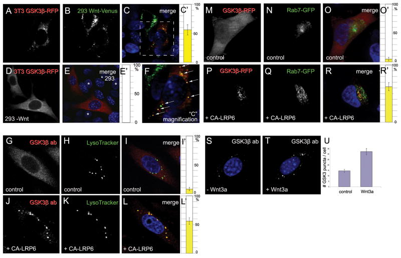
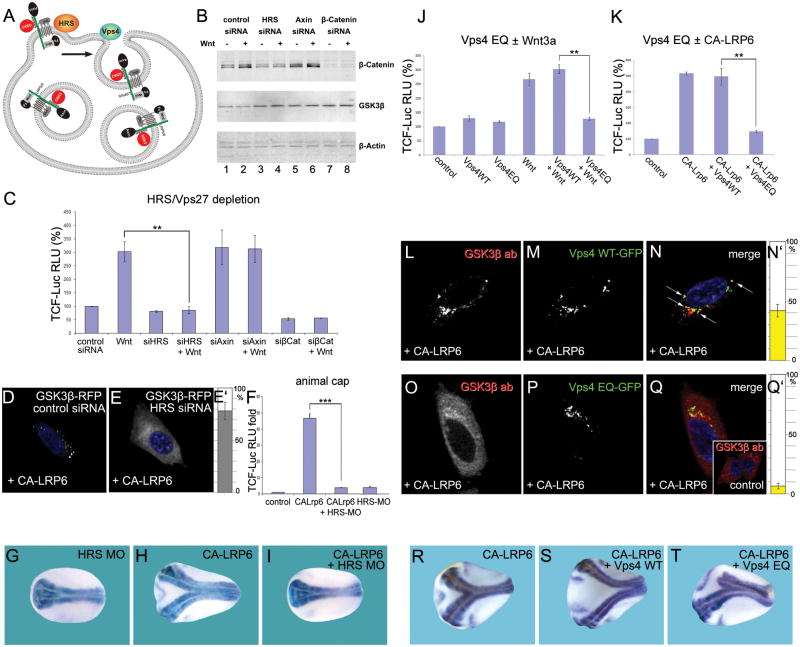
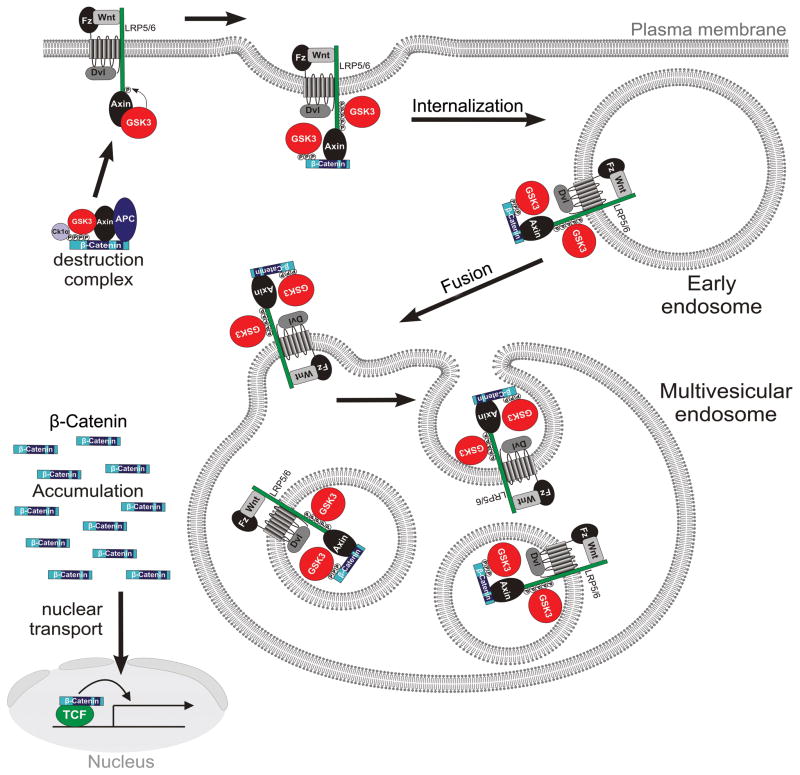
Canonical Wnt signaling requires inhibition of Glycogen Synthase Kinase 3 (GSK3) activity, but the molecular mechanism by which this is achieved remains unclear. Here, we report that Wnt signaling triggers the sequestration of GSK3 from the cytosol into multivesicular bodies (MVBs), so that this enzyme becomes separated from its many cytosolic substrates. Endocytosed Wnt colocalized with GSK3 in acidic vesicles positive for endosomal markers. After Wnt addition, endogenous GSK3 activity decreased in the cytosol, and GSK3 became protected from protease treatment inside membrane-bounded organelles. Cryoimmunoelectron microscopy showed that these corresponded to MVBs. Two proteins essential for MVB formation, HRS/Vps27 and Vps4, were required for Wnt signaling. The sequestration of GSK3 extended the half-life of many other proteins in addition to β-Catenin, including an artificial Wnt-regulated reporter protein containing GSK3 phosphorylation sites. We conclude that multivesicular endosomes are essential components of the Wnt signal-transduction pathway.


Figures

Figure 1
Figure 1. GSK3β is Translocated to Acidic Vesicles upon Wnt Signaling
(A–F) In co-culture experiments, xWnt8-Venus (green) secreted by 293T cells caused the relocalization of GSK3β-RFP (red) in 3T3 cells that endocytosed Wnt. Arrows in the enlargement in panel F indicate xWnt8-Venus endosomes that sequester GSK3-RFP. In controls lacking xWnt8-Venus, GSK3-RFP localization was cytoplasmic (D and E); asterisks indicate untransfected control 293 cell nuclei. (G–L′) GSK3β antigen relocalized to LysoTracker-positive endosomes upon activation of Wnt pathway by CA-LRP6 in 3T3 cells; note that endogenous GSK3β was depleted from cytoplasm. (M–R′) Rab7-GFP (green) and GSK3β-RFP (red) co-localized in CA-LRP6 signalosomes (in 62±7% of GSK3-positive puncta, n=80 HeLa cells); note GSK3 sequestration from cytosol. (S–U) Endogenous GSK3β puncta (white) were larger and more numerous after 4 h of Wnt3a addition to untransfected 3T3-cells permeabilized with Digitonin. Data are represented as mean ± SEM. Also see Figures S1 and S2.
Figure 2
Figure 2. Wnt Signaling Causes Sequestration of GSK3 inside Multivesicular Endosomes
(A) GSK3 kinase activity was decreased by 66±5% by Wnt3a treatment, and was recovered after membrane solubilization with 0.1% Triton X-100 in Digitonin-permeabilized L-cells. LiCl inhibition shows that the radioactive assay was specific for GSK3. Data from two independent experiments using untransfected L-cells. GSK3β and α-Tubulin provide loading controls. (B) GSK3β becomes protected from Proteinase K after Wnt3a treatment, but only in the absence of Triton X-100 (lanes 3–6). Similar results were obtained in 5 independent experiments (untransfected L-cells). All samples were permeabilized with Digitonin, which causes leakage of 37% of the endogenous GSK3 (3 independent experiments). (C and D) Cryoimmuno electron microscopy demonstrating relocation of endogenous GSK3β into MVBs (arrows) after Wnt3a treatment in untransfected 3T3 cells. (E–G) GSK3-GFP localized in MVBs (white arrows) in CA-LRP6 transfected HeLa cells, but was cytosolic in control cells lacking CA-LRP6 transfection. (H–J) Rab5-QL-DsRed forms giant endosomes (arrows), while GSK3β-GFP remains uniformly distributed in the cytosol (n=100). (K–M) In the presence of CA-LRP6, GSK3-GFP is translocated inside Rab5-QL giant multivesicular endosomes (see arrows) in 77±9%, n=80, of co-transfected cells. Note that GSK3 becomes depleted from cytosol. Data are represented as mean ± SEM.
Figure 3
Figure 3. Components of the ESCRT Machinery Are Required for Wnt Signaling
(A) HRS and Vps4 are proteins required for intraluminal vesicle formation in MVBs. GSK3 is indicated in red. (B) HRS siRNA inhibits Wnt3a-induced accumulation of β-Catenin. (C) TCF-luciferase reporter gene assays showing that Wnt signaling requires HRS (brackets). (D–E′) HRS is required for sequestration of GSK3-RFP in CA-LRP6 signalosomes (78±8%, n=150). (F) Wnt signaling induced by CA-LRP6 mRNA in Xenopus animal caps was blocked by HRS morpholino (brackets). (G–I) Induction of secondary axis in Xenopus embryos by CA-LRP6 mRNA (80 pg) requires HRS (neural tissue visualized with Sox2 probe). (J and K) Expression of Vps4-EQ, but not Vps-WT, inhibited signaling by Wnt3a or CA-LRP6 (brackets) in 293T cells. (L–N′) CA-LRP6 signalosomes sequestered endogenous GSK3β and partly co-localized with Vps4-WT-GFP, a multivesicular endosome marker (43±5% of vesicles, arrows, in n=80 cells). (O–Q′) Overexpression of Vps4-EQ-GFP inhibits sequestration of endogenous GSK3β in CA-LRP6 signalosomes. (R–T) Head formation in axes induced by CA-LRP6 was inhibited by Vps4-EQ, but not by Vps4-WT mRNA. Data are represented as mean ± SEM. Also see Figures S3 and S4.
Figure 4
Figure 4. β-Catenin Is Required for GSK3 Localization in Multivesicular Endosomes
(A and B) β-Catenin siRNA inhibited GSK3β relocalization in CA-LRP6 signalosomes (in 86±7% of transfected HeLa cells, n=300). (C) HRS MO blocks the induction of TCF reporter expression (brackets) by β-Catenin mRNA (4 injections of 80 pg) in Xenopus animal cap explants, and this was partially rescued by human HRS mRNA. (D–F) Endogenous phospho-β-Catenin co-localizes with CA-LRP6-GFP signalosomes in HeLa cells (85±3% of transfected cells, n=56). (G–I′) Overexpression of a stabilized mutant of β-Catenin-GFP caused its accumulation both in the nucleus and in cytoplasmic particles that sequester GSK3-RFP from the cytoplasm (75±11%, n=60). (J–L) Wild-type β-Catenin-GFP becomes localized inside giant endosomes induced by Rab5-QL-DsRed. 79±10% of giant MVBs contained β-Catenin-GFP, n=80. (M–O) Axis induction by β-Catenin mRNA (80 pg) in Xenopus was blocked by co-injection of HRS-MO, and partially rescued by 10 pg human HRS mRNA. (P–R) Vps4-EQ, but not Vps4-WT, mRNA inhibited secondary axis formation by β-Catenin mRNA. (S–X) Nuclear function of β-Catenin in Xenopus embryos. Wild-type, but not β-Catenin depleted embryos, contain neural tissue marked by Sox2 (n=44 and n=25). Microinjection of DN-GSK3 mRNA (150 pg, 4 times at 4-cell) dorsalized in a β-Catenin dependent manner (n=38 and n=28). DN-Tcf3 mRNA (200 pg, 4 times) blocked dorsalization by DN-GSK3 (n=36), while the fusion construct β-Catenin-DN-xTcf3 (30 pg) rescued the ventralizing effects of β-Catenin MO (n=26). Arrowheads indicate position of the blastopore. (Y-Aa) Epistatic experiment showing that β-Catenin-DN-xTcf3 fusion protein does not require MVB formation to induce secondary axes in Xenopus embryos. (Ba–Da) Epistatic experiment showing that the downstream target of Wnt signaling Siamois is not affected by MVB inhibition. Data are represented as mean ± SEM. Also see Figure S5.
Figure 5
Figure 5. Wnt Prolongs the Half-Life of Cellular Proteins via GSK3 Inhibition
(A) 20% of human proteins contain three or more putative GSK3 sites primed by various kinases. (B) Radioactive pulse-chase experiment showing that the half-life of total proteins was increased by Wnt3a treatment of 293T cells. (C) Wnt3a treatment stabilized many proteins after 6 hours of chase in autoradiographs, even after inhibiting protein synthesis with Cycloheximide (see Figure S6A); Coomassie Brilliant Blue (CBB) staining provides equal loading control. (D–H) β-Catenin was required for protein stabilization by Wnt3a. Densitometric tracings show that proteins stabilized by Wnt are within the 35 to 150 kDa range (note m.w. standards). (I–M) Depletion of GSK3α/β or their chemical inhibition by BIO resulted in protein stabilization similar to Wnt3a, while proteasomal inhibition by MG132 stabilized a wider range of proteins than Wnt3a or GSK3 inhibition. Also see Figure S6 and Tables S1, S2, and S3.
Figure 6
Figure 6. A Wnt-Regulated Reporter Protein Generated by Introducing Multiple GSK3 Phosphorylation Sites
(A and B) Sequences of the GFP-GSK3-MAPK biosensor construct and its GSK3 phosphorylation-resistant mutant. This protein is predicted to be primed by MAPK/Erk and further phosphorylated by GSK3. (C) Western blot showing stabilization of GFP-GSK3-MAPK and endogenous β-Catenin by Wnt3a treatment (6 hours) or DN-GSK3 transfection in transfected 3T3 cells. α-Tubulin served as loading control. (D) When GSK3 sites were mutated, GFP was stabilized and did not respond to Wnt. (E–H) GFP fluorescence in 3T3 cells transfected with GFP reporter proteins. Note that fluorescence was increased by Wnt3a treatment or mutation of GSK3 sites. Also see Figure S7.
Figure 7
Figure 7. Model of Canonical Wnt Signaling through the Sequestration of GSK3 inside Multivesicular Endosomes
Binding of GSK3 (in red) to the Wnt receptor complex (including phospho-LRP6, phospho-β-Catenin, and other GSK3 substrates such as Dvl, Axin and APC) sequesters GSK3 inside small intraluminal MVB vesicles, causing its cytosolic substrates such as β-Catenin (in blue) and many other proteins to become stabilized (see text). The initial GSK3 molecules are recruited to the receptor complex bound to Axin, ensuring that the GSK3 fraction bound to the destruction complex is depleted first. Diagram modified from Zeng et al. (2008).

Comment in

Similar articles

Cited by

References

    1. Angers S, Moon RT. Proximal events in Wnt signal transduction. Nat Rev Mol Cell Biol. 2009;10:468–477. - PubMed
    1. Bilic J, Huang YL, Davidson G, Zimmermann T, Cruciat CM, Bienz M, Niehrs C. Wnt induces LRP6 Signalosomes and promotes Dishevelled-dependent LRP6 phosphorylation. Science. 2007;316:1619–1622. - PubMed
    1. Bishop N, Woodman P. ATPase-defective mammalian VPS4 localizes to aberrant endosomes and impairs cholesterol trafficking. Mol Biol Cell. 2000;11:37583–37590. - PMC - PubMed
    1. Blitzer JT, Nusse R. A critical role for endocytosis in Wnt signaling. BMC Cell Biol. 2006;7:28. - PMC - PubMed
    1. Bucci C, Thomsen P, Nicoziani P, McCarthy J, van Deurs B. Rab7: a key to lysosomes biogenesis. Mol Biol Cell. 2000;11:467–480. - PMC - PubMed

Publication types