. 2010 Dec 14;33(6):863-77.
doi: 10.1016/j.immuni.2010.11.027.

The ubiquitin E3 ligase RAUL negatively regulates type i interferon through ubiquitination of the transcription factors IRF7 and IRF3

Affiliations

The ubiquitin E3 ligase RAUL negatively regulates type i interferon through ubiquitination of the transcription factors IRF7 and IRF3

Yanxing Yu et al. Immunity. .

Abstract

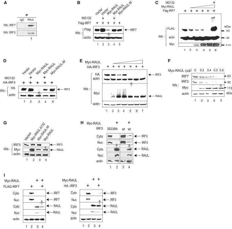
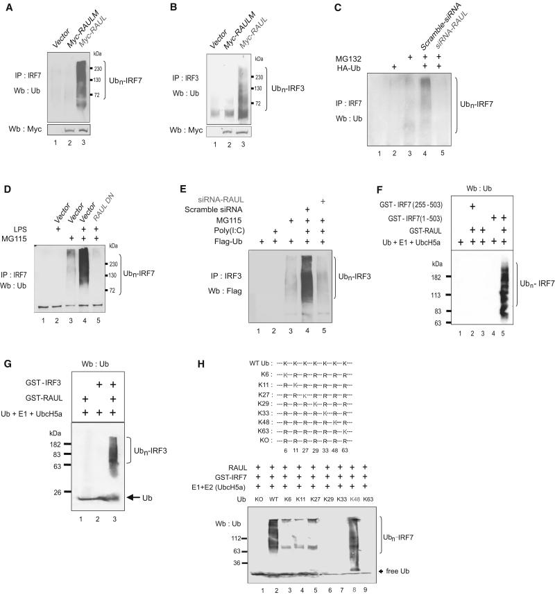
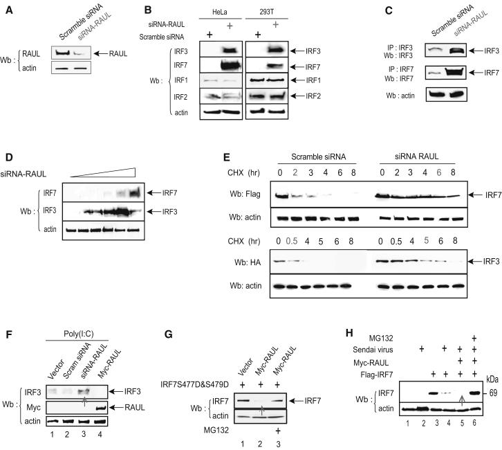
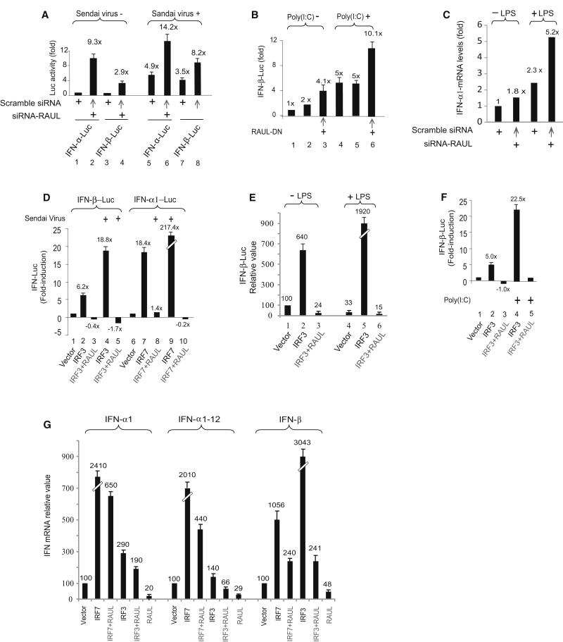
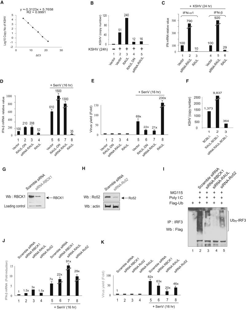
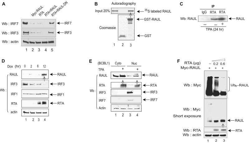
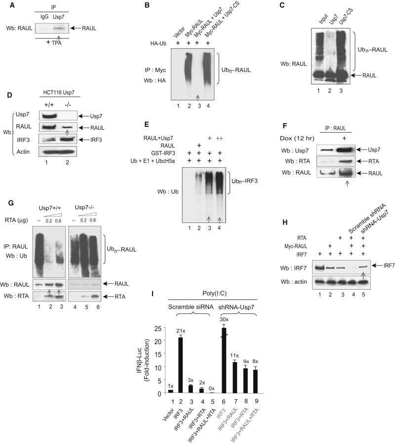
In the course of combating infectious agents, type I interferon (IFN) needs a timely downregulation mechanism to avoid detrimental overreaction. Here we showed a mechanism for restraining type I IFN responses, which relied on a HECT domain ubiquitin (Ub) E3 ligase, RAUL. RAUL limited type I IFN production by directly catalyzing lysine 48-linked polyubiquitination of both interferon regulatory factor 7 (IRF7) and IRF3 followed by proteasome-dependent degradation. Suppression of RAUL by dominant-negative RAUL or siRNA augmented both basal and virus-induced production of type I IFN, which resulted in reduced viral replication. The Kaposi's sarcoma-associated herpes virus immediate-early lytic cycle trigger protein RTA recruited this mechanism to augment its countermeasures against the host antiviral response. These results unveil a previously unrecognized "brake mechanism" for type I IFN that maintains proper low amounts of type I IFN under physiological conditions and restrains its magnitude when the antiviral response intensifies.


Figures

Figure 1
Figure 1. RAUL directs IRF7 and IRF3 for proteolysis
(A): Interaction between endogenous RAUL and IRF7 or IRF3. Lysates of DC2.4 mouse bone marrow dendritic cells treated with the proteasome inhibitor MG132 were immunoprecipitated with anti-RAUL and immunoblotted with anti-IRF7 or anti-IRF3. (B, C, D and E): RAUL, but not its calalytically inactive mutant, causes loss of IRF7 and IRF3 proteins. Flag-IRF7 or HA-IRF3 was cotransfected with Myc-RAUL or Myc-RAUL-M (point mutant C1051A) into 293T cells in the presence of MG132 (0.5 μM for 12 hr). The amounts of IRF7 and IRF3 proteins were evaluated by immunoblot. (C and E): Dose response with increasing amounts of Myc-RAUL. (F): Enforced RAUL expression causes loss of endogenous IRF7 and IRF3 proteins. The indicated increasing amounts of Myc-RAUL plasmid were transfected into 293T cells, and the endogenous IRF7 or IRF3 protein were assessed by IRF7 or IRF3 immunoblotting. (G): RAUL-mediated IRF3 proteolysis is dependent on its substrate binding and catalytic domains. WT RAUL or its substrate binding domain deletion mutant (Myc-RAULΔ132, also designated as Myc-RAUL-DN) or its catalytically inactive mutant (Myc-RAUL-M) were transfected into 293T cells and endogenous IRF3 protein was evaluated by IRF3 immunoblotting. (H): RAUL-mediated loss of IRF3 occurs independently of IRF3 phosphorylation status. Myc-RAUL and WT IRF3 or its phosphorylation-deficient mutant IRF3S339A were cotransfected into 293T cells and cytoplasmic and nuclear portions were isolated and amounts of IRF7, IRF3 and RAUL were determined by immunoblotting. (I): Subcellular distribution pattern. 293T cells were transfected with Myc-RAUL together with Flag-IRF7 or HA-IRF3 plasmids. Cytoplasmic and nuclear fractions were isolated and IRF7, IRF3 and RAUL proteins were detected by immunoblot with Flag, HA or RAUL antibodies, respectively.
Figure 2
Figure 2. RAUL ubiquitinates IRF7 and IRF3 in vivo and in vitro
(A and B): RAUL mediates ubiquitination of endogenous IRF7 and IRF3. DG75 B-lymphocyte cells were electroporated withWT Myc-RAUL or its catalytically inactive mutant (Myc-RAUL-M) plasmids in the presence of proteasome inhibitor MG115 and lysates were immunoprecipitated with anti-IRF7 and IRF3 and ubiquitinated forms of IRF7 and IRF3 were determined by Ub immunoblotting. (C): RAUL is required for endogenous IRF7 ubiquitination. Mouse bone marrow dendritic cells DC2.4 were electroporated with siRNA-RAUL or scramble siRNA together with HA-Ub plasmid and the indicated cells were treated with MG132. The cell lysates were immunoprecipitated with anti-IRF7 followed by Ub immunoblotting. (D): RAUL is required for ubiquitination of endogenous IRF7 following TLR4 activation. Mouse primary bone marrow dendritic cells were isolated (Tseng et al., 2001) and electroporated with dominant negative (Myc-RAUL-DN) plasmid or control vector and the indicated cells were treated with LPS (100 ng/ml for 9 hr) and proteasome inhibitor MG115. Conjugated IRF7 was determined by immunoprecipitation with anti-IRF7 followed by Ub immunoblotting. (E): RAUL is required for ubiquitination of IRF3 following TLR3 activation. 293-TLR3 cells were transfected with Flag-Ub plasmids and indicated cells were treated with poly I:C (25μg/ml for 9 hr) or MG115 (0.5μM for 12 hr). Conjugated IRF3 was evaluated by immunoprecipitation with anti-IRF3 followed by immunoblot with anti-Flag. (F and G): RAUL catalyzes the conjugation of polyUb chains on IRF7 and IRF3 in vitro. Purified GST-RAUL protein was incubated with various combinations of WT or truncated GST-IRF7 or GST-IRF3 plus E1, UbcH5a (E2) and Ub at 37° C for 1 hr. Following the ubiquitination reaction, the samples were subjected to immunoblot with Ub antibody for detection of polyubiquitinated forms of IRF7 and IRF3. Only a low catalytic concentration of GST-RAUL (20 nM, as an E3 source) was used compared to 400nM of target proteins to minimize interference from RAUL autoubiquitination (lane 3). (H): Determining IRF7 and IRF3 Ub chain structure by single lysine mutant assay in vitro. Illustration of Ub lysine mutants (upper panel). PurifiedWT Ub or its single lysine mutants, plus E1, E2 (Ubc5a), GST-IRF7 and RAUL were employed in in vitro cell-free Ub assays and ubiquitinated forms of the proteins were evaluated by Ub immunoblotting (lower panel).
Figure 3
Figure 3. Silencing RAUL increases IRF7 and IRF3 expression
(A): Immunoblot of lysates with RAUL specific antibody from 293T cells transiently expressing siRNA-RAUL or scramble siRNA. (B and C): Reducing RAUL specifically induces high amounts of endogenous IRF7 and IRF3 proteins in both cell lines and primary cells. (B): Immunoblot with anti-IRF7 or anti-IRF3 of lysates from cells transiently expressing siRNA-RAUL or scramble siRNA in Hela or 293T cells. (C): Isolated primary T-lymphocytes from 5 week-old Rag2−/− 5c.c7 TCR transgenic knockout mice were activated with pigeon cytochrome C (PCC) and expanded for 4 days. After activation, more than 90% of the cell population was TCR-specific transgenic T-lymphocyte cells, which were then transfected with siRNA RAUL or scrambled siRNA. Endogenous IRF3 and IRF7 proteins were determined by IRF3 or IRF7 immunoprecipitation followed by immunoblot. (D): siRNA-mediated reduction of RAUL increases accumulation of both the IRF7 and IRF3 proteins in a dose-dependent manner. 293T cells were transfected with two-fold increasing amounts of siRNA-RAUL, and endogenous IRF7 or IRF3 protein was determined by IRF7 or IRF3 immunoblot. (E): Knocking down RAUL extends the half-lives of IRF7 and IRF3 proteins. Flag-IRF7 or HA-IRF3 were cotransfected with siRNA-RAUL or scrambled siRNA into 293T cells, and the protein synthesis inhibitor cyclohexamide (CHX) was added at a concentration of 30 μg/ml at 36 hr for the indicated times. The IRF7 and IRF3 proteins were determined by Flag or HA immunoblot. (F): Silencing RAUL in 293-TLR3 cells increases the amount of IRF3 protein. Immunoblot analysis of IRF3 protein from 293-TLR3 cells transiently expressing siRNA-RAUL or Myc-RAUL. Poly I:C was used to stimulate TLR3 activation. (G): RAUL also regulates a constitutively active form of IRF7. Immunoblot analysis of lysates with IRF7 antibody from 293T cells transiently coexpressing Myc-RAUL and IRF7S477D and S479D in the presence and absence of MG132. H): Enforced RAUL expression augments the Sendai virus-mediated loss of IRF7 protein. Myc-RAUL and Flag-IRF7 plasmids as indicated were transfected into 293T cells Sendai virus infection (16 hr) with or without treatment with MG132. IRF7 protein was evaluated by immunoblot with a Flag antibody.
Figure 4
Figure 4. RAUL negatively regulates IRF7 and IRF3-mediated type 1 IFN production
(A, B and C): Suppression of RAUL by siRNA or dominant negative augments type 1 IFN activity. (A): Luciferase assay of 293T cells transiently expressing siRNA-RAUL together with IFN-α1 or IFN-β luciferase reporter plasmids. The cells were infected with Sendai virus for 16 hr before harvest. The basal IFN-α and IFN-β activities were arbitrarily set to 1.0. (B): Luciferase assay of IFN-β promoter activity from 293-TLR3 cells transiently expressing dominant negative RAUL poly I:C (25 μg/ml) for 9 hr to activate IRF3. (C): Silencing of RAUL increases IFN-α1 mRNA in dendritic cells. DC 2.4 cells were electroporated with siRNA-RAUL or scrambled siRNA in the presence or absence of LPS. Total RNA was isolated and subjected to SYBR green Real time RT-PCR with primers for IFN-α1. (D, E and F): Enforced RAUL expression abolishes IRF7 and IRF3-mediated type I IFN induction under Sendai virus or LPS or poly I:C stimulation. The indicated cells were cotransfected with IFN-α1 or IFN-β luciferase reporters, IRF7 or IRF3 and RAUL and the indicated cells were infected with Sendai virus (16 hr) or stimulated by LPS (100 ng/ml, for 9 hr) or poly I:C. IFN promoter activation was determined by luciferase assay. G: RAUL inhibits IRF7 or IRF3-mediated type I IFN mRNA production. 293T cells were cotransfected with IRF7, IRF3 and RAUL plasmids as indicated. Total RNA was isolated and subjected to SYBR green Real time RT-PCR with primers for IFN-α1, IFNα-1-12 and IFN-β, respectively, to assess type 1 IFN mRNA output.
Figure 5
Figure 5. RAUL regulates the IFN-mediated antiviral response
(A): A SYBR Real-time-PCR based quantitative approach for determining KSHV viral load. The standard curve was constructed using KSHV RAP DNA as target by the comparative Ct method. The viral load value, normalized to reference (β-actin) is given by 2 −ΔΔCt in the Applied Biosystems 7500 Real Time PCR system expressed as copy number per 100 ng of infected cell genomic DNA. (B, C, D and E): Role of RAUL in KSHV and Sendai virus-infected mouse embryonic fibroblast cells (MEFs). MEF cells were electroporated with WT RAUL,dominant RAUL-DN or siRNA-RAUL and the cells were infected with KSHV or Sendai virus for the indicated times. The cell lysates were assayed for viral DNA copy number (KSHV) per 100 ng of genomic DNA or relative viral RNA load (Sendai virus) (B and E) as well as for type 1 IFN mRNA production by Real time RT-PCR (C and D). (F):Role of RAUL in KSHV-infected BCBL1 cells. Lysates from BCBL1 derived cell lines stably expressing either RAUL or siRNA-RAUL were assayed for KSHV viral DNA copy number per 100 ng genomic DNA. (G and H): Comparison of the contribution of RBCK1, RAUL and Ro52 to endogenous IRF3 ubiquitination. Immunoblot of lysates with RBCK1 (G) or Ro52 (H) specific antibody from 293T cells transiently expressing siRNA-RBCK1 or siRNA-Ro52 or scramble siRNA. (I): Comparison of siRNA silencing to reduce polyubiquitination of IRF3 in activated cells. 293T-TLR3 cells were transfected with siRNA against RBCK1 or RAUL or Ro52 together with Flag-Ub and the cells were treated with poly I:C to induce polyUb-conjugation of IRF3 in the presence of the proteasome inhibitor MG115. The conjugation of endogenous IRF3 was detected by immunoprecipitation with IRF3 antibody and immunoblot with Flag antibody. (J and K): Relative roles of RBCK1, RAUL and Ro52 in viral replication. 293T cells were transfected with siRNA against RBCK1 or RAUL or Ro52, and then infected with Sendai virus for the indicated times. The cell lysates were assayed for IFN-β mRNA production (J) and for viral RNA load of Sendai virus by real-time RT-PCR (K).
Figure 6
Figure 6. RTA recruits and enhances RAUL
(A): RTA enhances the ability of RAUL to degrade IRF7 and IRF3. 293T cells were cotransfected with RTA, Myc-RAUL or Myc-RAUL-DN, IRF7 and IRF3 as indicated and the amounts of IRF7 and IRF3 proteins were detected by IRF7 or IRF3 immunoblot. (B and C):RTA binds to RAUL in vitro and vivo. (B): A GST-pull down assay was carried out with 35S-labeled in vitro translated RAUL and GST-RTA proteins. (C): Cell lysates from BCBL1 PEL cells were immunoprecipitated with anti-RTA followed by RAUL immunoblot to detect endogenous RTA-RAUL interaction with TPA. (D and E): Induction of RTA stabilizes RAUL protein. (D): TREx BCBL1 cells treated with doxycycline (Dox) for the indicated times to induce RTA expression, and RAUL, IRF3, IRF1 and RTA proteins were monitored by immunoblot using the indicated antibodies. (E): BCBL1 PEL cells were treated with TPA to induce RTA expression and cytoplasmic and nuclear fractions were isolated. The RAUL, IRF7, IRF3 and RTA proteins were evaluated by immunoblot using the indicated antibodies. (F):RTA mediates RAUL deubiquitination. RTA and Myc-RAUL plasmids were cotransfected into 293T cells and ubiquitinated forms of RAUL were determined by immunoblot with anti-Myc. To show both ubiquitinated and nonubiquitinated forms of RAUL, a short-exposure film was included (lower panel).
Figure 7
Figure 7. RTA mediates RAUL deubiquitination through Usp7
(A): Endogenous RAUL and Usp7 interact. BCBL1 cells were treated with TPA for 24 hr and cell lysates were immunoprecipitated with anti-Usp7 followed by RAUL immunoblot to detect bound protein. (B and C): Usp7 deubiquitinates RAUL in vivo and in vitro. 293T cells were transfected with Myc-RAUL, WT Usp7, Usp7-cs and HA-Ub. The cells were treated with MG132 for 6 hr before harvest. Polyubiquitinated forms of RAUL were detected by immunoprecipitation with anti-Myc followed by HA immunoblot. (C): Purified RAUL protein (400 nM) plus E1, E2 (UbcH5a) and Ub were incubated at 37°C for 1 hr to catalyze self-ubiquitination. The Ub-RAUL protein was then purified by immunoprecipitation with anti-RAUL antibody and incubated with purified Usp7 protein or its catalytically inactive mutant (Usp7-cs) in deubiquitination buffer at 37°C for 2 hr. The resulting reactions were subjected to immunoblot with RAUL. (D): Usp7 upregulates RAUL protein amounts. Colon cancer cell lines HCT116 Usp7+/+ or Usp7−/− cells were immunblotted with the indicated antibodies to evaluate both RAUL and its target substrate IRF3. (E): Usp7 enhances the E3 activity of RAUL toward IRF3 in vitro. Immunoprecipitated purified RAUL protein from either RAUL expressing 293T cells or RAUL+Usp7 expressing 293T cells were incubated with its substrate GST-IRF3, plus E1, UbcH5a and Ub in vitro at 37°C for 1 hr. Conjugated IRF3 was determined by Ub immunoblot. (F): RTA increases the recruitment of Usp7 to RAUL complexes. TRExBCBL1 cells were treated with Doxycyclin (Dox) for 12 hr to activate RTA expression. Endogenous RTA-Usp7-RAUL interaction was evaluated by immunoblot with the indicated antibodies. (G):RTA promotes RAUL deubiquitination. HCT116 (Usp7+/+ or Usp7−/−) cells were electroporated with the indicated amounts of RTA plasmid and RAUL self-ubiquitition was determined by Ub immunoblotting. Unconjugated RAUL and RTA were measured by RAUL or RTA immunoblotting. (H): RTA enhances the ability of RAUL to degrade IRF7 via Usp7. 293T cells were cotransfected with IRF7, Myc-RAUL, RTA and shRNA-Usp7 as indicated, and IRF7 protein amounts were evaluated by IRF7 immunoblotting. (I): RTA and RAUL cooperate in suppressing IRF3-mediated IFNβ reporter activity. 293-TLR3 cells were transfected with the indicated combinations of IRF3, RAUL, RTA, shRNA- Usp7 and IFNβ-LUC plasmids and the cells were treated with poly I:C for 9 hr to mimic TLR3 activation. IRF3-mediated IFN-β reporter activity in the cells that were pretreated shRNA-RAUL or scrambled IFN-β activation was determined by luciferase assays.

Comment in

Similar articles

Cited by

References

    1. Barrington RE, Subler MA, Rands E, Omer CA, Miller PJ, Hundley JE, Koester SK, Troyer DA, Bearss DJ, Conner MW, et al. A farnesyltransferase inhibitor induces tumor regression in transgenic mice harboring multiple oncogenic mutations by mediating alterations in both cell cycle control and apoptosis. Mol Cell Biol. 1998;18:85–92. - PMC - PubMed
    1. Barro M, Patton JT. Rotavirus NSP1 inhibits expression of type I interferon by antagonizing the function of interferon regulatory factors IRF3, IRF5, and IRF7. J Virol. 2007;81:4473–4481. - PMC - PubMed
    1. Chomvarin C, Siripornmongcolchai T, Chaicumpar K, Limpaiboon T, Wongkham C, Yutanawiboonchai W. Evaluation of polymerase chain reaction, conventional and MRSA screen latex agglutination methods for detection of methicillin-resistant, - borderline and -susceptible Staphylococcus aureus. Southeast Asian J Trop Med Public Health. 2004;35:879–885. - PubMed
    1. Cummins JM, Rago C, Kohli M, Kinzler KW, Lengauer C, Vogelstein B. Tumour suppression: disruption of HAUSP gene stabilizes p53. Nature. 2004;428:1. following 486. - PubMed
    1. Doyle S, Vaidya S, O’Connell R, Dadgostar H, Dempsey P, Wu T, Rao G, Sun R, Haberland M, Modlin R, Cheng G. IRF3 mediates a TLR3/TLR4-specific antiviral gene program. Immunity. 2002;17:251–263. - PubMed

Publication types

MeSH terms