. 2011 Feb 5;410(1):64-75.
doi: 10.1016/j.virol.2010.10.029. Epub 2010 Nov 19.

Epstein-Barr Virus nuclear antigen 1 (EBNA1) confers resistance to apoptosis in EBV-positive B-lymphoma cells through up-regulation of survivin

Affiliations

Epstein-Barr Virus nuclear antigen 1 (EBNA1) confers resistance to apoptosis in EBV-positive B-lymphoma cells through up-regulation of survivin

Jie Lu et al. Virology. .

Abstract

Resistance to apoptosis is an important component of the overall mechanism which drives the tumorigenic process. EBV is a ubiquitous human gamma-herpesvirus which preferentially establishes latent infection in viral infected B-lymphocytes. EBNA1 is typically expressed in most forms of EBV-positive malignancies and is important for replication of the latent episome in concert with replication of the host cells. Here, we investigate the effects of EBNA1 on survivin up-regulation in EBV-infected human B-lymphoma cells. We present evidence which demonstrates that EBNA1 forms a complex with Sp1 or Sp1-like proteins bound to their cis-element at the survivin promoter. This enhances the activity of the complex and up-regulates survivin. Knockdown of survivin and EBNA1 showed enhanced apoptosis in infected cells and thus supports a role for EBNA1 in suppressing apoptosis in EBV-infected cells. Here, we suggest that EBV encoded EBNA1 can contribute to the oncogenic process by up-regulating the apoptosis suppressor protein, survivin in EBV-associated B-lymphoma cells.


Figures

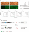
Fig. 1
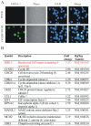
Fig. 1. Genes up-regulated in BJAB cells expressing EBNA1 by using gene array analysis
(A) Immunofluorescence assay for EBNA1 in BJAB cells. (B) The genes shown were up-regulated greater than 2.2 fold in BJAB cells expressing EBNA1. Data was standardized from BJAB-EBNA1 and BJAB-Vector sets by using GEArray Expression Analysis Suite.
Fig. 2
Fig. 2. The transcription and protein levels of survivin were induced by EBNA1
(A, B) Quantitation of survivin transcripts was measured by semi-quantitative RT-PCR. Total RNA was isolated from 20 million of EBV non-infected BL41 and infected BL41/B958 cells or BJAB, EBNA1-expressing BJAB cell by using TRIzol reagent. A total of 2 μg of RNA was used to synthesize cDNA. Real-time PCR was performed by using the Power SYBR green PCR master mix with GAPDH as control, Error bars show standard deviations; (C) Western blot analysis of endogenous survivin in the EBV infected BL41 and BJAB cells expressing EBNA1. GAPDH was used for internal control.
Fig. 3
Fig. 3. EBNA1 activated the survivin promoter in a dose-dependent manner
A fixed amount (2 μg) of the reporter plasmids was transfected or cotransfected with pA3M-EBNA1 (1, 2, 5, 10, 15 and 20μg [number 1 to 6]) into 10 million (A) DG75 cells and (B) 293 cells, individually. At 24 h posttransfection, cell lysate of each transfection was harvested for the luciferase assay. The promoter activity was presented as the fold activation relative to the reporter-alone control. Definitely, EBNA can activate the survivin promoter by approximately 3 to 6 fold compared to that of the vector control in DG75 cells as well as in 293cells in dose-dependent manner. The means and standard deviations from three independent experiments were shown. Western blots showed the expression of myc-tagged EBNA1 in cotransfected cells. The blot was stripped and reprobed with anti-GAPDH antibody to show equal protein loading.
Fig. 4
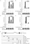
Fig. 4. Cis-acting elements within the survivin promoter are important for EBNA1 mediated induction of survivin expression
(A to D) A fixed amount of the wild type (WT) or mutation type (MT) of survivin promoter reporter plasmid was transfected or cotransfected into 10 million 293 cells with pA3M-EBNA1 expression vector. At 24 h posttransfection, cell lysate of each transfection was harvested for a luciferase assay. Western blots show the expression of EBNA1 in the cotransfected cells. The deletion of GC/Sp1 box (SurPΔGC/Sp1) in the survivin promoter resulted in an almost complete loss of promoter activity in the presence of EBNA1. The blot was stripped and reprobed with anti-GAPDH antibody to show equal protein loading. +, presence; −. absence. (E) Schematic showing the survivin WT and MT promoter used in this study. The putative domains include an NKx 2.5, Sp1 binding sites, GC/Sp1 domain and p53 conserved sequence. Numbering is from the initiating ATG.
Fig. 5
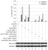
Fig. 5. Reporter Assay of the survivin promoter with GC/Sp1 and p53 mutation in Saos-2 cells
A fixed amount of the WT or MT reporter plasmid was transfected into 10 million Saos-2 cells in the presence of pA3M-EBNA1 expression vector. At 24 h posttransfection, cell lysate of each transfection was harvested for a luciferase assay. Obviously, mutation of p53 responsive element had no significant effects on the survivin promoter activity compared to the control pGL3B-SurP with EBNA1. +, presence; −, absence. Western blots show the expression of EBNA1 in cotransfected cells. The blot was stripped and reprobed with anti-GAPDH antibody to show equal protein loading.
Fig. 6
Fig. 6. Sp1 binds to the survivin promoter in complex with EBNA1 at its canonical binding site
Electrophoretic mobility shift assays were performed as described in material and method. (A) Binding of Sp1 site in the survivin promoter with EBNA1 protein as well as Sp1. Lane 1 is probe alone (bottom), Lane 2 is probe with nuclear extract containing Sp1 showing faster mobility of the probe (asterisks) which was abolished by adding an excess (200-fold) of unlabeled specific probe (lane 3). The shift in mobility was unaffected with similar amounts (200-fold) of nonspecific unlabeled competitor (lane 4). Adding anti-Sp1 antibody changed the mobility of Sp1 (lane 5). Lane 7 contains probe with nuclear extracts of pA3M-EBNA1 shows a change in the mobility of the probe due to the binding of Sp1 to EBNA1 (asterisks). Further, to add anti-myc antibody changed the mobility of the entire complex, supershifting it (lane 6, asterisks). Lane 8, which contains Sp1 and α-myc antibodies with probe, demonstrates that nonspecific binding did not occur. Lane 9 is vector control only but contains the cell essential factor Sp1, This is confirmed when the cold Sp1 probe is added and the shift is disappeared (lane 10). The EBNA1 (lane 11) still has some shift comparing the Sp1 (lane 2), but when cold Sp1 probe is added, EBNA1 doesn’t show obvious shift comparing to EBNA1 (lane 12). (B) Western blots to detect proteins from nuclear extracts. (C) LCL1 cells were harvested; cross-linked chromatin was precipitated with OT1x antibody or normal mouse IgG. Western blot shows the coimmunoprecipitation using OT1x antibody against EBNA1. The recovered DNA was quantitated by Real-time PCR as described for Fig. 6D. Shown are the results from three independent experiments. Error bars indicate standard deviations.
Fig. 7
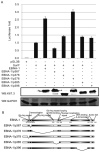
Fig. 7. The EBNA1 transactivation domain is required for activation of the survivin promoter
(A) A fixed amount of the WT or MT EBNA1 expression vector was transfected into 10 million 293 cells in the presence of survivin promoter reporter plasmid. At 24 h posttransfection, cell lysate of each transfection was harvested for a luciferase assay. Results of luciferase assay showed that EBNA1/p367 had a significant decrease in its ability to activate the survivin promoter. All the transfections were done multiple times, and the results shown represent the means of the data from three independent experiment. Western blots show the expression of EBNA1 and truncations in the cotransfected cells. The blot was stripped and reprobed with anti-GAPDH antibody to show equal protein loading. +, presence; −, absence. (B) Schematic showing the EBNA1 truncation used in this study.
Fig. 8
Fig. 8. Survivin expression is up-regulated in EBV-associated lymphoma tissue
Immunohistochemistry assays for survivin and EBNA1 were performed in EBV positive and negative tissues specimens. Paraffin-embedded EBV positive and negative diffuse large B-cell lymphoma (DLBL) tissue were deparaffinized, rehydrated, and washed with phosphate buffered saline. Following antigen retrieval, samples were blocked with BSA prior to incubation with primary antibodies overnight at 4°C. Survivin was detected by using mouse monoclonal anti-survivin antibody followed by detection using anti-mouse Alexa Fluor 488 (Panel, A) and EBNA1 was detected by using rabbit polyclonal anti-EBNA1 antibody K67.3 followed by anti-rabbit Alexa Fluor 594 (Panel, B). The merge panel shows that most of the survivin co-expression with EBNA1 (Panel, D).
Fig. 9
Fig. 9. Depletion of survivin by shRNA increases the proportion of cell undergoing apoptosis
(A) LCL1 cells were infected with a lentivirus encoding survivin shRNA and analyzed by fluorescence microscopy after 48 h for the expression of GFP. (B) Western blotting showing expression of EBNA 1 and survivin in EBV non-infected cell line and EBV infected cell line. GAPDH level was used for internal control. (C) Flow cytometry analysis of different treated cells. LCL1 Cells were treated with survivin shRNA and then washed, fixed, stained with propidium iodide (PI), and analyzed for their DNA content by flow cytometry. LCl1 cells infected with the sh-SV lentivirus had an increase in cell apoptosis compared to the LCL1 infected sh-C cells, LCL1 cells with a lentivirus-delivered shRNA that knocks down EBNA1 expression in these cells (LCL1-sh-E1) led to a decrease in apoptosis compared to LCL1-sh-SV. sh-SV, survivin shRNA; sh-C, control shRNA; sh-E1, EBNA1 shRNA. (D) Hypothetical model showing EBV encoded EBNA1 contributes to up-regulation of survivin and resistance to apoptosis in EBV-associated B-lymphoma cells.

Similar articles

Cited by

References

    1. Altieri DC. Validating survivin as a cancer therapeutic target. Nat Rev Cancer. 2003;3(1):46–54. - PubMed
    1. Altieri DC. New wirings in the survivin networks. Oncogene. 2008;27(48):6276–84. - PMC - PubMed
    1. Ambrosini G, Adida C, Altieri DC. A novel anti-apoptosis gene, survivin, expressed in cancer and lymphoma. Nat Med. 1997;3(8):917–21. - PubMed
    1. Boidot R, Vegran F, Jacob D, Chevrier S, Cadouot M, Feron O, Solary E, Lizard-Nacol S. The transcription factor GATA-1 is overexpressed in breast carcinomas and contributes to survivin upregulation via a promoter polymorphism. Oncogene. 2010;29(17):2577–84. - PubMed
    1. Borbely AA, Murvai M, Konya J, Beck Z, Gergely L, Li F, Veress G. Effects of human papillomavirus type 16 oncoproteins on survivin gene expression. J Gen Virol. 2006;87:287–94. Pt 2. - PubMed

Publication types

MeSH terms

Substances