. 2010 Aug 27;5(8):e12445.
doi: 10.1371/journal.pone.0012445.

Epithelial to mesenchymal transition is mechanistically linked with stem cell signatures in prostate cancer cells

Affiliations

Epithelial to mesenchymal transition is mechanistically linked with stem cell signatures in prostate cancer cells

Dejuan Kong et al. PLoS One. .

Abstract

Background: Current management of patients diagnosed with prostate cancer (PCa) is very effective; however, tumor recurrence with Castrate Resistant Prostate Cancer (CRPC) and subsequent metastasis lead to poor survival outcome, suggesting that there is a dire need for novel mechanistic understanding of tumor recurrence, which would be critical for designing novel therapies. The recurrence and the metastasis of PCa are tightly linked with the biology of prostate cancer stem cells or cancer-initiating cells that is reminiscent of the acquisition of Epithelial to Mesenchymal Transition (EMT) phenotype. Increasing evidence suggests that EMT-type cells share many biological characteristics with cancer stem-like cells.

Methodology/principal findings: In this study, we found that PCa cells with EMT phenotype displayed stem-like cell features characterized by increased expression of Sox2, Nanog, Oct4, Lin28B and/or Notch1, consistent with enhanced clonogenic and sphere (prostasphere)-forming ability and tumorigenecity in mice, which was associated with decreased expression of miR-200 and/or let-7 family. Reversal of EMT by re-expression of miR-200 inhibited prostasphere-forming ability of EMT-type cells and reduced the expression of Notch1 and Lin28B. Down-regulation of Lin28B increased let-7 expression, which was consistent with repressed self-renewal capability.

Conclusions/significance: These results suggest that miR-200 played a pivotal role in linking the characteristics of cancer stem-like cells with EMT-like cell signatures in PCa. Selective elimination of cancer stem-like cells by reversing the EMT phenotype to Mesenchymal-Epithelial Transition (MET) phenotype using novel agents would be useful for the prevention of tumor recurrence especially by eliminating those cells that are the "Root Cause" of tumor development and recurrence.


Conflict of interest statement

Competing Interests: The authors have declared that no competing interests exist.

Figures

Figure 1
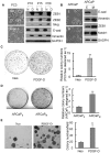
Figure 1. Cells with EMT signatures displayed increased Clonogenic ability.
(A) Photographs of cells were shown: PC3 Neo cells displayed rounded epithelial cell shape and PC3 PDGF-D cells exhibited a fibroblastic-type phenotype (left panel, original magnification, 200 X). Western blot analysis showed the expression of transcription repressors associated with EMT, and mesenchymal as well as epithelial markers in PC3 Neo and PC3 PDGF-D cells (right panel, passage 10 to 20). (B) The morphology of ARCaPM and ARCaPE cells was shown (left panel).Western blot analysis indicated the increased ZEB1, vimentin and Notch1 expression and decreased E-cadherin expression in ARCaPM cells compared with ARCaPE (right panel). (C) and (D) Photographs of colonies from PC3 Neo and PC3 PDGF-D or ARCaPM and ARCaPE were shown (left panel). The colony numbers were counted and the data was presented as relative colony numbers of PC3 Neo or ARCaPE designed as 1 (right panel). (E) The colonies grown on soft agar were photographed (left panel, bar, 200 µm). The colony numbers were counted under a phase contrast microscope. Data was presented as colony numbers per field (right panel). **, p<0.01 compared to Neo or ARCaPE cells (N: PC3 Neo cells, D: PC3 PDGF-D cells,p10: passage 10, E-cad: E-cadherin).
Figure 2
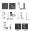
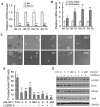
Figure 2. Prostaspheres were increased in cells having EMT phenotype.
(A) Prostaspheres from PC3 Neo and PC3 PDGF-D cells were photographed and shown (bar, 100 µm). (B) The numbers of prostaspheres were counted under microscope. (C) Prostaspheres were photographed and the size of prostaspheres in diameter was measured using software image analysis program Scion Image. (D) The prostaspheres (P1) were collected and re-plated at a density of one cell per well in 96-well plate with ultra low attachment. After 12 days of incubation, the numbers of prostaspheres (P2) were counted under a phase contrast microscope. (E) The numbers of prostaspheres from ARCaPE and ARCaPM cells were counted under microscope. (F) Prostaspheres from ARCaPE and ARCaPM cells were photographed and shown (bar, 100 µm). **, p<0.01 compared to Neo or ARCaPE cells (Neo: PC3 Neo cells, PDGF-D: PC3 PDGF-D cells).
Figure 3
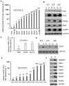
Figure 3. Gene expression profiling of stem cell markers in PC3 PDGF-D cells with EMT signatures.
(A) The expression of genes at the mRNA levels for Oct4, Nanog, Sox family genes and Lin28B in PC3 Neo and PC3 PDGF-D cells were determined by using real time RT-PCR. (B) The results from Western blot showed that the expressions of Oct4 Sox2, Nanog, Stat3 and Lin28B were significantly increased in PC3 PDGF-D cells. (C) Real time RT-PCR was used to quantify the mRNA expression of EED, EZH2 and Suz12a. Relative mRNA levels were normalized to GAPDH. (D) The results from Western blot showed that the expression of EZH2 was significantly increased in PC3 PDGF-D cells. (E) The mRNA levels of Notch signaling factors in PC3 Neo and PC3 PDGF-D cells were determined by using real time RT-PCR. (F) The results from Western blot showed that the expressions of Notch and Notch ligands were significantly increased in PC3 PDGF-D cells. GAPDH was used for protein loading control. (N: PC3 Neo cells, D: PC3 PDGF-D cells,p10: passage 10).
Figure 4
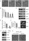
Figure 4. MiR-200 repressed the self-renewal capacity by regulating Notch1 and Lin28B expression.
PC3 PDGF-D cells were transfected with pre-miR-200. 3 days after transfection, cells were split and transfected repeatedly with pre-miR-200 every 3–4 days for 14 days. (A) Photographs of cells are shown: transfection of PC3 PDGF-D cells with pre-miR-200 for 14 days, miR-200b and miR-200c reversed EMT cells to MET cell morphology. (B) The cells were collected for generation of prostaspheres after 14-day transfection with pre-miR-200. MiR-200b and miR-200c reduced the number of prostaspheres. (C) Western Blot showing that Notch1, Lin28B and ZEB1 expressions were down-regulated in PC3 PDGF-D cells transfected with miR-200b and miR-200c. (D) The level of miR-200c significantly decreased in ARCaPM by using real time RT-PCR. (E) Transfection of pre-miR-200c after 6 days reduced the prostasphere-forming ability in ARCaPM cells (Bar:100 µm). (F) Western blot showed that re-expression of miR-200c by transfection of pre-miR-200c increased the E-cadherin expression and repressed expression of ZEB1 and Notch1 in ARCaPM cells, which consistent with the change from mesenchymal to epithelial cell morphology (G). *, p<0.05 compared to control; **, p<0.01 compared to control. (Con: control, 200a: pre-miR-200a, 200b: pre-miR-200b, 200c: pre-miR-200c, E-cad: E-cadherin).
Figure 5
Figure 5. Inactivation of Notch1 repressed the colonogenic and prostasphere-forming capacity.
(A) Conserved, predicted binding sites for the seed sequences of miR-200b and mir-200c in the 3′UTR of Notch1 mRNA. (B) MiR-200b and miR-200c inhibited the Notch1 3′UTR luciferase activity. **, p<0.01 compared to Neo control cells; #, p<0.01 compared to PDGF-D control cells. (C) DAPT treatment, a γ-secretase inhibitor, reduced the clonogenic ability in PC3 PDGF-D cells (upper panel).Western Blot showing that DAPT treatment reduced active form of Notch1 in dose-dependent manner (lower panel). (D) and (E) showing that transfection of Notch1 siRNA after 9 days repressed the prostasphere-forming capacity in PC3 PDGF-D cells (Bar:100 µm), which is consistent with downregulation of Notch1 expression (F). **, p<0.01 compared to control. (Neo: PC3 Neo cells, PDGF-D: PC3 PDGF-D cells, Con: control, 200a: pre-miR-200a, 200b: pre-miR-200b, 200c: pre-miR-200c).
Figure 6
Figure 6. Lin28B-mediated repression of let-7 was partially responsible for regulating self-renewal in PC3 PDGF-D cells.
(A) The levels for let-7 family in PC3 Neo and PC3 PDGF-D cells were determined by using real time RT-PCR. (B) Real time RT-PCR was used to quantify the expression of let-7 in PC3 PDGF-D cells transfected with Lin28B siRNA for 3 days. (C) Photomicrographs showing prostaspheres generated from PC3 PDGF-D cells transfected with pre-let-7 or combination of pre-let-7 and Lin28B siRNA 14 days after transfection (bar:100 µm). (D) The cells were collected for prostasphere-forming assay after 14-day transfection. Let-7a, let-7b, let-7d and combination of Let-7a, let-7b, let-7d as well as co-transfection with let-7 and Lin28B siRNA decreased the number of prostaspheres. (E) The results from Western blot showed the expression of Lin28B, Sox2, Nanog, and Oct4 in PC3 PDGF-D cells transfected with pre-let-7 or co-transfected with pre-let-7 and Lin28B siRNA after 9-day transfection. *, p<0.05 compared to control; **, p<0.01 compared to control. (Con: control, a: pre-let-7a, abd: combination of pre-let-7a, pre-let-7a, pre-let-7d, al: combination of pre-let-7a and Lin28B siRNA, Lin28B-si: Lin28B siRNA, Neo: PC3 Neo cells, PDGF-D: PC3 PDGF-D cells).
Figure 7
Figure 7. Sox2, Nanog, Oct4 and Lin28B regulated Prostasphere-forming ability of PC3 PDGF-D cells.
(A) Single cell suspensions of PC3 PDGF-D transfected with Sox2, Nanog, Oct4 and Lin28B siRNA and incubated for 24 h were plated on ultra low adherent wells of 6-well plate at 2000 cells/well. After 3 days, the numbers of prostaspheres were counted under microscope. Transfection of Sox2, Nanog, Oct4 or Lin28B siRNA reduced the prostasphere-forming capacity (B) Prostaspheres were photographed and the size of prostaspheres in diameter was measured using software image analysis program Scion Image. (C). Western blot analysis showed that levels of Sox2, Nanog, Oct4 and Lin28B in PC3 PDGF-D cells transfected with Sox2, Nanog, Oct4 and Lin28B siRNA compared with transfection with control siRNA (*, p <0.05, **, p<0.01 compared to control, Con: control).
Figure 8
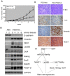
Figure 8. The cells with EMT phenotype promoted tumor growth.
(A) Tumor growth curve showing that over-expression of PDGF-D could promote tumor growth in SCID mice much faster than PC3 Neo cells. (B) Western blot analysis of tumor lysates showing the expression of EMT and stem cell makers. (C) H&E evaluation of the tumors from both the groups showed high grade carcinoma associated with tumor apoptosis and necrosis. The results from the immunostaining showed much intense staining for vimentin and Notch1 and less intense staining for E-cadherin. (D) Regulatory model showing mechanistic link between EMT and stem cells: EMT induced by different factors characterized by increased expression of ZEB1, which causes loss of miR-200 family. Loss of miR-200b and miR-200c lead to increased ZEB1, Notch1 and Lin28B expression, resulting in decreased expression of let-7. Downregulation of let-7 leads to the up-regulation of Sox2, Nanog and Oct4, which together with Notch1 and Lin28B contribute to stem cell signatures. (*, p <0.05, **, p<0.01 compared to PC3 Neo, E-cad: E-cadherin).

Similar articles

Cited by

References

    1. Visvader JE, Lindeman GJ. Cancer stem cells in solid tumours: accumulating evidence and unresolved questions. Nat Rev Cancer. 2008;8:755–768. - PubMed
    1. Wang X, Kruithof-de JM, Economides KD, Walker D, Yu H, et al. A luminal epithelial stem cell that is a cell of origin for prostate cancer. Nature. 2009;461:495–500. - PMC - PubMed
    1. Roudier MP, True LD, Higano CS, Vesselle H, Ellis W, et al. Phenotypic heterogeneity of end-stage prostate carcinoma metastatic to bone. Hum Pathol. 2003;34:646–653. - PubMed
    1. Aoi T, Yae K, Nakagawa M, Ichisaka T, Okita K, et al. Generation of pluripotent stem cells from adult mouse liver and stomach cells. Science. 2008;321:699–702. - PubMed
    1. Yu J, Vodyanik MA, Smuga-Otto K, ntosiewicz-Bourget J, Frane JL, et al. Induced pluripotent stem cell lines derived from human somatic cells. Science. 2007;318:1917–1920. - PubMed

Publication types

MeSH terms