

Signaling kinase AMPK activates stress-promoted transcription via histone H2B phosphorylation
- PMID: 20647423
- PMCID: PMC3922052
- DOI: 10.1126/science.1191241
Signaling kinase AMPK activates stress-promoted transcription via histone H2B phosphorylation
Abstract
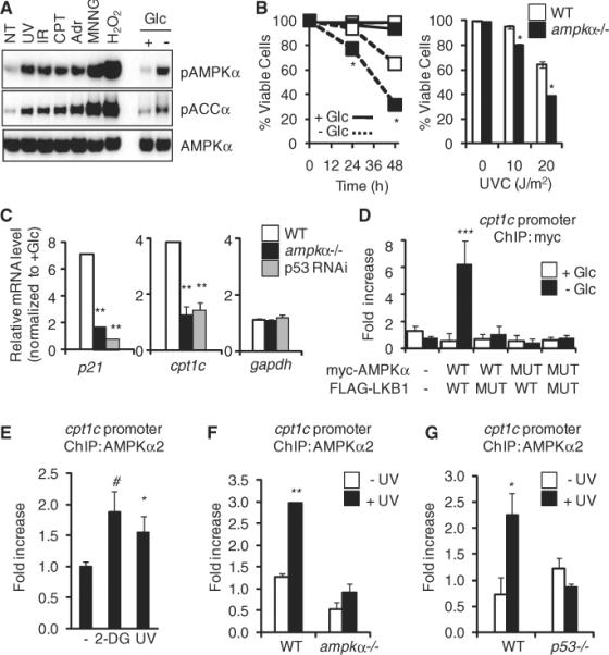
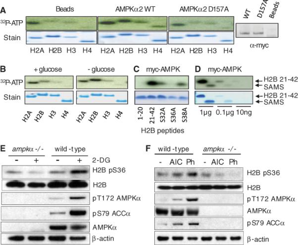
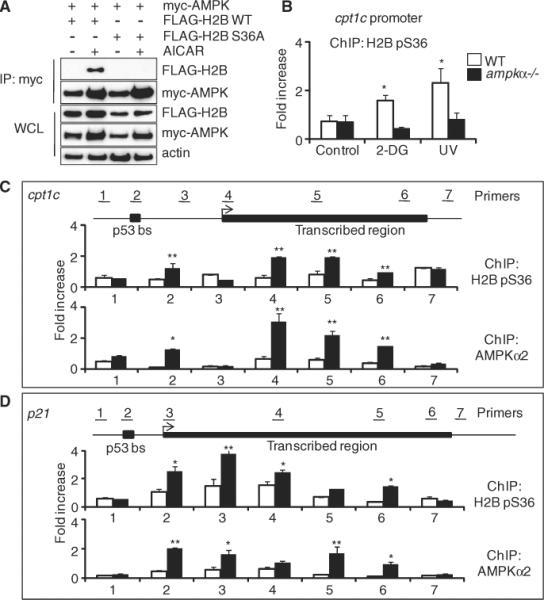
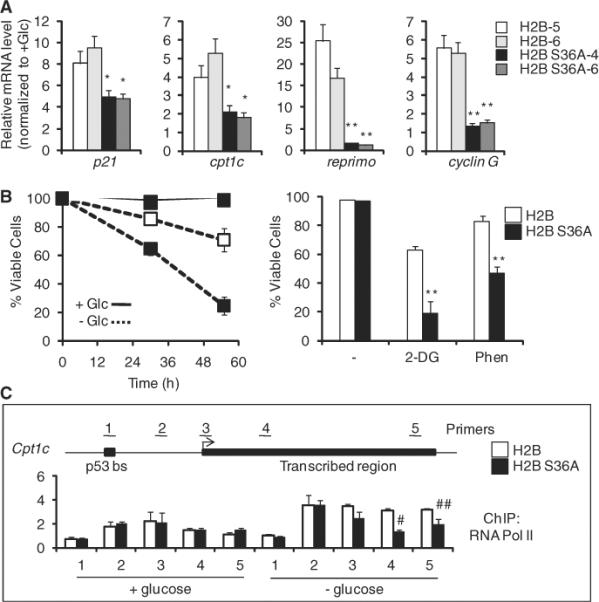
The mammalian adenosine monophosphate-activated protein kinase (AMPK) is a serine-threonine kinase protein complex that is a central regulator of cellular energy homeostasis. However, the mechanisms by which AMPK mediates cellular responses to metabolic stress remain unclear. We found that AMPK activates transcription through direct association with chromatin and phosphorylation of histone H2B at serine 36. AMPK recruitment and H2B Ser36 phosphorylation colocalized within genes activated by AMPK-dependent pathways, both in promoters and in transcribed regions. Ectopic expression of H2B in which Ser36 was substituted by alanine reduced transcription and RNA polymerase II association to AMPK-dependent genes, and lowered cell survival in response to stress. Our results place AMPK-dependent H2B Ser36 phosphorylation in a direct transcriptional and chromatin regulatory pathway leading to cellular adaptation to stress.
Figures
Comment in
-
Transcription. Targeting the core of transcription.Science. 2010 Sep 3;329(5996):1158-9. doi: 10.1126/science.1195447. Science. 2010. PMID: 20813944 No abstract available.
Similar articles
-
Transcription. Targeting the core of transcription.Science. 2010 Sep 3;329(5996):1158-9. doi: 10.1126/science.1195447. Science. 2010. PMID: 20813944 No abstract available.
-
DNA-dependent protein kinase regulates lysosomal AMP-dependent protein kinase activation and autophagy.Autophagy. 2020 Oct;16(10):1871-1888. doi: 10.1080/15548627.2019.1710430. Epub 2020 Jan 26. Autophagy. 2020. PMID: 31983282 Free PMC article.
-
AMPK regulates histone H2B O-GlcNAcylation.Nucleic Acids Res. 2014 May;42(9):5594-604. doi: 10.1093/nar/gku236. Epub 2014 Apr 1. Nucleic Acids Res. 2014. PMID: 24692660 Free PMC article.
-
AMP-activated protein kinase and cancer.Acta Physiol (Oxf). 2009 May;196(1):55-63. doi: 10.1111/j.1748-1716.2009.01980.x. Epub 2009 Feb 25. Acta Physiol (Oxf). 2009. PMID: 19243571 Review.
-
AMPK/Snf1 signaling regulates histone acetylation: Impact on gene expression and epigenetic functions.Cell Signal. 2016 Aug;28(8):887-95. doi: 10.1016/j.cellsig.2016.03.009. Epub 2016 Mar 20. Cell Signal. 2016. PMID: 27010499 Review.
Cited by
-
Role of diet in prostate cancer: the epigenetic link.Oncogene. 2015 Sep 3;34(36):4683-91. doi: 10.1038/onc.2014.422. Epub 2014 Dec 22. Oncogene. 2015. PMID: 25531313 Free PMC article. Review.
-
Small RNA and Freeze Survival: The Cryoprotective Functions of MicroRNA in the Frozen Muscle Tissue of the Grey Tree Frog.Metabolites. 2024 Jul 17;14(7):387. doi: 10.3390/metabo14070387. Metabolites. 2024. PMID: 39057710 Free PMC article.
-
Nuclear CaMKII enhances histone H3 phosphorylation and remodels chromatin during cardiac hypertrophy.Nucleic Acids Res. 2013 Sep;41(16):7656-72. doi: 10.1093/nar/gkt500. Epub 2013 Jun 26. Nucleic Acids Res. 2013. Retraction in: Nucleic Acids Res. 2023 Mar 21;51(5):2500. doi: 10.1093/nar/gkad112. PMID: 23804765 Free PMC article. Retracted.
-
Autophagy in Cancer: A Metabolic Perspective.Subcell Biochem. 2022;100:143-172. doi: 10.1007/978-3-031-07634-3_5. Subcell Biochem. 2022. PMID: 36301494 Review.
-
Histone variant-specific post-translational modifications.Semin Cell Dev Biol. 2023 Feb 15;135:73-84. doi: 10.1016/j.semcdb.2022.02.012. Epub 2022 Mar 9. Semin Cell Dev Biol. 2023. PMID: 35277331 Free PMC article. Review.
References
Publication types
MeSH terms
Substances
Grants and funding
LinkOut - more resources
Full Text Sources
Other Literature Sources
Molecular Biology Databases
