. 2010 May 11;5(5):e10573.
doi: 10.1371/journal.pone.0010573.

Antagonism of host antiviral responses by Kaposi's sarcoma-associated herpesvirus tegument protein ORF45

Affiliations

Antagonism of host antiviral responses by Kaposi's sarcoma-associated herpesvirus tegument protein ORF45

Fan Xiu Zhu et al. PLoS One. .

Abstract

Virus infection of a cell generally evokes an immune response by the host to defeat the intruder in its effort. Many viruses have developed an array of strategies to evade or antagonize host antiviral responses. Kaposi's sarcoma-associated herpesvirus (KSHV) is demonstrated in this report to be able to prevent activation of host antiviral defense mechanisms upon infection. Cells infected with wild-type KSHV were permissive for superinfection with vesicular stomatitis virus (VSV), suggesting that KSHV virions fail to induce host antiviral responses. We previously showed that ORF45, a KSHV immediate-early protein as well as a tegument protein of virions, interacts with IRF-7 and inhibits virus-mediated type I interferon induction by blocking IRF-7 phosphorylation and nuclear translocation (Zhu et al., Proc. Natl. Acad. Sci. USA. 99:5573-5578, 2002). Here, using an ORF45-null recombinant virus, we demonstrate a profound role of ORF45 in inhibiting host antiviral responses. Infection of cells with an ORF45-null mutant recombinant KSHV (BAC-stop45) triggered an immune response that resisted VSV super-infection, concomitantly associated with appreciable increases in transcription of type I IFN and downstream anti-viral effector genes. Gain-of-function analysis showed that ectopic expression of ORF45 in human fibroblast cells by a lentivirus vector decreased the antiviral responses of the cells. shRNA-mediated silencing of IRF-7, that predominantly regulates both the early and late phase induction of type I IFNs, clearly indicated its critical contribution to the innate antiviral responses generated against incoming KSHV particles. Thus ORF45 through its targeting of the crucial IRF-7 regulated type I IFN antiviral responses significantly contributes to the KSHV survival immediately following a primary infection allowing for progression onto subsequent stages in its life-cycle.


Conflict of interest statement

Competing Interests: The authors have declared that no competing interests exist.

Figures

Figure 1
Figure 1. Evaluation of antiviral state subsequent to KSHV primary infection.
(A) Human foreskin fibroblast (HFF) monolayers were mock-infected or infected with intact/UV-inactivated KSHV virions (5 viral genomes/cell) or UV-inactivated HSV-1 (multiplicity of infection [MOI] of 5) or treated with IFNα (1000 units/ml). After 16 h, 100 plaque forming units (Pfu) of vesicular stomatitis virus (VSV) was added to each well and then overlayed with 1% methylcellulose in DMEM. Cells were fixed and stained with crystal violet 24 h later for convenient visualization of VSV induced plaques. (B) Confocal immunoflorescence revealing dual infection of HFF cells with KSHV and VSV. HFF cells grown on coverslips were infected first with intact KSHV virions and subsequently superinfected with VSV. Cells were fixed, permeabilized and subjected to a double labeled immunoflorescence (IFA) with mouse monoclonal antibody against KSHV LANA (nuclear green florescence) and rabbit polyclonal antibody against VSV glycoprotein (cytoplasmic red florescennce). The right panel shows the merged image.
Figure 2
Figure 2. Induction of IFN signaling by KSHV soluble KSHV K8.1 glycoprotein but not by KSHV virions.
Subconfluent 293T cells grown in 24-well plates were transfected with pGL-IFNA1 (a luciferase report vector driven by the IFNA1 promoter), an IRF-7 expressing plasmid (pCR3.1-IRF-7) cloned into a pCR3.1 vector and a pRL-TK reporter (Renilla luciferase as an internal control) plasmid. At 24 h post-transfection, cells were infected with the indicated viruses at the indicated MOI or treated with purified soluble KSHV K8.1 or gB glycoproteins at increasing concentrations of 1 and 5 µg/ml, respectively. Six hours following infection, cell lysates were prepared and activation of the IFNA1 promoter transcription was evaluated using a dual luciferase assay kit.
Figure 3
Figure 3. KSHV tegument protein ORF45 is delivered into cells during de novo viral infection.
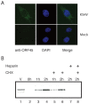
(A) HFF monolayers were mock infected or infected with gradient-purified KSHV virions. At one hour post-infection, these cells were subjected to an immunoflorescence (IFA) by staining with mouse monoclonal antibody (2D4A5) against ORF45 followed by subsequent treatment with Alexa-488 conjugated anti-mouse secondary antibody and counterstaining with DAPI. The DAPI panel shows the nucleus counterstained with DAPI. (B) 293T cells were infected with gradient-purified KSHV virions in the absence (lanes 3, 4) or the presence of cycloheximide (CHX) (lanes 5 and 6) or heparin (lanes 7 and 8). After 1 or 2 h post-infection, the unbound viruses were washed away with low pH buffer. Cell extracts were made and protein concentration was measured using a Bradford assay kit (Bio-Rad). About 50 µg of the protein extract was run on 4–12% Bis-Tris gels and expression levels of ORF45 were detected by a western blot using a specific mouse monoclonal anti-ORF45 antibody. The viral inoculum lysate (lane 1) and mock-infected cell lysate (lane 2) served as controls.
Figure 4
Figure 4. Induction of an antiviral state by the ORF45-null recombinant KSHV virus.
(A) Human foreskin fibroblast (HFF) monolayers were mock infected (Mock) or infected with the KSHV recombinant viruses, including the wild-type BAC36, UV-irradiated BAC36 (BAC36-UV), BAC-rev45, UV-irradiated BAC-rev45 (BAC-rev45-UV), BAC-stop45, or UV-irradiated BAC-stop45 (BAC-stop45-UV) at 5 genomes/cell. HFF cells infected with UV-inactivated HSV-1 (HSV-UV) at an MOI of 5 or treated with IFNα (1000 units/ml) were also included as controls. The following day, the cells were superinfected with 100 plaque forming units (Pfu) of VSV. Media were collected 24 h later and VSV titers were determined by a standard plaque assay. Data are the average of the results from three experiments. Error bars represent standard deviation. (B) HFF monolayers mock infected or infected with BAC36 (wild-type, wt), BAC-rev45 or BAC-stop45 viruses were super infected as above with VSV. Twenty-four hours postinfection, cell lysates were collected and analyzed by a Western blot for expression levels of KSHV latent nuclear antigen (LANA), KSHV ORF45, VSV G glycoprotein and β-actin (loading control) employing specific antibodies.
Figure 5
Figure 5. Evaluation of type I interferon (IFN) and downstream IFN stimulatory gene (ISG) transcription.
HFF cells seeded in 6-well plates were infected with the KSHV wild-type (BAC36) or the ORF45-null recombinant (stop45) viruses. Six hours post-infection, cells were lysed with Trizol reagent and total RNAs isolated. Residual DNA contamination was eliminated by subsequent treatment with Turbo DNase I and the RNA was subsequently reverse transcribed to cDNA. The cDNA samples were then subjected to an absolute real-time PCR based quantification with specific primers for IFNA1, IFNB and selected downstream antiviral effector genes (ISG56, MxA, PKR and OAS). The amounts of mRNA were quantitated based on comparison with the standard templates of cloned cDNAs of known copy number following which the expression levels were normalized to GAPDH.
Figure 6
Figure 6. Ectopic expression of KSHV ORF45 in lentivirus transduced HFF cells permits VSV superinfection.
HFF cells seeded in 24-well plates were mock-transduced or transduced with the lentiviruses expressing ORF45 (lenti-ORF45+) or not expressing ORF45 (lenti-empty and lenti-ORF45- [where the ORF45 sequence was inserted in the opposite direction]) as controls. (A) Cell lysates of lentivirus transduced cells were analyzed with Western blot with antibodies against ORF45 and GFP to ensure the success of transduction. The same blot was also probed with β-actin antibody for an equal loading control. (B) At 24 h post-tranduction, cells were superinfected with VSV, overlayed with 1% methylcellulose in media and stained with 0.1% crystal violet 24 h later for visulalization of plaques. Images were taken under 12x dissection microscopy. Some of the plaques in the mock transduced and the Lenti-ORF45+ transduced cell monolayers are indicated by arrows for easy visualization. (C) Quantitation of VSV super-infection. Parallel experiments were performed as above. After VSV superinfection, each well was supplemented with 1 ml fresh DMEM instead of being overlayed with methylcellulose. Media were collected 24 h later and VSV titers in the supernatant medium were determined by a standard plaque assay.
Figure 7
Figure 7. Interferon regulatory factor-7 (IRF-7) critically contributes to the immune response following primary KSHV infection.
(A) Knockdown of IRF-7 expression by short-hairpin (shRNA)-mediated silencing. Five shRNA constructs (clone #s 1-5) each consisting of specific shRNA sequences in pLKO.1-puro plasmids against different target sites of IRF-7 mRNA along with a non-targeting “control” shRNA that activates the RNAi pathway without targeting any known human gene were transduced into HFF cells in the presence of polybrene. Transduced HFF cells were selected with puromycin (2 µg/ml) to allow for the generation of HFF monolayers expressing stable and long term downregulation of IRF-7. The efficiency of the different shRNA clones in knocking down expression of IRF-7 as compared to the “control” was tested by a Western blot (WB) performed on cell extracts with a rabbit polyclonal antibody against IRF-7 and subsequent probing with β-actin (equal loading control). Endogenous expression levels of IRF-7 in mock transfected HFF cells was also checked. (B) HFF monolayers expressing efficient down regulation of IRF-7 (clone #s 1, 3 and 4) along with the “control” transduced HFF cells were infected with the wild-type (BAC36) and the ORF45-null (BAC-stop45) recombinant viruses and subsequently super infected with VSV. Twenty four hours post VSV infection, cell lysates were collected and by a Western blot were analyzed for expression levels of KSHV latent nuclear antigen (LANA), KSHV ORF45, VSV G glycoprotein and β-actin (loading control) employing specific antibodies.

Similar articles

Cited by

References

    1. Chang Y, Cesarman E, Pessin MS, Culpepper J, Knowles DM, et al. Identification of herpesvirus-loke DNA sequences in AIDS-associated Kaposi's sarcoma. Science. 1994;266:1865–1869. - PubMed
    1. Russo JJ, Bohenzky RA, Chien MC, Chen Y, Yan M, et al. Nucleotide sequence of the Kaposis sarcoma-associated herpesvirus (HHV-8). Proc Natl Acad Sci USA. 1996;93:14862–14867. - PMC - PubMed
    1. Cesarman E, Chang Y, Moore PS, Said JW, Knowles DM. Kaposi's sarcoma-associated herpesvirus-like DNA sequences in AIDS-related body-cavity-based lymphomas. N Engl J Med. 1995;332:1186–1191. - PubMed
    1. Soulier J, Grollet L, Okshendler E, Cacoub P, Cazals-Hatem D, et al. Kaposi's sarcoma-associated herpesvirus-like DNA sequences in multicentric Castleman's disease. Blood. 1995;86:1276–1280. - PubMed
    1. Goodbourn S, Didcock L, Randall RE. Interferons: cell signaling, immune modulation, antiviral response and virus countermeasures. J Gen Virol. 2000;81:2341–2364. - PubMed

Publication types

MeSH terms

Substances