. 2010 Jul;30(14):3542-52.
doi: 10.1128/MCB.01292-09. Epub 2010 May 17.

Sti1 regulation of Hsp70 and Hsp90 is critical for curing of Saccharomyces cerevisiae [PSI+] prions by Hsp104

Affiliations

Sti1 regulation of Hsp70 and Hsp90 is critical for curing of Saccharomyces cerevisiae [PSI+] prions by Hsp104

Michael Reidy et al. Mol Cell Biol. 2010 Jul.

Abstract

Although propagation of Saccharomyces cerevisiae prions requires Hsp104 protein disaggregating activity, overproducing Hsp104 "cures" cells of [PSI(+)] prions. Earlier evidence suggests that the Hsp70 mutant Ssa1-21 impairs [PSI(+)] by a related mechanism. Here, we confirm this link by finding that deletion of STI1 both suppresses Ssa1-21 impairment of [PSI(+)] and blocks Hsp104 curing of [PSI(+)]. Hsp104's tetratricopeptide repeat (TPR) interaction motif was dispensable for curing; however, cells expressing Sti1 defective in Hsp70 or Hsp90 interaction cured less efficiently, and the Hsp90 inhibitor radicicol abolished curing, implying that Sti1 acts in curing through Hsp70 and Hsp90 interactions. Accordingly, strains lacking constitutive or inducible Hsp90 isoforms cured at reduced rates. We confirm an earlier finding that elevating free ubiquitin levels enhances curing, but it did not overcome inhibition of curing caused by Hsp90 defects, suggesting that Hsp90 machinery is important for the contribution of ubiquitin to curing. We also find curing associated with cell division. Our findings point to crucial roles of Hsp70, Sti1, and Hsp90 for efficient curing by overexpressed Hsp104 and provide evidence supporting the earlier suggestion that destruction of prions by protein disaggregation does not adequately explain the curing.


Figures
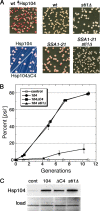
FIG. 1.
FIG. 1.
Sti1 mediates Ssa1-21 inhibition of [PSI+] and is critical for curing of [PSI+] by Hsp104 overexpression. (A) The upper left panel shows wild-type (wt) cells that overexpressed Hsp104 for ∼5 generations and then were spread onto 1/2YPD plates and grown for 2 to 3 days at 30°C. White colonies are [PSI+], and red colonies arose from cells that lost [PSI+]. The lower left panel shows hsp104Δ cells expressing Hsp104 or Hsp104ΔC4 (as indicated) from a plasmid, which produces roughly 3-fold more Hsp104 than chromosomally expressed Hsp104, grown similarly but on defined medium with limiting (8 mg/liter) adenine. Instability of [PSI+] caused by the modestly elevated Hsp104 is detectable as red sectoring in otherwise white colonies. Overall color and sectoring frequency of cells expressing the different Hsp104 proteins are indistinguishable. Wild-type and SSA1-21 mutant cells (center panels) or the same combined with deletion of STI1 (sti1Δ; right panels) were spread onto 1/2YPD and incubated as described above to illustrate [PSI+] phenotypes. All panels are representative 2.5-cm squares from plates, except those on the lower left, which are magnified ×2 relative to others. Images were made by scanning plates by the use of an HP ScanJet 6300C document scanner. Color and contrast of the assembly of images were optimized using Adobe Photoshop 7.0 software. (B) Appearance of [psi] cells in growing cultures is shown as a function of generations (monitored as doubling of OD600). All cultures were grown in medium containing 100 μM CuSO4 to induce expression from the CUP1 promoter and then spread onto 1/2YPD plates and incubated as described above. Wild-type cells carried plasmids without HSP104 (control), with wild-type HSP104 (104), or with HSP104ΔC4 (104ΔC4). The sti1Δ cells over-express wild-type HSP104. To maintain continuous growth, cultures were diluted into fresh medium when the OD600 reached 1 to 2. No [psi] colonies appeared among over 900 colonies from the control cultures at any of the time points. Values are averages of the results for three independent experiments ± standard error of the mean (SEM). (C) Western analysis of the abundance of Hsp104 and Hsp104ΔC4 in cells used for the experiment in panel B from cultures grown for roughly 10 generations. Bottom panel (load) shows a portion of the membrane used for the blot stained by amido black as a loading and transfer control.
FIG. 2.
FIG. 2.
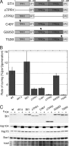
Both TPR domains of Sti1 are important for prion curing by overexpressed Hsp104. (A) Domain structure of Sti1 and mutants used in this study. Numbers above the diagram indicate the amino acid residue position; asterisks indicate the locations of point mutations indicated on the left. Deleted regions are indicated with horizontal lines. DP, aspartate-proline region. (B) Rates of prion curing were determined in cultures overexpressing Hsp104 for four or five generations. Strains to the right of the wild type (wt; carries empty vector) are sti1Δ with an empty vector or a vector encoding the indicated Sti1 protein. Values are averages of the results for three independent experiments ± standard deviation. (C) Western analysis of abundance of proteins indicated on the left in cells from experiments shown in panel B that were grown for four generations. All strains were exposed to copper and carry either an empty vector (−) or pMR26 for expression of Hsp104 (+), as indicated above each lane. Lower panel (load) is a portion of the blotted membrane stained by amido black as a loading and transfer control.
FIG. 3.
FIG. 3.
Curing of cells coexpressing Ssa1 or Sse1. Curing was done as described in the legend for Fig. 1, using cells carrying plasmids with HSP104 (104), SSA1 (A1), and SSE1 (E1) or the corresponding empty vectors (ev). Western analysis probing for the chaperones indicated on the left was done using cultures grown for four generations. Numbers below the image indicate rates of prion curing (%[psi] per generation) for the strains labeled directly above.
FIG. 4.
FIG. 4.
Sti1 mutations that suppress Hsp104-mediated curing affect interaction with Hsp90. Cells lacking chromosomal STI1 and carrying empty vector (cont) or plasmids expressing His6-tagged Sti1, Sti1(C49Y), Sti1(G325D), or Sti1(T526I) were grown as indicated in Materials and Methods. Sti1-containing complexes were purified from lysates by using metal affinity chromatography. Complexes were separated by SDS-PAGE, blotted, and probed with antibodies specific to Sti1 (top panel), an antibody raised against the C terminus of yeast Hsp90 that reacts with both Hsc82 and Hsp82 (Hsc82 + Hsp82) and a commercially available antibody raised against human Hsp90α that recognizes yeast Hsp82 only (Hsp82). The bottom panel (CB) shows a portion of a Coomassie brilliant blue-stained gel of the purified complexes, with molecular size markers indicated. Identical volumes of samples were loaded in this gel and in the gels used for the blots. The arrowhead indicates the position of Sti1.
FIG. 5.
FIG. 5.
Hsp90 function is important for curing of [PSI+] by overexpressed Hsp104. (A) Sensitivity of strains (all are [PSI+]) to the Hsp90-specific inhibitor radicicol. Overnight YPAD cultures were diluted to an OD600 of 1.0, and 5-fold serial dilutions were spotted onto plates without and with (+rad) 25 μg/ml radicicol. Plates were scanned after incubation for 3 days at 30°C. (B) Radicicol blocks curing of [PSI+] by overexpressed Hsp104. Wild-type cells with plasmid for copper-inducible overexpression of Hsp104 were grown under inducing conditions (100 μM CuSO4) for 1.5 generations, at which time (“add” arrow) the overexpressing culture was split. One culture (closed circles) was untreated, while radicicol (25 μg/ml final concentration) was added to the other (closed triangles). After growing for another 3.5 generations (“remove” arrow), the radicicol-treated culture was washed, suspended in fresh radicicol-free medium, and grown for another 2.5 generations. Cultures grown without CuSO4 and either lacking (open circles) or containing (open triangles) radicicol were monitored in parallel. Values are averages of the results for three independent experiments ± standard deviation. (C) Western analysis of chaperones (indicated on left) in cells used in a replicate of the experiment shown in panel B. The “load” image shows a portion of the blotted membrane stained by amido black. (D) Western analysis of Hsp90 and other chaperones in hsc82Δ and hsp82Δ cells used in curing experiments. The blot in the top image is probed with an antibody that recognizes both isoforms of Hsp90 (Hsc82 + Hsp82), while the panel beneath it is an identical blot probed with a different antibody reactive only to the inducible Hsp82 isoform (see also Fig. 3). The lower panels show blots probed for Hsp104, Hsp70, and Sse1, as indicated. All cultures contained 100 μM CuSO4; control cells (cont) carry the empty vector.
FIG. 6.
FIG. 6.
Ub effects on curing of [PSI+] by Hsp104 overexpression. Rates of curing of cultures grown for four to six generations were determined as described in the legend for Fig. 1. Strains (indicated at bottom) carry empty vector (−) or the same plasmid with UBI4 (+), which encodes Ub. Values are averages of the results for three independent experiments ± standard deviation. Student's t test values are as follows: wild type, P = 0.0014; others, P = 0.1.
FIG. 7.
FIG. 7.
Curing of [PSI+] by Hsp104 overexpression is associated with cell division. Curing of a wild-type culture was done as described in the legend for Fig. 1 except that the culture was harvested 5 h after inducing Hsp104 expression and split into two flasks, one containing identical medium (control; circles) and the other into a similar medium lacking a required nutrient (leucine or histidine; squares). After incubation for another 20 h, the depleted nutrient was added back to the second culture (arrow). (A) Growth of cultures is shown as log of CFU per ml as a function of time. (B) The proportion of [psi] colonies in the same culture aliquots used to monitor growth is shown. Values in panels A and B are averages from the results for three independent experiments, (two with leucine depleted, one with histidine depleted) ± standard deviation. (C) Western analysis of Hsp104 abundance in cells of the same aliquots of starved (+) and unstarved (−) cultures removed at the indicated time points from the experiment shown in panels A and B.
FIG. 8.
FIG. 8.
Curing of [PSI+] by guanidine inactivation of Hsp104. Cells grown in YPAD were diluted into similar medium containing 3 mM guanidine hydrochloride and grown continuously for roughly 12 generations. Aliquots of cells were removed periodically to determine the proportion of [PSI+] cells as described in the legend for Fig. 1. The proportion of [PSI+] cells remaining is shown as a function of generations.

Similar articles

Cited by

References

    1. Abbas-Terki, T., O. Donze, P. A. Briand, and D. Picard. 2001. Hsp104 interacts with Hsp90 cochaperones in respiring yeast. Mol. Cell. Biol. 21:7569-7575. - PMC - PubMed
    1. Acebron, S. P., I. Martin, U. del Castillo, F. Moro, and A. Muga. 2009. DnaK-mediated association of ClpB to protein aggregates. A bichaperone network at the aggregate surface. FEBS Lett. 583:2991-2996. - PubMed
    1. Allen, K. D., T. A. Chernova, E. P. Tennant, K. D. Wilkinson, and Y. O. Chernoff. 2007. Effects of ubiquitin system alterations on the formation and loss of a yeast prion. J. Biol. Chem. 282:3004-3013. - PubMed
    1. Allen, K. D., R. D. Wegrzyn, T. A. Chernova, S. Muller, G. P. Newnam, P. A. Winslett, K. B. Wittich, K. D. Wilkinson, and Y. O. Chernoff. 2005. Hsp70 chaperones as modulators of prion life cycle: novel effects of Ssa and Ssb on the Saccharomyces cerevisiae prion [PSI+]. Genetics 169:1227-1242. - PMC - PubMed
    1. Bailleul, P. A., G. P. Newnam, J. N. Steenbergen, and Y. O. Chernoff. 1999. Genetic study of interactions between the cytoskeletal assembly protein Sla1 and prion-forming domain of the release factor Sup35 (eRF3) in Saccharomyces cerevisiae. Genetics 153:81-94. - PMC - PubMed

Publication types

MeSH terms

LinkOut - more resources