

Host hindrance to HIV-1 replication in monocytes and macrophages
- PMID: 20374633
- PMCID: PMC2868797
- DOI: 10.1186/1742-4690-7-31
Host hindrance to HIV-1 replication in monocytes and macrophages
Abstract
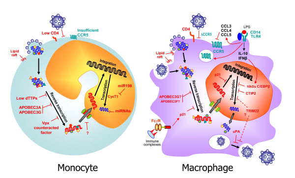
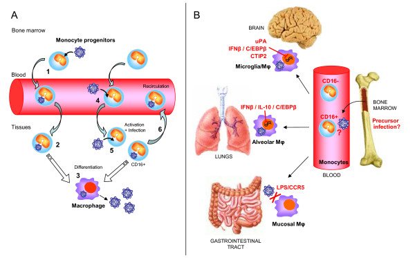
Monocytes and macrophages are targets of HIV-1 infection and play critical roles in multiple aspects of viral pathogenesis. HIV-1 can replicate in blood monocytes, although only a minor proportion of circulating monocytes harbor viral DNA. Resident macrophages in tissues can be infected and function as viral reservoirs. However, their susceptibility to infection, and their capacity to actively replicate the virus, varies greatly depending on the tissue localization and cytokine environment. The susceptibility of monocytes to HIV-1 infection in vitro depends on their differentiation status. Monocytes are refractory to infection and become permissive upon differentiation into macrophages. In addition, the capacity of monocyte-derived macrophages to sustain viral replication varies between individuals. Host determinants regulate HIV-1 replication in monocytes and macrophages, limiting several steps of the viral life-cycle, from viral entry to virus release. Some host factors responsible for HIV-1 restriction are shared with T lymphocytes, but several anti-viral mechanisms are specific to either monocytes or macrophages. Whilst a number of these mechanisms have been identified in monocytes or in monocyte-derived macrophages in vitro, some of them have also been implicated in the regulation of HIV-1 infection in vivo, in particular in the brain and the lung where macrophages are the main cell type infected by HIV-1. This review focuses on cellular factors that have been reported to interfere with HIV-1 infection in monocytes and macrophages, and examines the evidences supporting their role in vivo, highlighting unique aspects of HIV-1 restriction in these two cell types.
Figures
Similar articles
-
MicroRNA-mediated restriction of HIV-1 in resting CD4+ T cells and monocytes.Viruses. 2012 Sep;4(9):1390-409. doi: 10.3390/v4091390. Epub 2012 Aug 29. Viruses. 2012. PMID: 23170164 Free PMC article. Review.
-
C5a and C5a(desArg) enhance the susceptibility of monocyte-derived macrophages to HIV infection.J Immunol. 2001 Mar 1;166(5):3410-5. doi: 10.4049/jimmunol.166.5.3410. J Immunol. 2001. PMID: 11207298
-
Quantitation of Productively Infected Monocytes and Macrophages of Simian Immunodeficiency Virus-Infected Macaques.J Virol. 2016 May 27;90(12):5643-5656. doi: 10.1128/JVI.00290-16. Print 2016 Jun 15. J Virol. 2016. PMID: 27030272 Free PMC article.
-
Cellular resistance to HIV-1 infection in target cells coincides with a rapid induction of X-DING-CD4 mRNA: indication of the unique host innate response to virus regulated through function of the X-DING-CD4 gene.Innate Immun. 2012 Aug;18(4):563-70. doi: 10.1177/1753425911426893. Epub 2011 Oct 31. Innate Immun. 2012. PMID: 22042911 Free PMC article.
-
The HIV Reservoir in Monocytes and Macrophages.Front Immunol. 2019 Jun 26;10:1435. doi: 10.3389/fimmu.2019.01435. eCollection 2019. Front Immunol. 2019. PMID: 31297114 Free PMC article. Review.
Cited by
-
Nanoparticle based galectin-1 gene silencing, implications in methamphetamine regulation of HIV-1 infection in monocyte derived macrophages.J Neuroimmune Pharmacol. 2012 Sep;7(3):673-85. doi: 10.1007/s11481-012-9379-7. Epub 2012 Jun 12. J Neuroimmune Pharmacol. 2012. PMID: 22689223 Free PMC article.
-
HIV infection of non-classical cells in the brain.Retrovirology. 2023 Jan 13;20(1):1. doi: 10.1186/s12977-023-00616-9. Retrovirology. 2023. PMID: 36639783 Free PMC article. Review.
-
SERINC5 Is an Unconventional HIV Restriction Factor That Is Upregulated during Myeloid Cell Differentiation.J Innate Immun. 2020;12(5):399-409. doi: 10.1159/000504888. Epub 2020 Jan 14. J Innate Immun. 2020. PMID: 31935717 Free PMC article.
-
p21-mediated RNR2 repression restricts HIV-1 replication in macrophages by inhibiting dNTP biosynthesis pathway.Proc Natl Acad Sci U S A. 2013 Oct 15;110(42):E3997-4006. doi: 10.1073/pnas.1306719110. Epub 2013 Sep 30. Proc Natl Acad Sci U S A. 2013. PMID: 24082141 Free PMC article. Clinical Trial.
-
Impact of viral factors on subcellular distribution and RNA export activity of HIV-1 rev in astrocytes 1321N1.PLoS One. 2013 Sep 4;8(9):e72905. doi: 10.1371/journal.pone.0072905. eCollection 2013. PLoS One. 2013. PMID: 24023789 Free PMC article.
References
-
- Whitelaw DM. Observations on human monocyte kinetics after pulse labeling. Cell Tissue Kinet. 1972;5:311–7. - PubMed
-
- Hasegawa A, Liu H, Ling B, Borda JT, Alvarez X, Sugimoto C, Vinet-Oliphant H, Kim WK, Williams KC, Ribeiro RM, Lackner AA, Veazey RS, Kuroda MJ. The level of monocyte turnover predicts disease progression in the macaque model of AIDS. Blood. 2009;114(14):2917–25. doi: 10.1182/blood-2009-02-204263. - DOI - PMC - PubMed
Publication types
MeSH terms
LinkOut - more resources
Full Text Sources
Other Literature Sources
