

Docosahexaenoic acid supplementation increases prefrontal cortex activation during sustained attention in healthy boys: a placebo-controlled, dose-ranging, functional magnetic resonance imaging study
- PMID: 20130094
- PMCID: PMC2844685
- DOI: 10.3945/ajcn.2009.28549
Docosahexaenoic acid supplementation increases prefrontal cortex activation during sustained attention in healthy boys: a placebo-controlled, dose-ranging, functional magnetic resonance imaging study
Abstract
Background: Emerging evidence suggests that docosahexaenoic acid (DHA, 22:6n-3), the principal omega-3 (n-3) fatty acid in brain gray matter, positively regulates cortical metabolic function and cognitive development. However, the effects of DHA supplementation on functional cortical activity in human subjects are unknown.
Objective: The objective was to determine the effects of DHA supplementation on functional cortical activity during sustained attention in human subjects.
Design: Healthy boys aged 8-10 y (n = 33) were randomly assigned to receive placebo or 1 of 2 doses of DHA (400 or 1200 mg/d) for 8 wk. Relative changes in cortical activation patterns during sustained attention at baseline and endpoint were determined by functional magnetic resonance imaging.
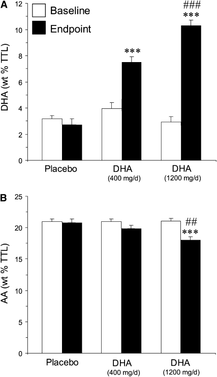
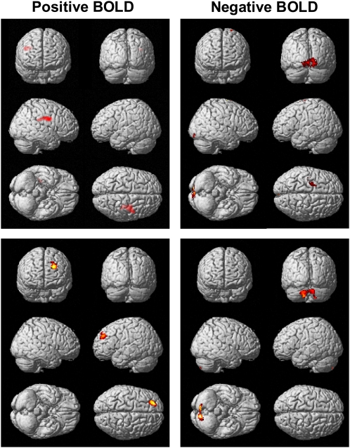
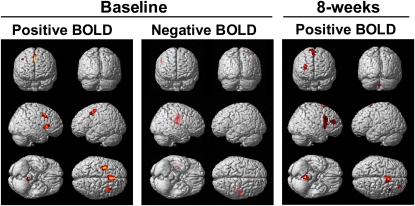
Results: At 8 wk, erythrocyte membrane DHA composition increased significantly from baseline in subjects who received low-dose (by 47%) or high-dose (by 70%) DHA but not in those who received placebo (-11%). During sustained attention, both DHA dose groups had significantly greater changes from baseline in activation of the dorsolateral prefrontal cortex than did the placebo group, and the low-dose and high-dose DHA groups had greater decreases in the occipital cortex and cerebellar cortex, respectively. Relative to low-dose DHA, high-dose DHA resulted in greater decreases in activation of bilateral cerebellum. The erythrocyte DHA composition was positively correlated with dorsolateral prefrontal cortex activation and was inversely correlated with reaction time, at baseline and endpoint.
Conclusion: Dietary DHA intake and associated elevations in erythrocyte DHA composition are associated with alterations in functional activity in cortical attention networks during sustained attention in healthy boys. This trial was registered at clinicaltrials.gov as NCT00662142.
Figures
Comment in
-
Do you need a supplement of docosahexaenoic acid or an n-3 long-chain polyunsaturated fatty acid?Am J Clin Nutr. 2010 Apr;91(4):827-8. doi: 10.3945/ajcn.2010.29387. Epub 2010 Mar 3. Am J Clin Nutr. 2010. PMID: 20200255 No abstract available.
Similar articles
-
Docosahexaenoic acid biostatus is associated with event-related functional connectivity in cortical attention networks of typically developing children.Nutr Neurosci. 2017 May;20(4):246-254. doi: 10.1179/1476830515Y.0000000046. Epub 2016 Mar 24. Nutr Neurosci. 2017. PMID: 26463682
-
Low docosahexaenoic acid status is associated with reduced indices in cortical integrity in the anterior cingulate of healthy male children: a 1H MRS Study.Nutr Neurosci. 2013 Jul;16(4):183-90. doi: 10.1179/1476830512Y.0000000045. Nutr Neurosci. 2013. PMID: 23582513 Free PMC article.
-
Prenatal supplementation with DHA improves attention at 5 y of age: a randomized controlled trial.Am J Clin Nutr. 2016 Oct;104(4):1075-1082. doi: 10.3945/ajcn.114.101071. Epub 2016 Sep 7. Am J Clin Nutr. 2016. PMID: 27604770 Free PMC article. Clinical Trial.
-
Effects of fish oil supplementation on prefrontal metabolite concentrations in adolescents with major depressive disorder: a preliminary 1H MRS study.Nutr Neurosci. 2016 May;19(4):145-55. doi: 10.1179/1476830514Y.0000000135. Epub 2016 Mar 2. Nutr Neurosci. 2016. PMID: 24915543 Free PMC article. Clinical Trial.
-
Distribution, interconversion, and dose response of n-3 fatty acids in humans.Am J Clin Nutr. 2006 Jun;83(6 Suppl):1467S-1476S. doi: 10.1093/ajcn/83.6.1467S. Am J Clin Nutr. 2006. PMID: 16841856 Review.
Cited by
-
Effect of Omega-3 Long Chain Polyunsaturated Fatty Acids (n-3 LCPUFA) Supplementation on Cognition in Children and Adolescents: A Systematic Literature Review with a Focus on n-3 LCPUFA Blood Values and Dose of DHA and EPA.Nutrients. 2020 Oct 12;12(10):3115. doi: 10.3390/nu12103115. Nutrients. 2020. PMID: 33053843 Free PMC article.
-
Determinants of Plasma Docosahexaenoic Acid Levels and Their Relationship to Neurological and Cognitive Functions in PKU Patients: A Double Blind Randomized Supplementation Study.Nutrients. 2018 Dec 7;10(12):1944. doi: 10.3390/nu10121944. Nutrients. 2018. PMID: 30544518 Free PMC article. Clinical Trial.
-
Relationship Between Polyunsaturated Fatty Acids and Psychopathology in the NEURAPRO Clinical Trial.Front Psychiatry. 2019 Jun 6;10:393. doi: 10.3389/fpsyt.2019.00393. eCollection 2019. Front Psychiatry. 2019. PMID: 31244693 Free PMC article.
-
Preventative strategies for early-onset bipolar disorder: towards a clinical staging model.CNS Drugs. 2010 Dec;24(12):983-96. doi: 10.2165/11539700-000000000-00000. CNS Drugs. 2010. PMID: 21090835 Review.
-
Detection and treatment of omega-3 fatty acid deficiency in psychiatric practice: Rationale and implementation.Lipids Health Dis. 2016 Feb 10;15:25. doi: 10.1186/s12944-016-0196-5. Lipids Health Dis. 2016. PMID: 26860589 Free PMC article. Review.
References
-
- Carver JD, Benford VJ, Han B, Cantor AB. The relationship between age and the fatty acid composition of cerebral cortex and erythrocytes in human subjects. Brain Res Bull 2001;56:79–85 - PubMed
-
- Anderson GJ, Neuringer M, Lin DS, Connor WE. Can prenatal N-3 fatty acid deficiency be completely reversed after birth? Effects on retinal and brain biochemistry and visual function in rhesus monkeys. Pediatr Res 2005;58:865–72 - PubMed
-
- Connor WE, Neuringer M, Lin DS. Dietary effects on brain fatty acid composition: the reversibility of n−3 fatty acid deficiency and turnover of docosahexaenoic acid in the brain, erythrocytes, and plasma of rhesus monkeys. J Lipid Res 1990;31:237–47 - PubMed
-
- Calderon F, Kim HY. Docosahexaenoic acid promotes neurite growth in hippocampal neurons. J Neurochem 2004;90:979–88 - PubMed
-
- Coti Bertrand P, O'Kusky JR, Innis SM. Maternal dietary (n−3) fatty acid deficiency alters neurogenesis in the embryonic rat brain. J Nutr 2006;136:1570–5 - PubMed
Publication types
MeSH terms
Substances
Associated data
Grants and funding
LinkOut - more resources
Full Text Sources
Other Literature Sources
Medical
