. 2010 Jan 29;6(1):e1000742.
doi: 10.1371/journal.ppat.1000742.

Upregulation of xCT by KSHV-encoded microRNAs facilitates KSHV dissemination and persistence in an environment of oxidative stress

Affiliations

Upregulation of xCT by KSHV-encoded microRNAs facilitates KSHV dissemination and persistence in an environment of oxidative stress

Zhiqiang Qin et al. PLoS Pathog. .

Abstract

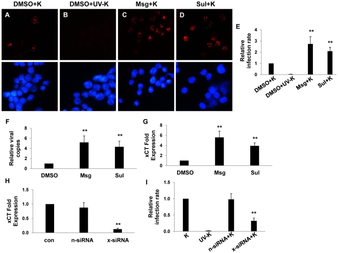
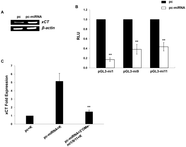
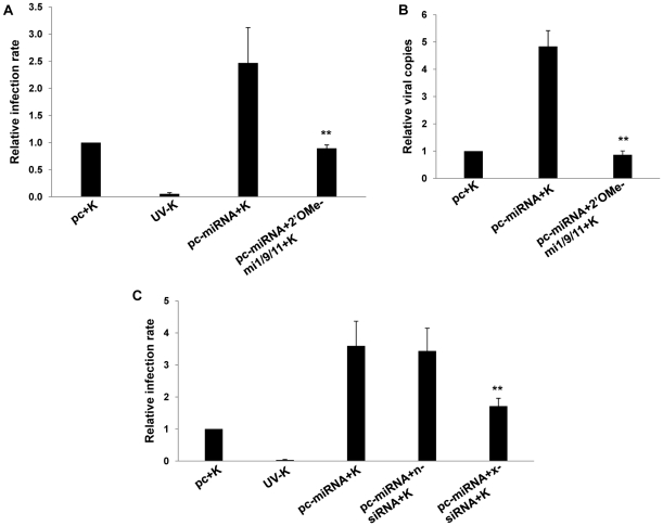
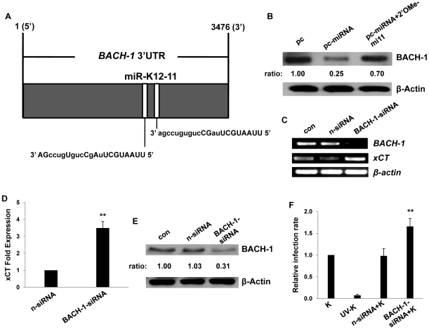
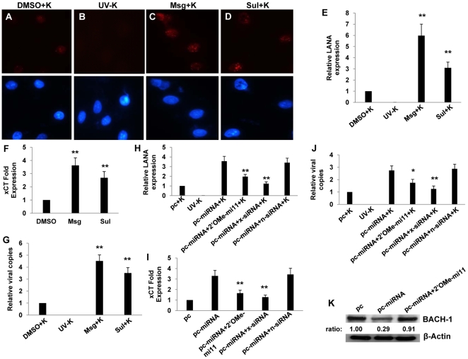
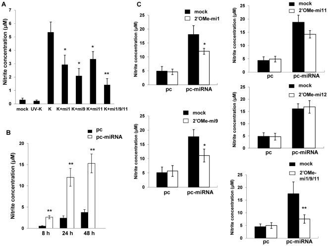
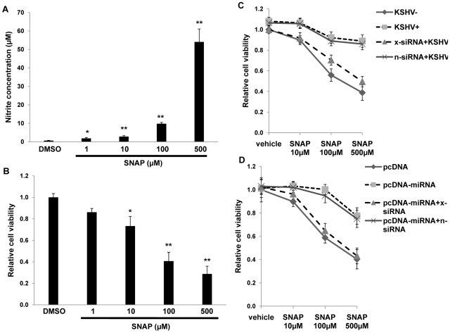
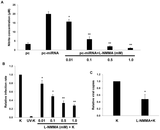
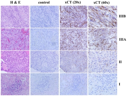
Upregulation of xCT, the inducible subunit of a membrane-bound amino acid transporter, replenishes intracellular glutathione stores to maintain cell viability in an environment of oxidative stress. xCT also serves as a fusion-entry receptor for the Kaposi's sarcoma-associated herpesvirus (KSHV), the causative agent of Kaposi's sarcoma (KS). Ongoing KSHV replication and infection of new cell targets is important for KS progression, but whether xCT regulation within the tumor microenvironment plays a role in KS pathogenesis has not been determined. Using gene transfer and whole virus infection experiments, we found that KSHV-encoded microRNAs (KSHV miRNAs) upregulate xCT expression by macrophages and endothelial cells, largely through miR-K12-11 suppression of BACH-1-a negative regulator of transcription recognizing antioxidant response elements within gene promoters. Correlative functional studies reveal that upregulation of xCT by KSHV miRNAs increases cell permissiveness for KSHV infection and protects infected cells from death induced by reactive nitrogen species (RNS). Interestingly, KSHV miRNAs simultaneously upregulate macrophage secretion of RNS, and biochemical inhibition of RNS secretion by macrophages significantly reduces their permissiveness for KSHV infection. The clinical relevance of these findings is supported by our demonstration of increased xCT expression within more advanced human KS tumors containing a larger number of KSHV-infected cells. Collectively, these data support a role for KSHV itself in promoting de novo KSHV infection and the survival of KSHV-infected, RNS-secreting cells in the tumor microenvironment through the induction of xCT.


Conflict of interest statement

The authors have declared that no competing interests exist.

Figures

Figure 1
Figure 1. xCT mediates KSHV infection of macrophages.
(AD) 264.7 (“RAW”) cells were first incubated with Monosodium glutamate (Msg), Sulfasalazine (Sul) or vehicle control (DMSO) for 12 h followed by purified KSHV (K). 16 h later, IFA employing anti-LANA monoclonal antibodies and secondary antibodies conjugated to Texas Red were performed to identify expression of LANA signified by the typical punctate intranuclear expression pattern. Nuclei were identified using DAPI (blue). Some cells were incubated with UV-inactivated virus (UV-K) for negative controls. Representative images from one of three independent experiments are shown. (E) Relative infection rate was determined for groups in A–D as outlined in Methods. (F) qPCR was used to determine relative intracellular KSHV DNA content normalized to the vehicle control group (relative viral copies) as explained in Methods. (G) qRT-PCR was used to determine relative xCT transcript expression. (H) qRT-PCR was used to determine xCT transcript expression relative to control cells for cells transfected with either control (n) or xCT-specific siRNA. (I) Relative infection rates were calculated for groups in (H) using LANA IFA. For all assays, error bars represent the S.E.M. for three independent experiments. * * = p<0.01.
Figure 2
Figure 2. KSHV miRNAs upregulate xCT expression by macrophages.
(A) RT-PCR was used to determine expression of xCT transcripts in RAW cells transfected with either control (pc) or miRNA-expressing vectors (pc-miRNA). β-actin was used as a loading control. (B) RAW cells were co-transfected with miRNA luciferase reporter constructs (pGL3-miX where X = complimentary sequence for the individual KSHV miRNAs noted) and either control or miRNA-expressing vectors. 48 h later, luciferase expression was determined for miRNA transfectants relative to controls (RLU). (C) Cells were transfected with control or miRNA-expressing vectors with or without 2'OMe RNA antagomirs targeting miR-K12-1, miR-K12-9, and miR-K12-11 (mi1/9/11). 48 h following subsequent incubation with KSHV (K), qRT-PCR was used to determine relative xCT transcript expression. For all assays, error bars represent the S.E.M. for three independent experiments. * * = p<0.01.
Figure 3
Figure 3. KSHV miRNA upregulation of xCT increases macrophage susceptibility to KSHV infection.
(A) RAW cells were transfected with control or miRNA-expressing vectors then incubated with purified KSHV (K). LANA IFA were performed 16 h later, and relative infection rates were determined as outlined in Methods. (B) qPCR was used to determine relative intracellular KSHV DNA content for groups in A. (C) Cells were co-transfected with control or miRNA-expressing vectors and either control (n) or xCT-specific siRNA then incubated with purified KSHV. 16 h later, LANA IFA were used to determine relative infection rates. For all assays, error bars represent the S.E.M. for three independent experiments. * * = p<0.01.
Figure 4
Figure 4. KSHV miRNAs upregulate xCT expression through repression of BACH-1.
(A) Potential KSHV miRNA binding sites were identified within the 3'UTR of murine BACH-1 using an ad-hoc scanning program as described in Methods. miR-K12-11 nucleotides with matching base pairs depicted in capital letters bind within positions 2318–2339 and 2530–2551. (B) RAW cells were transfected with 1 µg control or miRNA-expressing vectors with or without 300 pmol of 2'OMe RNA antagomirs targeting miR-K12-11. 48 h later, BACH-1 expression was quantified by Western blot. β-Actin was used for loading controls. Numbers represent immunoreactivity relative to control transfectants as quantified using Image-J software. (C–D) RT-PCR (C) and qRT-PCR (D) were used to quantify transcripts for BACH-1 (C) and xCT (C and D), respectively, in controls cells or cells transfected with either control (n) or BACH-1-specific siRNA. (E) Western blots were used to quantify BACH-1 protein expression in siRNA-transfected cells for groups in (C). Immunoreactivity was quantified as in (B). (F) Cells were transfected with control or BACH-1 siRNA as above and subsequently incubated with KSHV. Relative infection rates were determined 12 h later using LANA IFA. Error bars represent the S.E.M. for three independent experiments. * * = p<0.01.
Figure 5
Figure 5. miR-K12-11 suppresses BACH-1 expression and increases endothelial cell susceptibility to KSHV through upregulation of xCT.
(A–G) HUVEC were incubated with vehicle control (DMSO), Msg, or Sul for 12 h followed by purified KSHV (K) using an MOI∼0.5–1. 16 h later, LANA IFA were performed as previously described. (A–D) Representative images from one of three independent experiments are shown. (H–K) HUVEC were transfected with control or miRNA-expressing vectors along with a 2'OMe RNA antagomir for miR-K12-11 or either control (n) or xCT-specific siRNA. (E,H) Relative LANA expression was determined as described in Methods. (F,I) qRT-PCR was used to determine relative xCT transcript expression. (G,J) qPCR was used to determine relative intracellular KSHV DNA content normalized to controls. (K) Western blots were used to identify BACH-1 protein expression and immunoreactivity quantified as previously described. For all assays, error bars represent the S.E.M. for three independent experiments. * = p<0.05, * * = p<0.01 (For Fig. H–J, comparisons are relative to either pc-miRNA or pc-miRNA+K).
Figure 6
Figure 6. KSHV miRNAs induce reactive nitrogen species (RNS) secretion by macrophages.
(A) RAW cells were transfected with 2'OMe RNA antagomirs targeting miR-K12-1, miR-K12-9, and miR-K12-11 or all three together (mi1/9/11). Cells were subsequently incubated with purified KSHV (K) and nitrite quantified within culture supernatants as described in Methods. (B) Cells were transfected with control or miRNA-expressing vectors and nitrite quantified within culture supernatants at the times indicated. (C) Cells were co-transfected with either control or miRNA-expression constructs without (mock) or with specific 2'OMe RNA antagomirs. As an additional control, some cells were transfected with an antagomir targeting miR-K12-12 which is not expressed by the miRNA-expressing construct. For all assays, error bars represent the S.E.M. for three independent experiments. * = p<0.05, * * = p<0.01.
Figure 7
Figure 7. KSHV miRNAs enhance macrophage survival in an environment of oxidative stress through the upregulation of xCT.
(A) RAW cells were treated with the indicated concentrations of SNAP or vehicle control for 12 h prior to nitrite quantification within culture supernatants. (B) Relative cell viability was determined for groups in (A) as described in Methods. (C) Cells were transfected with either control (n) or xCT-specific (x) siRNA and incubated with UV-K (KSHV−) or KSHV (KSHV+) 48 h later. (D) Cells were co-transfected with xCT siRNA and either control or miRNA-expressing vectors for 48 h, then incubated for an additional 12 h with SNAP prior to viability determinations. For all assays, error bars represent the S.E.M. for three independent experiments. * = p<0.05, * * = p<0.01.
Figure 8
Figure 8. RNS inhibition reduces macrophage susceptibility to KSHV infection.
(A) RAW cells were first transfected with 1 µg of either control or miRNA-expressing vectors then incubated with L-NMMA for 12 h prior to nitrite quantification in culture supernatants. (B) Cells were incubated with L-NMMA for 12 h prior to their incubation with purified KSHV (K) for 2 h. Following an additional 12 h, LANA IFA were performed and relative infection rates determined as described in Methods. (C) qPCR was used to determine relative intracellular KSHV DNA content for KSHV-infected cells pre-treated with either vehicle or 0.5mM L-NMMA. In all assays, error bars represent the S.E.M. for three independent experiments. * = p<0.05, * * = p<0.01.
Figure 9
Figure 9. xCT expression within KS lesions correlates with tumor stage.
KS diagnosis and histopathologic staging were independently confirmed by a dermatopathologist using hematoxylin and eosin (H & E). Tumors were then processed for immunohistochemistry as described in Methods using either pre-immune sera (control) or xCT anti-sera. xCT expression is revealed by dark brown membrane-associated staining in contrast to blue nuclear staining. Representative images from all stages (I = patch, II = plaque, III = nodule) are shown, including two different stage III tumors (A and B). All images are shown at original magnification ×20 or 60.

Similar articles

Cited by

References

    1. Luppi M, Barozzi P, Schulz TF, Setti G, Staskus K, et al. Bone marrow failure associated with human herpesvirus 8 infection after transplantation. N Engl J Med. 2000;343:1378–1385. - PubMed
    1. Soulier J, Grollet L, Oksenhendler E, Cacoub P, Cazals-Hatem D, et al. Kaposi's sarcoma-associated herpesvirus-like DNA sequences in multicentric Castleman's disease. Blood. 1995;86:1276–1280. - PubMed
    1. Cesarman E, Chang Y, Moore PS, Said JW, Knowles DM. Kaposi's sarcoma-associated herpesvirus-like DNA sequences in AIDS-related body-cavity-based lymphomas. N Engl J Med. 1995;332:1186–1191. - PubMed
    1. Chang Y, Cesarman E, Pessin MS, Lee F, Culpepper J, et al. Identification of herpesvirus-like DNA sequences in AIDS-associated Kaposi's sarcoma. Science. 1994;266:1865–1869. - PubMed
    1. Engels EA, Biggar RJ, Hall HI, Cross H, Crutchfield A, et al. Cancer risk in people infected with human immunodeficiency virus in the United States. Int J Cancer. 2008;123:187–194. - PubMed

Publication types

MeSH terms

Substances