. 2010 Feb;120(2):457-71.
doi: 10.1172/JCI40483. Epub 2010 Jan 19.

Membrane-associated Hsp72 from tumor-derived exosomes mediates STAT3-dependent immunosuppressive function of mouse and human myeloid-derived suppressor cells

Affiliations

Membrane-associated Hsp72 from tumor-derived exosomes mediates STAT3-dependent immunosuppressive function of mouse and human myeloid-derived suppressor cells

Fanny Chalmin et al. J Clin Invest. 2010 Feb.

Abstract

Myeloid-derived suppressor cells (MDSCs) have been identified in humans and mice as a population of immature myeloid cells with the ability to suppress T cell activation. They accumulate in tumor-bearing mice and humans and have been shown to contribute to cancer development. Here, we have isolated tumor-derived exosomes (TDEs) from mouse cell lines and shown that an interaction between TDE-associated Hsp72 and MDSCs determines the suppressive activity of the MDSCs via activation of Stat3. In addition, tumor-derived soluble factors triggered MDSC expansion via activation of Erk. TDE-associated Hsp72 triggered Stat3 activation in MDSCs in a TLR2/MyD88-dependent manner through autocrine production of IL-6. Importantly, decreasing exosome production using dimethyl amiloride enhanced the in vivo antitumor efficacy of the chemotherapeutic drug cyclophosphamide in 3 different mouse tumor models. We also demonstrated that this mechanism is relevant in cancer patients, as TDEs from a human tumor cell line activated human MDSCs and triggered their suppressive function in an Hsp72/TLR2-dependent manner. Further, MDSCs from cancer patients treated with amiloride, a drug used to treat high blood pressure that also inhibits exosome formation, exhibited reduced suppressor functions. Collectively, our findings show in both mice and humans that Hsp72 expressed at the surface of TDEs restrains tumor immune surveillance by promoting MDSC suppressive functions.


Figures
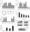
Figure 1
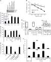
Figure 1. TDEs determine STAT3 activation, while TDSFs determine MDSC expansion.
Bone marrow from naive mice was cultured 3 days in complete medium (CM) alone or with TCSs, TDEs, or TDSFs. (A) The percentage of Gr1+CD11b+ precursor cells ± SD was determined by flow cytometry. (B) The percentage of Ki67+ cells ± SD in Gr1+CD11b+ cells was determined by flow cytometry. (C) pStat3 MFI in Gr1+CD11b+ cells was determined by flow cytometry. Data represent MFI ± SD (n = 3); inset shows representative FACS histogram. (D) Purified splenic MDSCs from naive mice were treated with increasing dosages of TDEs. pStat3 expression was determined by FACS analysis. Data represent MFI ± SD (n = 3). (E) Activation of Stat3 and Erk in MDSC clones stimulated by TDEs or TDSFs was determined by Western blotting. (F) GM-CSF production by tumor cells was determined by ELISA. (G) Activation of Erk by TDSFs plus anti–GM-CSF blocking Ab was assessed by Western blotting. *P < 0.05.
Figure 2
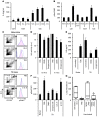
Figure 2. TDE-induced Stat3 activation determines MDSC suppressive functions.
(A) Expression of pStat3 in MDSCs from naive and tumor-bearing mice (TB) was determined by Western blotting (upper panel) and FACS (lower panel). (B) CFSE-labeled OT-1 cells loaded with SIINFEKL and cultured alone or with MDSCs isolated from TB or naive mice at different MDSC/OT-1 ratios. Percentage of OT-1 proliferating cells was determined by flow cytometry (n = 3). (C) Peptide-loaded, CFSE-labeled OT-1 cells were cultured alone or with MDSCs isolated from TB mice treated with PBS, JSI124, STA21, or Stat3 siRNA. CFSE dilution was determined by flow cytometry (n = 3). (D) Nude or WT mice were vaccinated or not with frozen/thawed CT26 cells 1 week before i.v. injection of CT26 cells admixed or not with MDSCs isolated from TB and previously treated with STA21 or Stat3 siRNA. 2 weeks later, lung metastasis numbers were evaluated (n = 5 mice per group). For box and whisker plots, bottoms and tops of boxes show the 25th and 75th percentiles, respectively, and middle bands show the median; whiskers show extrema. (E) Peptide-loaded, CFSE-labeled OT-1 cells were cultured alone or with bone marrow–derived MDSCs of naive mice previously treated alone or with tumor cell whole supernatant or TDE or TDSF fractions. CFSE dilution was determined by flow cytometry (n = 3 mice per group). (F) TB mice were injected or not with MDSCs. These MDSCs were either from TB or naive mice and stimulated with PBS, TDEs, or TDSFs. 2 days later, spleen cells were harvested and restimulated in vitro with CD3mAb+ dead tumor cells, then stained for intracellular CD4 and IFN-γ (upper panel) or CD8 IFN-γ (lower panel). *P < 0.05. Error bars represent mean + SD.
Figure 3
Figure 3. TDEs trigger pStat3 expression in MDSCs through autocrine production of IL-6.
MDSCs from naive mice were treated with PBS or with TDSFs or TDEs. (A) IL-6 concentration in the supernatant was determined by ELISA. (B) In some wells, rIL-6 or blocking anti–IL-6 mAb (aIL-6) were added. Data represent pStat3 MFI ± SD. (C) Naive mice were i.v. injected with PBS, EL4 TDEs, or rIL-6. EL4 TB mice were i.v. injected with PBS or IL-6 siRNA. 24 hours later, spleens were harvested. MDSC percentage (denoted) and pStat3 expression were determined by FACS on gated MDSCs. (D) EL4 cells were cultured in vitro for 24 hours and treated with PBS, DMA, or omeprazole. In vivo EL4 TB mice were injected daily with DMA for 1 week. Acetylcholine esterase activity was assayed in supernatant or sera. (E) IL-6 concentration in sera was determined by ELISA in naive mice, which received a single injection of PBS or EL4 TDEs (left), or in EL4 TB mice, which received 1 daily injection of PBS, DMA, or omeprazole for 1 week (right). (F) EL4 TB mice were injected with PBS alone or DMA, omeprazole, or IL-6 siRNA (si). Spleens were harvested 24 hours later, and pStat3 expression was determined by FACS. Data are shown as MFI ± SD. (G) Mice (n = 5) were vaccinated with frozen/thawed CT26 cells 1 week before i.v. injection of live CT26 admixed or not with MDSCs isolated from PBS- or DMA-treated CT26 TB mice. Twelve days later, lung metastasis number was evaluated. Each experiment was done in triplicate. For box and whisker plots, bottoms and tops of boxes show the 25th and 75th percentiles, respectively, and middle bands show the median; whiskers show extrema. *P < 0.05.
Figure 4
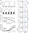
Figure 4. IL-6–induced pStat3 expression in MDSCs is dependent on the TLR2/MyD88 pathway.
Purified MDSCs from WT or TLR2-, TLR4-, MyD88-, and Trif-deficient C57BL/6 tumor-free mice were cultured for 24 hours in complete medium supplemented or not with TDEs. IL-6 concentration in the supernatant was determined by ELISA (A), and pStat3 expression in cells was determined by FACS analysis (B). Data represent mean ± SD. (C) WT C57BL/6 mice or TLR2-, TLR4-, MyD88-, and Trif-deficient mice were s.c. injected with 1 × 10⁶ EL4 cells. 3 weeks later, spleen cells were harvested, MDSC percentage was determined in spleen (denoted in left panels), and pStat3 expression was determined by FACS analysis on MDSC gated cells (right panel). (D) 2 × 10⁵ OT-1 cells were labeled with CFSE, loaded with 10 μg/ml of SIINFEKL, and cultured 3 days alone or with different ratios of MDSCs from EL4 tumor-bearing WT mice or from EL4 tumor-bearing TLR2-deficient mice. Percentage of OT-1 proliferating cells was determined by flow cytometry. Each experiment was done in duplicate (n = 3 mice per group). (E) WT or TLR2–/– mice were injected s.c. with EL4 cells, and tumor growth was monitored. *P < 0.05.
Figure 5
Figure 5. Hsp72 is expressed at the surface of TDEs and bound on TLR2.
(A) Endogenous TLR2 ligand expression was investigated by immunoblots in TDEs, whole-cell lysates, and TDSFs from EL4, TS/A, and CT26 tumor cells. CTRL, control. (B) TDEs from EL4, TS/A, and CT26 tumor cells and exosomes from control 3T3 cells were coated on beads and labeled with control isotype or anti-Hsp72 or anti-Hsc73 Abs. Then FACS analysis was performed to determine expression of Hsp72 and Hsp73 at the surface of exosomes. One representative FACS histogram (blue line) is represented with its isotype control (pink area). (C) Surface plasmon resonance studying the binding of Hsp72 to TLR2 protein. FSL-1 (a bacterial TLR2 ligand) was used as positive control; LPS was used as negative control. (D) Purified myeloid cells from naive WT or TLR2-deficient C57BL/6 mice were cultured in complete medium supplemented or not as indicated. pStat3 expression was determined by FACS analysis on MDSC gated cells. Data represent MFI ± SD. *P < 0.05.
Figure 6
Figure 6. pStat3 expression in MDSCs is dependent on Hsp72 on TDEs.
(A) Purified myeloid cells from naive WT or TLR2-deficient mice were either untreated or treated as indicated. (B) Naive mice were i.v. injected with TDEs from shRNA mock CT26 cells or Hsp72 shRNA CT26 clones H96 or H97. 18 hours later, spleen cells were harvested and pStat3 expression was determined by FACS. (C) Naive BALB/c mice were s.c. injected with 1 × 10⁶ H96, H97, or mock CT26 cells. 2 weeks later, spleens were harvested and pStat3 expression determined by FACS. For AC, pStat3 was determined by FACS analysis on MDSC gated cells. Data represent MFI ± SD. Experiments were performed in triplicate (n = 3 mice per group). Inset shows immunoblot of pStat3 expression in sorted MDSCs from mice bearing mock CT26 or Hsp72 shRNA CT26 clone H96. (D) Mice were vaccinated with frozen/thawed CT26 cells 1 week before i.v. injection of live CT26 cells admixed or not with MDSCs isolated from mice bearing shRNA mock-transfected or Hsp72 shRNA-transfected (clone H96) CT26 tumors. Twelve days later, lung metastasis numbers were evaluated. Experiments were performed in triplicate (n = 5 mice per group). For box and whisker plots, bottoms and tops of boxes show the 25th and 75th percentiles, respectively, and middle bands show the median; whiskers show extrema. *P < 0.05. Error bars represent mean + SD.
Figure 7
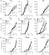
Figure 7. Exosome depletion restores the efficacy of immunotherapy.
Immunocompetent mice (A) were injected s.c. with 1 × 10⁶ CT26, TS/A, or EL4 cells. Mice were then either untreated or treated with 1 i.p. injection of 100 mg/kg cyclophosphamide (CTX), daily i.p. injection of DMA, or both. Mean tumor volume ± SD (n = 6 mice per group). (B) WT mice were injected as in A with CT26 and treated with intratumoral CpG 1668 injection (10 μg once a week) with or without DMA. (C) Nude mice were treated as in A. (D) BALB/c mice were injected s.c. with 1 × 10⁶ CT26 cells. Mice were then either untreated or treated with 1 i.p. injection of cyclophosphamide or 1 i.p. injection of cyclophosphamide plus repeated daily i.p. injections of DMA with or without an i.v. adoptive transfer of 5 × 10⁶ MDSCs from CT26 tumor-bearing mice. Data shown represent mean tumor volume ± SD (n = 5 mice per group). (E) BALB/c mice were injected s.c. with 1 × 10⁶ mock-transfected (left panel) or Hsp72 shRNA–transfected (clone H96, middle panel; clone H97, right panel) CT26 cells. Mice were then either untreated or treated with 1 i.p. injection of cyclophosphamide, i.p. injection of DMA twice a week, or both. Mean tumor volume ± SD (n = 6 mice per group). Experiments were performed in duplicate. *P < 0.05.
Figure 8
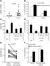
Figure 8. Exosomes produced by human cancer cell lines or metastatic cancer patients dictate Stat3 activation in MDSCs and their immunosuppressive function through TLR2 and Hsp72.
(A) The frequency of MDSCs, defined as HLA-DR CD33+ cells, is shown in the PBMCs of healthy volunteers (H.V.) (n = 11) and metastatic cancer patients (n = 18). Each plot is an individual measure, and the horizontal bar is the mean. (B) Immunosuppressive function of MDSCs from peripheral blood of healthy volunteers and metastatic cancer patients on stimulated T cell proliferation. T cell stimulation was induced by a mixture of anti-CD2, anti-CD3, and anti-CD28 beads (n = 10). (C) PBMCs from healthy volunteers were cultured for 24 hours in medium alone or medium containing TDEs from H23 cells with or without blocking TLR2 Abs or anti-Hsp72 polyclonal Abs (pAbs). pStat3 was determined by flow cytometry on MDSC gated cells (n = 10). (D) Immunosuppressive function of MDSCs from blood of healthy volunteers either untreated or treated with TDEs from H23 cells alone or with blocking TLR2 Abs or anti-Hsp72 pAbs (n = 8). (E) PBMCs from metastatic cancer patients were incubated overnight in serum-free medium supplemented with autologous serum or PBS. pStat3 expression in gated MDSC was determined by flow cytometry. pStat3 MFI ratio between PBS and serum condition was represented. The same patients were sampled before and after 3 weeks of amiloride treatment (n = 11). (F) Immunosuppressive function of MDSCs prepared from peripheral blood of metastatic cancer patients, treated with amiloride or not treated, on T cell proliferation stimulated as in B. *P < 0.05. Error bars represent mean + SD.

Similar articles

Cited by

References

    1. Gabrilovich DI, et al. The terminology issue for myeloid-derived suppressor cells. Cancer Res. 2007;67(1):425; author reply 426. - PMC - PubMed
    1. Ostrand-Rosenberg S, Sinha P. Myeloid-derived suppressor cells: linking inflammation and cancer. J Immunol. 2009;182(8):4499–4506. doi: 10.4049/jimmunol.0802740. - DOI - PMC - PubMed
    1. Filipazzi P, et al. Identification of a new subset of myeloid suppressor cells in peripheral blood of melanoma patients with modulation by a granulocyte-macrophage colony-stimulation factor-based antitumor vaccine. J Clin Oncol. 2007;25(18):2546–2553. doi: 10.1200/JCO.2006.08.5829. - DOI - PubMed
    1. Mirza N, et al. All-trans-retinoic acid improves differentiation of myeloid cells and immune response in cancer patients. Cancer Res. 2006;66(18):9299–9307. doi: 10.1158/0008-5472.CAN-06-1690. - DOI - PMC - PubMed
    1. Srivastava MK, et al. Lung cancer patients’ CD4(+) T cells are activated in vitro by MHC II cell-based vaccines despite the presence of myeloid-derived suppressor cells. . Cancer Immunol Immunother. 2008;57(10):1493–1504. doi: 10.1007/s00262-008-0490-9. - DOI - PMC - PubMed

Publication types