. 2010 Mar 5;285(10):7208-21.
doi: 10.1074/jbc.M109.018838. Epub 2010 Jan 4.

Kaposi sarcoma-associated herpesvirus latency-associated nuclear antigen inhibits interferon (IFN) beta expression by competing with IFN regulatory factor-3 for binding to IFNB promoter

Affiliations

Kaposi sarcoma-associated herpesvirus latency-associated nuclear antigen inhibits interferon (IFN) beta expression by competing with IFN regulatory factor-3 for binding to IFNB promoter

Nathalie Cloutier et al. J Biol Chem. .

Abstract

Host cells respond to viral infections by synthesizing and producing antiviral molecules such as type I interferons (IFN). The Kaposi sarcoma-associated herpesvirus (KSHV) encodes multiple proteins expressed during the lytic replication cycle that alter the antiviral response of the host. Considering that in Kaposi sarcoma lesions and primary effusion lymphoma cells KSHV is latent in the vast majority of cells, we were interested in determining whether latently expressed viral proteins have the ability to modulate IFN synthesis. The latency-associated nuclear antigen (LANA-1) is a large nuclear protein that plays a role in the establishment and maintenance of latent KSHV episome in the nucleus of infected cells. LANA-1 is also described to modulate the cellular transcription. Here, we report that LANA-1 inhibits IFN-beta transcription and synthesis by competing with the binding of interferon regulatory factor-3 (IRF3) to the IFNB promoter. Using mutants of LANA-1, we have identified the central acidic repeated region as the domain essential for interfering with the binding of IRF3 to the positive regulatory domains I-III of the IFNB promoter. In addition, the nuclear localization of LANA-1 proved essential for IFN-beta inhibition. Thus, LANA-1 interferes with the formation of IFN-beta enhanceosome by competing with the fixation of IRF3 and by inhibiting the expression of the CREB-binding protein. The ability of LANA-1 to inhibit IFNB gene expression highlights a new role for this protein in cellular gene modulation and immune evasion strategies.


Figures

FIGURE 1.
FIGURE 1.
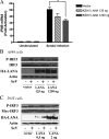
LANA-1 represses IFNB gene activation triggered by SeV, TBK1, poly(dA-dT), and IRF3–5D. A, HEK-293T cells were transfected with increasing amounts of pCMV3T-LANA-1 (100–1000 ng) and infected by SeV or co-transfected with expression vectors coding for TBK1 and IRF3–5D or transfected 24 h later with poly(dA-dT), as described under “Experimental Procedures.” Forty eight hours post-transfection, total RNA was isolated and processed for IFNB mRNA by RT-QPCR. Results are expressed as mean (triplicate) induction ± S.D. relative to pCMV3T-transfected cells after normalization of samples with GAPDH mRNA expression. Results are representative of three independent experiments. **, 0.001 < p < 0.01, ***, p < 0.0001 as determined using two-tailed t test. B, HEK-293T cells were co-transfected with HA-LANA-1 and TBK1 expression vectors. Forty eight hours after transfection, supernatants were collected, and 20 μl of each sample was used for IFN-β detection using HEK-Blue IFN-α/β cells. Secreted embryonic alkaline phosphatase levels were determined using a spectrophotometer at 650 nm and compared with a standard curve composed of a logarithmic scale between 0.0025 and 250 units/well of IFN-β protein. Results are representative of three experiments performed in triplicate. ***, p < 0.0001 as determined using two-tailed t test. C, HEK-293T were transfected with increasing amounts of pCMV3T-LANA-1 (0.5–5-20 ng) and co-transfected with 25 ng of TBK1 expression vector. Forty eight hours post-transfection, total RNA was isolated and processed for IFNB mRNA by RT-QPCR. Results are expressed as mean (triplicate) induction ± S.D. relative to pCMV3T-transfected cells after normalization of samples with GAPDH mRNA expression. Results are representative of three independent experiments. *, 0.01< p < 0.05 as determined using two-tailed t test. Ctrl, control. D, HEK-293T were transfected with increasing amounts of pCMV3T-LANA-1 (0.5–5-20 ng). Forty eight hours later, cells were lysed and boiled in Laemmli sample buffer and then resolved by SDS-PAGE. Western blots were carried out for the detection of LANA-1 and actin using specific antibodies. LANA-1 expression in transfected HEK-293T cells was compared with that of HEK-293T-E1 cells. E, HEK-293T-E1 and BCBL-1 cells were lysed and boiled in Laemmli sample buffer and then resolved by SDS-PAGE. Western blots were carried out for the detection of LANA-1 and actin using specific antibodies.
FIGURE 2.
FIGURE 2.
LANA-1 does not interfere with the phosphorylation of IRF3. A and B, A549 cells were transfected with increasing amounts of pCMV3T-LANA-1 vector (125–1250 ng). Thirty six hours later, cells were infected or not with SeV (20 HAU) for 8 h. A, total RNA was isolated and processed for IFNB mRNA by RT-QPCR. Results are expressed as mean (triplicate) induction ± S.D. relative to pCMV3T transfected cells after normalization of samples with GAPDH mRNA expression. *, 0.01 < p < 0.05 as determined using two-tailed t test. B, cell lysates were analyzed by Western blotting for P-IRF3, IRF3, LANA-1 (HA), and actin expression. C, HEK-293T were transfected with increasing amounts of pCMV3T-LANA-1 (0.5–2.0 μg) and Myc-IRF3. Thirty six hours later, cells were infected or not by SeV for 8 h. Cell lysates were analyzed by Western blotting for P-IRF3, IRF3 (Myc), LANA-1 (HA), and actin expression. Results are representative of three independent experiments.
FIGURE 3.
FIGURE 3.
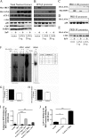
LANA-1 binds to the PRD-I–III region and interferes with the fixation of IRF3 to the IFNB promoter. A–E, HEK-293T cells were transfected with pCMV3T-LANA-1 (5–20 μg), Myc-IRF3, p50, and CBP vectors. Twenty four hours later, half of the cells were infected with SeV for 18 h. A, nuclear extracts were made and analyzed by Western blot (5% of input used in the binding reaction). The nuclear extracts were incubated with biotinylated oligonucleotides containing either the IFN-β (B), the PRD-I–III (C), PRD-II (D), or the PRD-IV (E) promoter sequences. Streptavidin beads were added for 40 min, washed three times, boiled in Laemmli sample buffer, and resolved by SDS-PAGE. Western blots were carried out for the detection of IRF3 (Myc), LANA-1 (HA), ATF-2, c-Jun, p50, and CBP using specific antibodies. PARP-1 was used as a control to demonstrate the equal amounts of nuclear proteins between the different samples. F, binding of IRF3 on the ISG15 promoter in cell extracts of HEC-1B cells transfected with 500 ng of FLAG-IRF3 and 2 μg of pCMV3T or pCMV3T-LANA-1 and infected or not by 200 HAU of SeV for 6 h was analyzed by EMSA. Specificity of binding was confirmed by homologous competition using excess (25 times) unlabeled ISG15 oligonucleotides. Supershift experiments were done using 2 μg of anti-FLAG (IRF3) antibody. * and ** denote different protein complexes on the ISG15 promoter. G and H, fraction of the cells used for EMSA (F) were kept to analyze the IFNB (G) and ISG15 (H) mRNA content by RT-QPCR. Results are expressed as mean (triplicate) induction ± S.D. relative to pCMV3T- transfected cells after normalization of samples with GAPDH mRNA expression. I and J, HEK-293T cells were transfected with 2 μg of pCMV3T-LANA-1 in a 10-cm dish. Twenty four hours later, half of the cells were infected with 20 HAU of SeV for 18 h. Nuclear extracts were isolated and measured using BCA assay, and 5 μg were used to determine the binding activity. I, endogenous IRF3 binding activity determined using the TransAMTM IRF3 ELISA (Active Motif). Results are expressed mean ± S.D. of IRF3 binding (OD 450 nm). J, LANA-1 binding on the IRF3 consensus binding site was determined using a modified TransAMTM ELISA by substituting the anti-IRF3 primary antibody for anti-LANA-1 diluted 1:1000. *, 0.01 < p < 0.05; **, 0.001 < p < 0.01; ***, p < 0.0001 as determined using two-tailed t test.
FIGURE 4.
FIGURE 4.
KSHV LANA-1 inhibits the activation of a reporter gene driven by the IFNB promoter or the PRD-I–III domain. A, HEK-293T cells were transfected with 50 ng of reporter-luciferase (IFN-β-luciferase and PRD-I–III-luciferase) and increasing amounts of pCMV3T-LANA-1 vector (100–500 ng). Twenty four hours later, cells were transfected again or not with poly(dA-dT) as IFN inducer for 24 h. The cells were then lysed in cell lysis buffer and luciferase activity was measured. These values were normalized with the protein concentration determined using BCA protein assay reagent. Results were expressed in fold activation of normalized luciferase units. *, 0.01< p < 0.05; **, 0.001 < p < 0.01 as determined using two-tailed t test. B, Western blot experiments on cell extracts were performed to show expression of LANA-1 (HA) and actin. RLU, relative luciferase units.
FIGURE 5.
FIGURE 5.
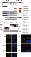
A, stick diagram of LANA-1 and the various deletion mutants generated. LANA-1 can be divided into three general regions. The N-terminal portion (aa 1–300) includes an NLS and a Pro-rich domain; the central repeated region (aa 338–922) and the C-terminal region (aa 996–1139) include an NLS and a chromosome-binding domain (aa 996–1130). The repeated region includes a DE repeat (aa 338–421), a QEP repeat (aa 441–551), a QERP repeat (aa 552–594), a QED (aa 595–751), a putative leucine zipper (aa 767–841), and a QELV repeat (aa 861–922). Seven different mutants (1–300, 854–1162, 888–1162, 1–996, 1–875, 319–1162, and 319–892) were generated and compared with LANA-1 WT (1–1162). P-RICH, proline-rich domain; CBD, C-terminal binding domain. B, cellular extracts were made in Laemmli sample buffer from HEK-293T cells transfected with vector, LANA-1 WT, or the seven LANA-1 mutant expression vectors. These cell extracts were analyzed by Western blot using anti-HA (LANA-1) and anti-actin antibodies. C, HEK-293T cells were transfected with the pCMV3T vector, LANA-1 WT or the seven LANA-1 mutant expression vectors and analyzed by immunofluorescence using anti-HA antibodies. Acetone-fixed cells were incubated with anti-HA (LANA-1 WT and mutants) followed by incubation with Alexa 488-conjugated secondary antibody (green). Nuclei were stained with 4′,6-diamidino-2-phenylindole (DAPI) (blue). The localization of the proteins is presented in the merge pictures (magnification ×60).
FIGURE 6.
FIGURE 6.
Mapping of LANA-1 domains responsible for IFNB inhibition. HEK-293T cells were co-transfected with pCMV3T vector or expression vectors encoding LANA-1 WT or mutants and TBK1. Forty eight hours post-transfection, total RNA was isolated and processed for IFNB mRNA by RT-QPCR. Results are expressed as mean (triplicate) induction ± S.D. relative to pCMV3T-transfected cells after normalization of samples with GAPDH mRNA expression. Results are representative of three independent experiments. Statistical analysis was done between TBK1-transfected cells and co-transfected with LANA-1 mutants and TBK1. **, 0.001< p < 0.01; ***, p < 0.0001 as determined using two-tailed t test.
FIGURE 7.
FIGURE 7.
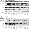
Central repeated domain of LANA-1 prevents IRF3 binding to the IFNB promoter. HEK-293T cells were transfected with 20 μg of LANA-1 WT or LANA-1 mutant vectors, Myc-IRF3, p50, and CBP vectors. Twenty four hours later, half of the cells were infected with SeV for 18 h. A, nuclear extracts were made and analyzed by Western blot (5% of input used in the binding reaction). B, nuclear extracts were incubated with biotinylated oligonucleotide containing the entire IFNB promoter sequence, and DNA-bound proteins were isolated using streptavidin beads and resuspended in Laemmli buffer. These samples were resolved by SDS-PAGE and analyzed by Western blot. Protein analyses were carried out for the detection of IRF3 (Myc), LANA-1 WT, or mutants (HA), c-Jun, p50, CBP, and PARP-1 using specific antibodies.
FIGURE 8.
FIGURE 8.
Knockdown of LANA-1 enhances IFNB gene activation in KSHV-infected cells. A, HEK-293T-E1 were transfected with 500 ng of shRNA against LANA-1 (pTER-shRNA LANA-1) or nonspecific shRNA (pTER-shRNA NS). B, HEK-293T-E1 were transfected with 500 ng of two other shRNAs against LANA-1 (pSUPER-shRNA LANA-1-N and pSUPER-shRNA LANA-1-C) or nonspecific shRNA (pSUPER-shRNA NS). A and B (top), 48 h later, cells were lysed and boiled in Laemmli sample buffer and then resolved by SDS-PAGE. Western blots were carried out for the detection of LANA-1 and actin using specific antibodies. A and B (bottom), 48 h post-transfection, cells were infected with SeV (10 HAU) for 18 h, and total RNA was isolated and processed for IFNB mRNA by RT-QPCR. Results are expressed as mean (triplicate) induction ± S.D. relative to pTER or pSUPER-transfected cells after normalization of samples with GAPDH mRNA expression and are representative of at least three independent experiments. *, p < 0.05 as determined using two-tailed t test between cells expressing shRNA LANA-1 and cells expressing nonspecific shRNA.

Similar articles

Cited by

References

    1. Cesarman E., Chang Y., Moore P. S., Said J. W., Knowles D. M. (1995) N. Engl. J. Med. 332, 1186–1191 - PubMed
    1. Zhong W., Wang H., Herndier B., Ganem D. (1996) Proc. Natl. Acad. Sci. U.S.A. 93, 6641–6646 - PMC - PubMed
    1. Ballestas M. E., Chatis P. A., Kaye K. M. (1999) Science 284, 641–644 - PubMed
    1. Krithivas A., Young D. B., Liao G., Greene D., Hayward S. D. (2000) J. Virol. 74, 9637–9645 - PMC - PubMed
    1. Lim C., Gwack Y., Hwang S., Kim S., Choe J. (2001) J. Biol. Chem. 276, 31016–31022 - PubMed

Publication types

MeSH terms

LinkOut - more resources