Compartmentalization of HIV-1 within the female genital tract is due to monotypic and low-diversity variants not distinct viral populations
- PMID: 19771165
- PMCID: PMC2741601
- DOI: 10.1371/journal.pone.0007122
Compartmentalization of HIV-1 within the female genital tract is due to monotypic and low-diversity variants not distinct viral populations
Abstract
Background: Compartmentalization of HIV-1 between the genital tract and blood was noted in half of 57 women included in 12 studies primarily using cell-free virus. To further understand differences between genital tract and blood viruses of women with chronic HIV-1 infection cell-free and cell-associated virus populations were sequenced from these tissues, reasoning that integrated viral DNA includes variants archived from earlier in infection, and provides a greater array of genotypes for comparisons.
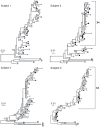
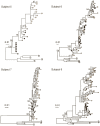
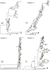
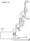
Methodology/principal findings: Multiple sequences from single-genome-amplification of HIV-1 RNA and DNA from the genital tract and blood of each woman were compared in a cross-sectional study. Maximum likelihood phylogenies were evaluated for evidence of compartmentalization using four statistical tests. Genital tract and blood HIV-1 appears compartmentalized in 7/13 women by >/=2 statistical analyses. These subjects' phylograms were characterized by low diversity genital-specific viral clades interspersed between clades containing both genital and blood sequences. Many of the genital-specific clades contained monotypic HIV-1 sequences. In 2/7 women, HIV-1 populations were significantly compartmentalized across all four statistical tests; both had low diversity genital tract-only clades. Collapsing monotypic variants into a single sequence diminished the prevalence and extent of compartmentalization. Viral sequences did not demonstrate tissue-specific signature amino acid residues, differential immune selection, or co-receptor usage.
Conclusions/significance: In women with chronic HIV-1 infection multiple identical sequences suggest proliferation of HIV-1-infected cells, and low diversity tissue-specific phylogenetic clades are consistent with bursts of viral replication. These monotypic and tissue-specific viruses provide statistical support for compartmentalization of HIV-1 between the female genital tract and blood. However, the intermingling of these clades with clades comprised of both genital and blood sequences and the absence of tissue-specific genetic features suggests compartmentalization between blood and genital tract may be due to viral replication and proliferation of infected cells, and questions whether HIV-1 in the female genital tract is distinct from blood.
Conflict of interest statement
Figures


Similar articles
-
Human immunodeficiency viruses appear compartmentalized to the female genital tract in cross-sectional analyses but genital lineages do not persist over time.J Infect Dis. 2013 Apr 15;207(8):1206-15. doi: 10.1093/infdis/jit016. Epub 2013 Jan 11. J Infect Dis. 2013. PMID: 23315326 Free PMC article.
-
Evidence for both Intermittent and Persistent Compartmentalization of HIV-1 in the Female Genital Tract.J Virol. 2019 May 1;93(10):e00311-19. doi: 10.1128/JVI.00311-19. Print 2019 May 15. J Virol. 2019. PMID: 30842323 Free PMC article.
-
Monotypic human immunodeficiency virus type 1 genotypes across the uterine cervix and in blood suggest proliferation of cells with provirus.J Virol. 2009 Jun;83(12):6020-8. doi: 10.1128/JVI.02664-08. Epub 2009 Apr 1. J Virol. 2009. PMID: 19339344 Free PMC article.
-
HIV compartmentalization: a review on a clinically important phenomenon.Curr HIV Res. 2012 Mar;10(2):133-42. doi: 10.2174/157016212799937245. Curr HIV Res. 2012. PMID: 22329519 Review.
-
Clinical parameters essential to methodology and interpretation of mucosal responses.Am J Reprod Immunol. 2011 Mar;65(3):352-60. doi: 10.1111/j.1600-0897.2010.00947.x. Epub 2011 Jan 12. Am J Reprod Immunol. 2011. PMID: 21223419 Free PMC article. Review.
Cited by
-
Simulating within host human immunodeficiency virus 1 genome evolution in the persistent reservoir.Virus Evol. 2020 Nov 23;6(2):veaa089. doi: 10.1093/ve/veaa089. eCollection 2020 Jul. Virus Evol. 2020. PMID: 34040795 Free PMC article.
-
Review: HIV-1 phylogeny during suppressive antiretroviral therapy.Curr Opin HIV AIDS. 2019 May;14(3):188-193. doi: 10.1097/COH.0000000000000535. Curr Opin HIV AIDS. 2019. PMID: 30882485 Free PMC article. Review.
-
Identification of Unequally Represented Founder Viruses Among Tissues in Very Early SIV Rectal Transmission.Front Microbiol. 2018 Mar 29;9:557. doi: 10.3389/fmicb.2018.00557. eCollection 2018. Front Microbiol. 2018. PMID: 29651274 Free PMC article.
-
Multiple independent lineages of HIV-1 persist in breast milk and plasma.AIDS. 2011 Jan 14;25(2):143-52. doi: 10.1097/QAD.0b013e328340fdaf. AIDS. 2011. PMID: 21173592 Free PMC article.
-
Viral linkage in HIV-1 seroconverters and their partners in an HIV-1 prevention clinical trial.PLoS One. 2011 Mar 2;6(3):e16986. doi: 10.1371/journal.pone.0016986. PLoS One. 2011. PMID: 21399681 Free PMC article. Clinical Trial.
References
-
- Diem K, Nickle DC, Motoshige A, Fox A, Ross S, et al. Male genital tract compartmentalization of human immunodeficiency virus type 1 (HIV). AIDS Res Hum Retroviruses. 2008;24:561–571. - PubMed
-
- Ghosn J, Viard JP, Katlama C, de Almeida M, Tubiana R, et al. Evidence of genotypic resistance diversity of archived and circulating viral strains in blood and semen of pre-treated HIV-infected men. Aids. 2004;18:447–457. - PubMed
-
- Gupta P, Leroux C, Patterson BK, Kingsley L, Rinaldo C, et al. Human immunodeficiency virus type 1 shedding pattern in semen correlates with the compartmentalization of viral Quasi species between blood and semen. J Infect Dis. 2000;182:79–87. - PubMed
-
- Coombs RW, Speck CE, Hughes JP, Lee W, Sampoleo R, et al. Association between culturable human immunodeficiency virus type-1 (HIV-1) in semen and HIV-1 RNA levels in semen and blood: evidence for compartmentalization of HIV-1 between semen and blood. J Infect Dis. 1998;177:320–330. - PubMed
Publication types
MeSH terms
Substances
Grants and funding
LinkOut - more resources
Full Text Sources
Medical
Molecular Biology Databases

