

Tissue-specific remodeling of the mitochondrial proteome in type 1 diabetic akita mice
- PMID: 19542201
- PMCID: PMC2731527
- DOI: 10.2337/db09-0259
Tissue-specific remodeling of the mitochondrial proteome in type 1 diabetic akita mice
Abstract
Objective: To elucidate the molecular basis for mitochondrial dysfunction, which has been implicated in the pathogenesis of diabetes complications.
Research design and methods: Mitochondrial matrix and membrane fractions were generated from liver, brain, heart, and kidney of wild-type and type 1 diabetic Akita mice. Comparative proteomics was performed using label-free proteome expression analysis. Mitochondrial state 3 respirations and ATP synthesis were measured, and mitochondrial morphology was evaluated by electron microscopy. Expression of genes that regulate mitochondrial biogenesis, substrate utilization, and oxidative phosphorylation (OXPHOS) were determined.
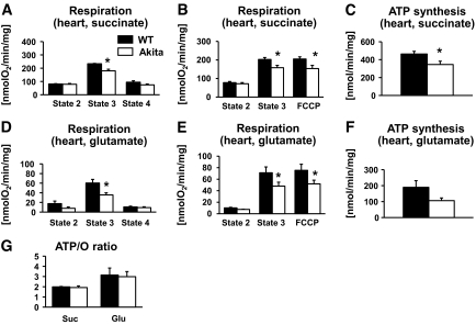
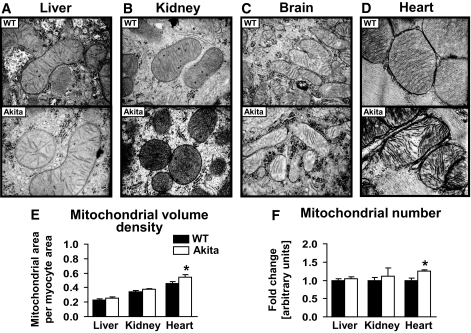
Results: In diabetic mice, fatty acid oxidation (FAO) proteins were less abundant in liver mitochondria, whereas FAO protein content was induced in mitochondria from all other tissues. Kidney mitochondria showed coordinate induction of tricarboxylic acid (TCA) cycle enzymes, whereas TCA cycle proteins were repressed in cardiac mitochondria. Levels of OXPHOS subunits were coordinately increased in liver mitochondria, whereas mitochondria of other tissues were unaffected. Mitochondrial respiration, ATP synthesis, and morphology were unaffected in liver and kidney mitochondria. In contrast, state 3 respirations, ATP synthesis, and mitochondrial cristae density were decreased in cardiac mitochondria and were accompanied by coordinate repression of OXPHOS and peroxisome proliferator-activated receptor (PPAR)-gamma coactivator (PGC)-1alpha transcripts.
Conclusions: Type 1 diabetes causes tissue-specific remodeling of the mitochondrial proteome. Preservation of mitochondrial function in kidney, brain, and liver, versus mitochondrial dysfunction in the heart, supports a central role for mitochondrial dysfunction in diabetic cardiomyopathy.
Figures


Similar articles
-
Quantitative proteomic and functional analysis of liver mitochondria from high fat diet (HFD) diabetic mice.Mol Cell Proteomics. 2013 Dec;12(12):3744-58. doi: 10.1074/mcp.M113.027441. Epub 2013 Sep 12. Mol Cell Proteomics. 2013. PMID: 24030101 Free PMC article.
-
Type 1 diabetic akita mouse hearts are insulin sensitive but manifest structurally abnormal mitochondria that remain coupled despite increased uncoupling protein 3.Diabetes. 2008 Nov;57(11):2924-32. doi: 10.2337/db08-0079. Epub 2008 Aug 4. Diabetes. 2008. PMID: 18678617 Free PMC article.
-
The diabetes medication canagliflozin promotes mitochondrial remodelling of adipocyte via the AMPK-Sirt1-Pgc-1α signalling pathway.Adipocyte. 2020 Dec;9(1):484-494. doi: 10.1080/21623945.2020.1807850. Adipocyte. 2020. PMID: 32835596 Free PMC article.
-
Combined defects in oxidative phosphorylation and fatty acid β-oxidation in mitochondrial disease.Biosci Rep. 2016 Feb 2;36(2):e00313. doi: 10.1042/BSR20150295. Biosci Rep. 2016. PMID: 26839416 Free PMC article. Review.
-
Alterations in the mitochondrial regulatory pathways constituted by the nuclear co-factors PGC-1alpha or PGC-1beta and mitofusin 2 in skeletal muscle in type 2 diabetes.Biochim Biophys Acta. 2010 Jun-Jul;1797(6-7):1028-33. doi: 10.1016/j.bbabio.2010.02.017. Epub 2010 Feb 20. Biochim Biophys Acta. 2010. PMID: 20175989 Review.
Cited by
-
Guidelines on models of diabetic heart disease.Am J Physiol Heart Circ Physiol. 2022 Jul 1;323(1):H176-H200. doi: 10.1152/ajpheart.00058.2022. Epub 2022 Jun 3. Am J Physiol Heart Circ Physiol. 2022. PMID: 35657616 Free PMC article. Review.
-
Lack of β, β-carotene-9', 10'-oxygenase 2 leads to hepatic mitochondrial dysfunction and cellular oxidative stress in mice.Mol Nutr Food Res. 2017 May;61(5):10.1002/mnfr.201600576. doi: 10.1002/mnfr.201600576. Epub 2017 Feb 9. Mol Nutr Food Res. 2017. PMID: 27991717 Free PMC article.
-
Mitochondrial dysfunction in diabetic neuropathy: a series of unfortunate metabolic events.Curr Diab Rep. 2015 Nov;15(11):89. doi: 10.1007/s11892-015-0671-9. Curr Diab Rep. 2015. PMID: 26370700 Review.
-
Insulin prevents aberrant mitochondrial phenotype in sensory neurons of type 1 diabetic rats.Exp Neurol. 2017 Nov;297:148-157. doi: 10.1016/j.expneurol.2017.08.005. Epub 2017 Aug 10. Exp Neurol. 2017. PMID: 28803751 Free PMC article.
-
Mitochondrial complex I defect and increased fatty acid oxidation enhance protein lysine acetylation in the diabetic heart.Cardiovasc Res. 2015 Sep 1;107(4):453-65. doi: 10.1093/cvr/cvv183. Epub 2015 Jun 22. Cardiovasc Res. 2015. PMID: 26101264 Free PMC article.
References
-
- Borch-Johnsen K: The prognosis of insulin-dependent diabetes mellitus. An epidemiological approach. Dan Med Bull 1989; 36: 336– 348 - PubMed
-
- Dorman JS, Laporte RE, Kuller LH, Cruickshanks KJ, Orchard TJ, Wagener DK, Becker DJ, Cavender DE, Drash AL: The Pittsburgh insulin-dependent diabetes mellitus (IDDM) morbidity and mortality study: mortality results. Diabetes 1984; 33: 271– 276 - PubMed
-
- Bugger H, Boudina S, Hu XX, Tuinei J, Zaha VG, Theobald HA, Yun UJ, McQueen AP, Wayment B, Litwin SE, Abel ED: Type 1 diabetic akita mouse hearts are insulin sensitive but manifest structurally abnormal mitochondria that remain coupled despite increased uncoupling protein 3. Diabetes 2008; 57: 2924– 2932 - PMC - PubMed
-
- Yoon JC, Puigserver P, Chen G, Donovan J, Wu Z, Rhee J, Adelmant G, Stafford J, Kahn CR, Granner DK, Newgard CB, Spiegelman BM: Control of hepatic gluconeogenesis through the transcriptional coactivator PGC-1. Nature 2001; 413: 131– 138 - PubMed
-
- de Cavanagh EM, Ferder L, Toblli JE, Piotrkowski B, Stella I, Fraga CG, Inserra F: Renal mitochondrial impairment is attenuated by AT1 blockade in experimental type I diabetes. Am J Physiol Heart Circ Physiol 2008; 294: H456– H465 - PubMed
Publication types
MeSH terms
Substances
Grants and funding
LinkOut - more resources
Full Text Sources
Medical
Molecular Biology Databases
