. 2009 Jun;15(6):682-9.
doi: 10.1038/nm.1954.

Inhibition of osteoblastic bone formation by nuclear factor-kappaB

Affiliations

Inhibition of osteoblastic bone formation by nuclear factor-kappaB

Jia Chang et al. Nat Med. 2009 Jun.

Abstract

An imbalance in bone formation relative to bone resorption results in the net bone loss that occurs in osteoporosis and inflammatory bone diseases. Although it is well known how bone resorption is stimulated, the molecular mechanisms that mediate impaired bone formation are poorly understood. Here we show that the time- and stage-specific inhibition of endogenous inhibitor of kappaB kinase (IKK)--nuclear factor-kappaB (NF-kappaB) in differentiated osteoblasts substantially increases trabecular bone mass and bone mineral density without affecting osteoclast activities in young mice. Moreover, inhibition of IKK-NF-kappaB in differentiated osteoblasts maintains bone formation, thereby preventing osteoporotic bone loss induced by ovariectomy in adult mice. Inhibition of IKK-NF-kappaB enhances the expression of Fos-related antigen-1 (Fra-1), an essential transcription factor involved in bone matrix formation in vitro and in vivo. Taken together, our results suggest that targeting IKK-NF-kappaB may help to promote bone formation in the treatment of osteoporosis and other bone diseases.


Figures
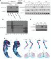
Fig. 1
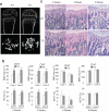
Fig. 1. Generation of transgenic mouse specifically expressing IKK-DN in differentiated osteoblasts
(a) Generation of Bblap2-dependent IKK DN construct. Cells were probed with anti-HA monoclonal antibodies. α-tubulin was used as a loading control. (b) Generation of Bblap2-IKK-DN transgenic mice. Genomic DNAs from tail tissues were probed with 32P-labeled human IKK-DN cDNA probes. N, Genomic DNAs from WT mice; TG, Bblap2-IKK-DN transgenic mouse; 5P, 5 copies of human IKK-DN cDNA; 10P, 10 copies of human IKK-DN cDNA. (c) IKK-DN was specifically expressed in bone tissues. Total RNA from long bones, brain, muscle and liver examined with RT-PCR. WT, wild type mouse; TG1, Bblap2-IKK-DN founder line 1; TG2, Bblap2-IKK-DN founder line 2. (d) IKK-DN was induced in differentiated osteoblasts. Differentiated cells were probed with anti-HA antibodies. α-tubulin was used as a loading control. TG1, Bblap2-IKK-DN transgenic mouse line 1; TG2, Bblap2-IKK-DN transgenic mouse line 2. (e) Normal skeleton of newborn WT and Bblap2-IKK-DN mice. Whole mounts of newborn skeletons were stained with alcian blue and alizarin red. 1, whole skeleton; 2, upper extremities; 3, hind limbs; 4, Rib.
Fig. 2
Fig. 2. The inhibition of NF-κB by IKK-DN in mature osteoblasts enhanced bone formation in young mice
(a) μCT images of the trabecular bone of femurs from WT and Bblap2-IKK-DN mice. (b) Enhanced bone formation in young Bblap2-IKK-DN mice. Morphometric properties of femurs from different ages of both WT and Bblap2-IKK-DN mice were measured by μCT. The results are the average values from 12-15 mice per group and presented as mean ± s.d. **P < 0.01. BMD, bone mineral density; BV/TV, trabecular bone volume per tissue volume; WT, wild type mice; TG, Bblap2-IKK-DN mice. (c) H&E analysis of femurs from WT and Bblap2-IKK-DN mice. Scale bar, 50 μm. (d) von Kossa staining of tibiae from WT and Bblap2-IKK-DN mice. Scale bar, 50 μm. (e) Osteoblast numbers in both WT and Bblap2-IKK-DN mice. The results are the average value from 12-15 mice per group and presented as mean ± s.d. (f) Enhanced bone formation in Bblap2-IKK-DN mice as determined by calcein double-labeling. Four-week-old mice were labeled with calcein. The results are the average value from 10 mice per group. **P < 0.01. Scale bar, 10 μm. (g) The expression of bone matrix genes was enhanced in young Bblap2-IKK-DN mice. Total RNAs were examined with Real-time RT-PCR. *P < 0.05; **P < 0.01. (h) Osteoclast numbers in both WT and Bblap2-IKK-DN mice. The results are the average value from 12-15 mice per group and presented as mean ± s.d. Oc.S/BS, osteoclast surface per bone surface; Oc.N/BPm (mm-1); osteoclast number per bone perimeter. ( i)Serum Trap5b in both WT and Bblap2-IKK-DN mice. The results are the average values from 12-15 mice per group and presented as mean ± s.d.
Fig. 3
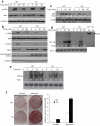
Fig. 3. The inhibition of NF-κB in mature osteoblasts enhances bone formation in a cell-autonomous fashion
(a) NF-κB signaling was intact in un-differentiated osteoblast progenitors. The phosphorylation and degradation of IκBα were examined by Western blot analysis. (b) IKK-DN inhibited IKKβ activities in differentiated osteoblast cells isolated from Bblap2-IKK-DN mice. Calvarial cells were induced to differentiate for 10 days. After induction, cells were treated with TNF. The phosphorylation and degradation of IκBα and p65 phosphorylation were examined by Western blot analysis. The induction of IKK-DN was probed with anti-IKKγ. (c) IKK-DN blocked the nuclear translocation of p65 induced by TNF. The level of nuclear p65 was examined by Western blot analysis. (d) IKK-DN blocked TNF-induced NF-κB-binding activities. (e) IKK-DN blocked NF-κB in differentiated osteoblasts. Calvarial cells from both WT and Bglap2-IKK-DN mice were induced to differentiate for the indicated periods and the nuclear proteins were isolated. Nuclear proteins were incubated 32P-labeled κB probed. (f) The inhibition of NF-κB in differentiated osteoblasts enhanced mineralization. The experiments were performed in duplicate. The results represent average value from three independent experiments. **P < 0.01. (g) The inhibition of NF-κB enhanced the expression of Runx2, Sp7, Ocn and Ibsp as determined by Real-time RT-PCR. **P < 0.01. (h) The inhibition of NF-κB in differentiated osteoblasts did not affect the expression of Tnfsf11 and Tnfrsf11b. (i) Over-expression of p65 in calvarial cells isolated from Bglap2-IKK-DN mice inhibited mineralization. p65 was examined by Western blot analysis. The mineralization was examined by Alizarin red staining. (j) p65 overcame IKK-DN effects on bone matrix gene expression. Cells were induced to differentiate and total RNAs were examined by Real-time RT-PCR. *P < 0.05; **P < 0.01.
Fig. 4
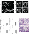
Fig. 4. The inhibition of NF-κB in mature osteoblasts prevents trabecular bone loss induced by OVX in adult mice
(a) The inhibition of NF-κB prevented trabecullar bone loss of femurs in adult mice as determined by μCT. (b) The inhibition of NF-κB prevented trabecullar bone loss of vertabrae as determined by μCT. (c and d) Quantitative measurement of bone loss in vertebrae and femurs by μCT. The results are average values from 12-15 mice per group and presented as mean ± s.d. **P < 0.01. (e) The inhibition of NF-κB prevented trabecular bone loss of femurs as determined by the histological analysis. Scale bar, 100 μm.
Fig. 5
Fig. 5. The inhibition of NF-κB in mature osteoblasts prevents bone loss by maintaining osteoblast functions in adult mice
(a) NF-κB in osteoblasts was activated during OVX-induced bone loss. 3-month-old WT and Bblap2-IKK-DN mice were OVXed or sham-operated. Femurs from mice were sectioned and stained with anti-active form of p65 and anti-HA. Scale bars at left panel, 50 μm; scale bars at right panel, 20 μm. (b) The inhibition of NF-κB enhanced osteoblast activities in osteoporosis. Serum Ocn was measured using an Ocn ELISA kit. The results are average values from 6-8 mice per group and presented as mean values ± s.d. *P < 0.05; **P < 0.01. (c) The inhibition of NF-κB enhanced bone formation in osteoporosis. The bone formation rate in mice was determined 4 weeks after operation. The results are average values from 6-8 mice per group and presented as mean values ± s.d. *P < 0.01. (d) The inhibition of NF-κB did not affect osteoblast numbers. Osteoblast numbers in mice were examined 4 weeks after operation. The results are average values from 6-8 mice per group and presented as mean values ± s.d. (e) The inhibition of NF-κB in osteoblasts did not affect osteoclast formation. Osteoclast numbers in mice were examined 4 weeks after operation. The results are average values from 6-8 mice per group and presented as mean values ± s.d. (f) The inhibition of NF-κB in osteoblasts did not inhibit bone resorption in osteoporosis. Mice were operated and sacrificed at 0, 1, 2, 3, 4, 6 and 8 weeks. Serum Trap5b levels were measured using a mouse TRAP™ assay kit. The results are average values from 6-8 mice per group and presented as mean values ± s.d.
Fig. 6
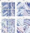
Fig. 6. The inhibition of NF-κB promoted osteoblast activities by inducing Fra-1 expression
(a) The inhibition of NF-κB enhanced JNK activities and Fra-1 expression in differentiated osteoblasts. Calvarial cells from both WT and Bblap2-IKK-DN mice were induced to differentiate and the JNK phosphorylation and Fra-1 expression were examined by Western blot analysis. α-tubulin was used as a loading control. (b) The inhibition of NF-κB did not affect p38 activation. (c) The inhibition of NF-κB in mature osteoblasts enhanced Fosl1 as determined by Real-time RT-PCR. **P < 0.01. (d) The inhibition of NF-κB in mature osteoblasts did not affect the expression of Fosb and deltaFosb as determined by RT-PCR. (e) The inhibition of NF-κB in mature osteoblasts did not affect the expression of Jun, Junb, Jund and Fos as determined by Real-time RT-PCR. (f) The inhibition of JNK suppressed Fosl1. Whole cell lysates were examined by Western blot analysis. (g) The knock-down of Fosl1 by shRNA. Whole cell lysates were probed with anti-Fra-1 antibodies. α-tubulin was used as a loading control. (h) The knock-down of Fosl1 significantly decreased mineralization. *P < 0.05; **P < 0.01. (i) The knock-down of Fosl1 significantly reduced the expression of bone matrix genes. *P < 0.05; **P < 0.01. (j) The inhibition of NF-κB in mature osteoblasts enhanced Fra-1 expression in OVX mice. The femoral sections from WT and Bblap2-IKK-DN mice after OVX or sham operation were stained with anti-Fra-1 antibodies. Scale bars, 50 μm.

Similar articles

Cited by

References

    1. Wagner EF, Karsenty G. Genetic control of skeletal development. Curr. Opin. Genet. Dev. 2001;11:527–532. - PubMed
    1. Khosla S, Westendorf JJ, Oursler MJ. Building bone to reverse osteoporosis and repair fractures. J Clin. Invest. 2008;118:421–428. - PMC - PubMed
    1. Zaidi M. Skeletal remodeling in health and disease. Nat Med. 2007;13:791–801. - PubMed
    1. Zelzer E, Olsen BR. Multiple roles of vascular endothelial growth factor (VEGF) in skeletal development, growth, and repair. Curr Top Dev Biol. 2005;65:169–187. - PubMed
    1. Kronenberg HM. Twist Genes Regulate Runx2 and Bone Formation. Dev. Cell. 2004;6:317–318. - PubMed

Publication types