. 2009 Jun 5;388(2):236-47.
doi: 10.1016/j.virol.2009.03.027. Epub 2009 Apr 24.

Epstein-Barr virus nuclear antigen 3C targets p53 and modulates its transcriptional and apoptotic activities

Affiliations

Epstein-Barr virus nuclear antigen 3C targets p53 and modulates its transcriptional and apoptotic activities

Fuming Yi et al. Virology. .

Abstract

The p53 tumor suppressor gene is one of the most commonly mutated genes in human cancers and the corresponding encoded protein induces apoptosis or cell-cycle arrest at the G1/S checkpoint in response to DNA damage. To date, previous studies have shown that antigens encoded by human tumor viruses such as SV40 large T antigen, adenovirus E1A and HPV E6 interact with p53 and disrupt its functional activity. In a similar fashion, we now show that EBNA3C, one of the EBV latent antigens essential for the B-cell immortalization in vitro, interacts directly with p53. Additionally, we mapped the interaction of EBNA3C with p53 to the C-terminal DNA-binding and the tetramerization domain of p53, and the region of EBNA3C responsible for binding to p53 was mapped to the N-terminal domain of EBNA3C (residues 130-190), previously shown to interact with a number of important cell-cycle components, specifically SCF(Skp2), cyclin A, and cMyc. Furthermore, we demonstrate that EBNA3C substantially represses the transcriptional activity of p53 in luciferase based reporter assays, and rescues apoptosis induced by ectopic p53 expression in SAOS-2 (p53(-/-)) cells. Interestingly, we also show that the DNA-binding ability of p53 is diminished in the presence of EBNA3C. Thus, the interaction between the p53 and EBNA3C provides new insights into the mechanism(s) by which the EBNA3C oncoprotein can alter cellular gene expression in EBV associated human cancers.


Figures

Figure 1
Figure 1
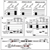
EBNA3C forms a stable complex with p53 in vitro. (A) GST-p53 fusion protein was expressed in E. coli and purified with glutathione Sepharose beads. Full-length EBNA3C was labeled with 35S methionine by in vitro translation and incubated with either GST control or GST-p53 beads normalized by Coomassie staining. 5% of in vitro translation (IVT) input was used for comparison. Precipitated proteins were resolved by SDS-PAGE, and bands were visualized with a phosphorimager screen. Relative Density was quantified using Storm 850 imaging system. (B–C) Either GST control or GST-p53 beads were incubated with lysates prepared from either 50 million B) lymphoblastoid cell lines (LCL1 and LCL2) or C) BJAB cells and BJAB cells stably expressing EBNA3C (two clones – BJAB EBNA3C#7 and BJAB EBNA3C#10). Approximately 5% of the lysed cells were saved as input and precipitated protein complexes were resolved by 7% SDS-PAGE. EBNA3C was detected by western blot with the specific monoclonal antibody (A10) followed by an infrared tagged secondary antibody and scanned using Odyssey imager. All panels are representative gels from similar repeat experiments.
Figure 2
Figure 2
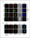
EBNA3C interacts with p53 in vivo. (A–B) Either 10 million HEK 293 cells or (C–D) 20 million DG75 cells were co-transfected with flag-tagged EBNA3C and myc-tagged p53. Cells were harvested at 36h post-transfection and approximately 5% of the lysed cells were saved as input and remaining were immunoprecipitated (IP) with 1.5 µg of appropriate antibody. Lysates and IP complexes were resolved by SDS-PAGE and immuno blotted with the indicated antibodies. The same blots were stripped and reprobed with appropriate antibodies. PC: preclear; prior to set up IP, cell lysates were precleard with normal mouse serum with protein A/G beads. C) Either 50 million BJAB cells or LCLs (LCL1 and 2) were collected at exponential growth phase and lysed in RIPA buffer. Protein complexes were immunoprecipitated (IP) with 2.0 µg p53 specific antibody (DO-1, Santa Cruz). Samples were resolved by 7% SDS-PAGE and western blotting for the indicated proteins was done by stripping and reprobing the same membrane. PC: preclear. All panels are representative gels from similar repeat experiments.
Figure 3
Figure 3
N-terminal domain of EBNA3C binds to C-terminal domain of p53. (A–C) 35S-radiolabeled either full-length (C) or different EBNA3C truncated fragments (A) or full-length p53 (B) was in vitro translated using a T7 TNT translation kit. All 35S-radiolabeled in vitro translated proteins in binding buffer were precleared by rotating with GST-beads for 1h at 4°C. The binding reaction mixture was set up with either bacterially purified GST control or the indicated GST fusion proteins. Reactions were resolved by appropriate SDS-PAGE, exposed to phosphorimager plate, and scanned on a Storm 850 imaging system. The amount of protein bound in each GST-pulldown sample was quantified with ImageQuant software (Molecular Dynamics). Coomassie staining of SDS-PAGE resolved purified GST proteins is shown (B and C, bottom panels). All panels are representative gels from similar repeat experiments. (D) Schematics illustrate different structural domains of EBNA3C and p53. Red boxes indicate the respective binding domain(s) of two proteins.
Figure 4
Figure 4
EBNA3C colocalizes with p53 in EBV positive cell lines. A) EBV negative burkitt lymphoma cell line, BJAB, and two EBV transformed cell lines - LCL1 and LCL2 were air-dried onto slides and fixed using a 1:1 mixture of acetone and methanol. Endogenously expressed p53 was detected using mouse monoclonal antibody (DO-1, 1:200 dilution) respectively, followed by anti-mouse Alexa Fluor 594 (red), and EBNA3C was detected using EBNA3C–reactive human serum (1:150 dilution) followed by anti-human Alexa Fluor 488 (green). EBV negative BJAB cells were used as EBNA3C null control cell lines. B) 10 million BJAB cells were transfected with GFP-tagged EBNA3C truncated mutants – residues 1–365 (top panels) and residues 366–620 (bottom panels). Endogenous p53 was detected using DO-1 antibody as B. The nuclei were counterstained using DAPI (blue). The images were sequentially captured using an Olympus confocal microscope. All panels are representative pictures from similar repeat experiments.
Figure 5
Figure 5
EBNA3C represses p53 mediated transcriptional activity. (A–B) Approximately 0.4 × 10⁶ SAOS-2 (p53−/−) cells were cotransfected with 0.25 µg of the promoter construct containing p53 responsive element and 0.5 µg of myc-tagged p53 plus either vector control or A) increasing amount of flag-tagged EBNA3C expressing constructs (0, 0.25, 0.5, 1.0 µg respectively) B) flag-tagged different truncated mutants of EBNA3C using Lipofectamine 2000 (Invitrogen). At 24 h post-transfection, cells were harvested and lysed for luciferase assays. Total amount of proteins were normalized by Bradford assay. Increasing amounts of EBNA3C show proportional increment in luciferase activity. The representative plot is a mean of two independent experiments. Error bar represents standard deviation (SD). Bottoms panels indicate the fractions of the cell lysates were resolved by SDS-PAGE to demonstrate the expression levels of p53 and EBNA3C. GAPDH blot was done for loading control.
Figure 6
Figure 6
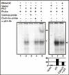
EBNA3C reduces DNA-binding ability of p53. A probe containing the p53 binding sequence (5’-AGGAAGAAGACTGGGCATGTCTGGGCA-3’) was labeled by Klenow fill-in reaction with [α-32P]dCTP and used for EMSA in presence and absence of EBNA3C. Lanes 1 and 6, probe incubated with nuclear extracts (NE) isolated from SAOS-2 (p53−/−) cells transfected with emprty vector control; lane 2 and 7, probe incubated with myc-tagged p53 expressing NE, showing band shift due to p53 binding (*); lane 3, probe with 200-fold molar excess of cold specific competitor, which abolished completely the p53 specific shift; lane 4, probe with 200fold molar excess of cold mutant probe competitor showing no effect on p53 specific shift; lane 5, probe incubated with p53 expressing NE and monoclonal antibody against p53 (DO-1), which super-shifted the p53 specific probe (**); lane 8, probe incubated with NE expressing both p53 and EBNA3C, reduced the p53 specific band intensity most likely due to formation of a stable inhibitory complex between p53 and EBNA3C in vivo (arrowhead).
Figure 7
Figure 7
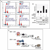
EBNA3C blocks p53 induced apoptosis in p53 null cell line SAOS-2. (A–B) SAOS-2 (p53−/−) cells were transiently transfected either with vector control, or constructs expressing untagged wild-type EBNA3C and p53 alone, or p53 and EBNA3C together. After 48 h of transfection, cells were harvested, fixed and levels of apoptotic cells in each samples were analyzed by flow cytometry (Becton-Dickinson). Total 20,000 events were analyzed for each sample. Data were analyzed using the ModFIT model program (Verity Software House). The representative plot is a mean of three independent experiments. Error bar represents SD. EBNA3C (top panel) and p53 (middle panel) protein expression levels were judged by western blot. Equal protein loading was analyzed GAPDH (bottom panel) blot. (C) A schematic representation of p53 mediated transcriptional regulation in EBV negative (-) and positive (+) cells. In response to genotoxic stress, p53 achieves its antiproliferative properties through its action as a DNA-binding transcriptional activator, to induce expression of numerous downstream target genes, involved in cell-cycle arrest and apoptosis. In EBV positive cells, EBNA3C potentially inhibits p53 mediated transcriptional activity via forming a stable complex with p53. p53 RE: p53 responsive element.

Similar articles

Cited by

References

    1. Adimoolam S, Ford JM. p53 and regulation of DNA damage recognition during nucleotide excision repair. DNA Repair. 2003;2(9):947–954. - PubMed
    1. Adler V, Pincus MR, Minamoto T, Fuchs SY, Bluth MJ, Brandt-Rauf PW, Friedman FK, Robinson RC, Chen JM, Wang XW, Harris CC, Ronai Z. Conformation-dependent phosphorylation of p53. Proc Natl Acad Sci U S A. 1997;94(5):1686–1691. - PMC - PubMed
    1. Aiello L, Guilfoyle R, Huebner K, Weinmann R. Adenovirus 5 DNA sequences present and RNA sequences transcribed in transformed human embryo kidney cells (HEK-Ad-5 or 293) Virology. 1979;94(2):460–469. - PubMed
    1. Ambinder RF. Human lymphotropic viruses associated with lymphoid malignancy: Epstein-Barr and HTLV-1. Hematol Oncol Clin North Am. 1990;4(4):821–833. - PubMed
    1. Anderson ME, Woelker B, Reed M, Wang P, Tegtmeyer P. Reciprocal interference between the sequence-specific core and nonspecific C-terminal DNA binding domains of p53: implications for regulation. Mol Cell Biol. 1997;17(11):6255–6264. - PMC - PubMed

Publication types

MeSH terms

Substances