. 2009 Apr 15;23(8):912-27.
doi: 10.1101/gad.1782209.

Mechanisms that regulate localization of a DNA double-strand break to the nuclear periphery

Affiliations

Mechanisms that regulate localization of a DNA double-strand break to the nuclear periphery

Pranav Oza et al. Genes Dev. .

Abstract

DNA double-strand breaks (DSBs) are among the most deleterious forms of DNA lesions in cells. Here we induced site-specific DSBs in yeast cells and monitored chromatin dynamics surrounding the DSB using Chromosome Conformation Capture (3C). We find that formation of a DSB within G1 cells is not sufficient to alter chromosome dynamics. However, DSBs formed within an asynchronous cell population result in large decreases in both intra- and interchromosomal interactions. Using live cell microscopy, we find that changes in chromosome dynamics correlate with relocalization of the DSB to the nuclear periphery. Sequestration to the periphery requires the nuclear envelope protein, Mps3p, and Mps3p-dependent tethering delays recombinational repair of a DSB and enhances gross chromosomal rearrangements. Furthermore, we show that components of the telomerase machinery are recruited to a DSB and that telomerase recruitment is required for its peripheral localization. Based on these findings, we propose that sequestration of unrepaired or slowly repaired DSBs to the nuclear periphery reflects a competition between alternative repair pathways.


Figures

Figure 1.
Figure 1.
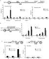
A single DNA DSB causes a decrease in chromosome interaction frequencies. (A,B) 3C interactions of the DSB region with ∼100 kb of surrounding region in the absence of a DSB and at 1 h (A) and 2 h (B) after galactose addition. The DSB (HO-cs) is located on an ∼6-kb EcoRI restriction fragment (see Supplemental Fig. S4). Using a primer at the 3′ end of the DSB-containing EcoRI fragment, we determined its interactions with the surrounding region by PCR using specific primers for each fragment. Primers are shown as small arrows below the schematic. Interactions were normalized to a region on chromosome VI to adjust for experimental variation. (C) The decrease in 3C interactions is not a property of the MAT sequences and does not occur in the absence of a DSB. A DSB was introduced at the LEU2 locus on chromosome III in the strain YMV45 in which the HO recognition site at MAT and the donors are deleted. 3C interactions were determined using the same primer pairs as in A albeit in the absence of a DSB at MAT. (D) Formation of a DSB leads to chromosome-wide decreases in interaction frequencies. Using the same 3C template as in B, 3C analysis was performed using an EcoRI fragment that is ∼106 kb from the DSB. Interactions of this fragment were determined with EcoRI fragments close to the DSB by PCR with primers specific for each fragment. The interaction frequencies were normalized to frequencies in the absence of a DSB. The primer at the 4.5-kb EcoRI site measures the interactions with the fragment containing the DSB. The primer at the −2-kb EcoRI site measures the interactions of the fragment immediately adjacent to the DSB site that is ∼4 kb in size. Hence, this interaction represents the interactions of the region from −2 kb to −6 kb from the DSB. (E) Interchromosomal 3C interactions with the DSB region also decrease. 3C interactions of the EcoRI fragments next to and bearing the DSB—(2) and (3), respectively—were determined with EcoRI fragments from the chromosome III subtelomeric region, the left arm of chromosome I, and the right arm of chromosome XV. The control primer (1) is the same one used in D. Interactions were normalized to a region on chromosome VI to control for experimental variation. In all the panels, error bars represent one standard deviation from the mean for three separate PCRs. Results were confirmed with at least two independent 3C templates.
Figure 2.
Figure 2.
The DSB-dependent decreases in 3C interaction frequencies requires RAD51, RAD52, and MPS3. (A) 3C interactions in G1-arrested cells. CY1276 cells (MATa “donorless,” isogenic to JKM179) were arrested in the G1 phase of the cell cycle by treatment with 4 μM α-factor prior to DSB induction with galactose. 3C analysis was carried out as in Figure 1B using a primer on the DSB-containing fragment and various primers specific for the neighboring fragments. G1 arrest was monitored by flow cytometry, and DSB formation was confirmed by Southern blotting (Supplemental Fig. S3). (B,C) Recombination proteins are required for the decrease in interactions of the DSB seen by 3C. 3C interactions in asynchronous rad51Δ (B) and rad52Δ (C) strains were analyzed as in Figure 1B. (D) The Mps3p N-terminal domain is required for DSB-induced changes in 3C interaction frequencies. 3C interactions in asynchronous wild-type or mps3Δ75-150 cells were analyzed as in Figure 1B. For each panel, representative experiments of at least three independent data sets are shown. Error bars represent one standard deviation from the mean for three separate PCRs. DSB formation was shown to occur normally in each of these mutants (Supplemental Fig. S8).
Figure 3.
Figure 3.
DSBs are sequestered at the nuclear periphery by Mps3p. (A) Schematic of ∼256 LacOR sites integrated at ARS313, which is located on the arm of chromosome III ∼6 kb from the HO cleavage site at MAT. Following induction of the DSB with 2% galactose, the percentage of wild-type and mps3Δ75-150 cells showing localization of this chromosome to zone 1, the outermost region of the nucleus, was determined by epifluorescence microscopy. Localization to other zones is presented in Supplemental Table S1. The red horizontal bar at 33% corresponds to a random distribution. (B) Cells were arrested in mitosis using 15 μg/mL nocodazole and 30 μg/mL benomyl for 3 h at 25°C prior to induction or repression of HO for 2 h with 2% galactose or 2% glucose, respectively. The distribution of ARS313 spots in zones 1, 2, and 3 are indicated. Confidence values (p) for the χ2 test were calculated for each data set between random and test distributions. In addition, a χ2 test was also used to compare the distributions obtained between samples grown in glucose (expected) and galactose (observed); for wild type P = 1.9 × 10⁻⁴ and for mps3Δ75-150 P = 7.8 × 10⁻⁶. The number of cells examined in each data set is n. (C) rad52Δ and rad54Δ cells containing the HO cleavage site at MAT and LacOR at ARS313 were grown and analyzed as in A. (D) Addition of 2% galactose induced an HO-cleavage at MAT on chromosome III in wild-type and mps3Δ75-150 cells containing the MATa-inc donor sequence at ARG5,6 on chromosome V (top two schematics). Localization was monitored as in A using LacOR sites integrated at ARS313 (top schematic) or iYER066W (middle schematic), respectively, as indicated in the schematic. (Bottom schematic) The “donorless” strain (SLJ2826) contains ∼256 LacOR sites on chromosome V but lacks MATa-inc. (E) Wild-type and mps3Δ75-150 cells containing ∼256 LacOR sites and an HO cleavage site integrated on the arm of chromosome VII were analyzed as in A.
Figure 4.
Figure 4.
Mps3p interacts with chromatin surrounding a DSB. (A) ChIP analysis of Mps3p was conducted in MPS3-13Myc, mps3Δ75-150-13Myc, and rad51Δ MPS3-13Myc strains using polyclonal anti-myc antiserum (9E10; Santa Cruz Biotechnologies). DSBs were induced in these derivatives of the MATα donorless strain (CY915) as well as the isogenic MATa “switching” strain (CY924), and samples were collected before and 1 and 2 h after induction of a DSB. Immunoprecipitated (IP) DNAs were amplified by real-time PCR using primer pairs for regions either 1 kb or 10 kb distal to the HO recognition site. The percent immunoprecipitated (IP/Input) values were normalized to the percent immunoprecipitated for the control ACT1 ORF. To compare between each of the two independent experiments shown, the percent immunoprecipitated values were normalized to the time 0 samples to yield the fold IP values plotted on the Y-axis. Primers used for ChIP analysis in this and subsequent figures are represented in the schematics accompanying each panel. The “alpha” represents a primer set ∼0.5 kb to the left of the DSB (in the Yalpha region), and MAT 1.0, 2.5, 10.0 represent primers 1.0, 2.5, and 10.0 kb from the DSB. Primer sequences are available on request. (B) Mps3p is recruited to a DSB in the presence of a donor. ChIP analysis was carried out as above for Mps3p in the Ectopic Donor strain (yJK17). yJK17 contains an HO recognition site at MATα on chromosome III, deletions of HML and HMR, and an incleavable MATa-inc locus integrated at arg5,6 on chromosome V that can act as a donor. For ChIP detection, we used amplicons in the unique Yalpha region next to the DSB and a region 2.5 kb from MAT that are not common with the donor. Primer sequences are available on request. (C) Mps3p is recruited to a DSB at a location other than MAT. DSB was induced in the SSA strains YMV2 and YMV45 where the HO-cs is present within the LEU2 gene on chromosome III. The DSB is repaired by SSA after ∼30 kb and ∼5 kb of resection, respectively. Unique primers were designed next to the DSB, and ChIP was carried out and analyzed as above. All ChIPs plotted on the same panel were always carried out simultaneously. Error bars represent one standard error of the mean for three independent experiments.
Figure 5.
Figure 5.
The N-terminal region of Mps3p impairs ectopic recombination. (A) Mps3p does not influence the rate of recombinational repair between a DSB at MAT and HMLα. Cells (JKM154) were grown in raffinose to OD 0.5, a DSB was induced at MAT by addition of 2% galactose for 1 h (t = 0), and then HO expression was repressed by addition of 2% glucose (t = 1). Strand invasion and subsequent extension of the joint at HMLα was monitored at each time point after glucose addition by real-time PCR analysis with the DNA primers P2 and P3 that are depicted in the schematic. Values were normalized to the ACT1 ORF. Fraction strand invasion value was calculated using the level of the P1–P2 amplicon prior to DSB induction as a value of 1.0. (B) Mps3p inhibits the rate of recombinational repair between a DSB at MAT and an ectopic donor. A DSB at MAT was induced in the Ectopic Donor bearing strain yJK17, and strand invasion was monitored by PCR using primers P1 and P4. (Bottom panel) Primer P4 is located in a region of heterology outside the MAT locus, and due to the large size of the amplicon, PCR with radioactive nucleotides had to be used. Products were quantified by PhosphorImager analysis using ImageQuant ver1.3 for Mac and values were normalized to the levels of ACT1 ORF determined by real-time PCR analysis. Fraction strand invasion was calculated as in A. The experiment shown is representative of four independent experiments. In all experiments, DSB formation was equivalent between wild-type and mps3Δ75-150 strains.
Figure 6.
Figure 6.
The telomerase machinery is recruited to an unrepaired or slowly repaired DSB. (A) Cdc13p is recruited to a DSB in the donorless strain. ChIP analysis of Cdc13p recruitment was conducted in a “donorless” CDC13-13Myc strain as described in Figure 4. (B,C) Cdc13p is recruited to a DSB that is slowly repaired. Cdc13-myc recruitment was studied in the strain with the Ectopic Donor yJK17 (B) and in the SSA strain YMV2 (C) in a manner similar to A. (D) Telomerase is recruited to a DSB. ChIPs for Est2p-13Myc show that it is recruited to the DSB locus in a donorless cell with kinetics that mirror those of Cdc13p. All ChIPs plotted on the same panel were always carried out simultaneously. Error bars represent one standard error of the mean for at least three independent experiments.
Figure 7.
Figure 7.
Recruitment of Mps3p to a DSB requires Cdc13p (A) Recruitment of Cdc13p requires Rad51p and Mre11p. ChIPs for Cdc13-13myc were carried out in isogenic donorless rad51Δ and mre11Δ strains. DSB induction was shown to be equivalent by real-time PCR analysis of DNA extracted in parallel at each time point (data not shown). (B) Optimal recruitment of Mps3p to a DSB is dependent on Cdc13p. ChIP was carried out for Mps3-13myc in a cdc13-1 strain that carries a ts allele of CDC13. Cells were maintained at 22°C throughout their lifetime. ChIP experiments were carried out by inducing a DSB 1 h after shifting cells to 37°C in a water bath. Equal DSB induction was confirmed by real-time PCR. (C) Cdc13p recruitment to a DSB is not dependent on the recruitment of the DSB to the periphery. CDC13-13Myc ChIP was carried out in a donorless strain lacking the N-terminal region of Mps3p. All ChIPs plotted on the same panel were always carried out simultaneously. Error bars represent one standard error of the mean for at least three independent experiments.

Similar articles

Cited by

References

    1. Antoniacci L.M., Kenna M.A., Uetz P., Fields S., Skibbens R.V. The spindle pole body assembly component Mps3p/Nep98p functions in sister chromatid cohesion. J. Biol. Chem. 2004;279:49542–49550. - PubMed
    1. Antoniacci L.M., Kenna M.A., Skibbens R.V. The nuclear envelope and spindle pole body-associated Mps3 protein bind telomere regulators and function in telomere clustering. Cell Cycle. 2007;6:75–79. - PubMed
    1. Aten J.A., Stap J., Krawczyk P.M., van Oven C.H., Hoebe R.A., Essers J., Kanaar R. Dynamics of DNA double-strand breaks revealed by clustering of damaged chromosome domains. Science. 2004;303:92–95. - PubMed
    1. Aylon Y., Liefshitz B., Kupiec M. The CDK regulates repair of double-strand breaks by homologous recombination during the cell cycle. EMBO J. 2004;23:4868–4875. - PMC - PubMed
    1. Bakkenist C.J., Kastan M.B. DNA damage activates ATM through intermolecular autophosphorylation and dimer dissociation. Nature. 2003;421:499–506. - PubMed

Publication types

MeSH terms

LinkOut - more resources