. 2009 Apr 17;30(4):588-98.
doi: 10.1016/j.immuni.2009.02.009. Epub 2009 Apr 9.

Autophagy is an essential component of Drosophila immunity against vesicular stomatitis virus

Affiliations

Autophagy is an essential component of Drosophila immunity against vesicular stomatitis virus

Spencer Shelly et al. Immunity. .

Abstract

Intrinsic innate immune mechanisms are the first line of defense against pathogens and exist to control infection autonomously in infected cells. Here, we showed that autophagy, an intrinsic mechanism that can degrade cytoplasmic components, played a direct antiviral role against the mammalian viral pathogen vesicular stomatitis virus (VSV) in the model organism Drosophila. We found that the surface glycoprotein, VSV-G, was likely the pathogen-associated molecular pattern (PAMP) that initiated this cell-autonomous response. Once activated, autophagy decreased viral replication, and repression of autophagy led to increased viral replication and pathogenesis in cells and animals. Lastly, we showed that the antiviral response was controlled by the phosphatidylinositol 3-kinase (PI3K)-Akt-signaling pathway, which normally regulates autophagy in response to nutrient availability. Altogether, these data uncover an intrinsic antiviral program that links viral recognition to the evolutionarily conserved nutrient-signaling and autophagy pathways.


Figures
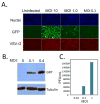
Figure 1
Figure 1. VSV infects Drosophila cells
A. Schneider cells were infected with VSV-GFP at the indicated multiplicity of infection (MOI) for the 20 hours. Cells were processed for immunofluorescence and imaged using an automated microscope (ImagXpress Micro). Infected cells express GFP and the viral glycoprotein, VSV-G and are counterstained with Hoechst 33342 to observe nuclei. B. Viral antigen production at the indicated MOI at 24 hours post infection was measured by immunoblot against the virally produced antigen GFP or the cellular control tubulin. These data show a representative experiment; similar findings were made in at least three repetitions. C. Viral titers from cells infected with the indicated MOI of VSV at 24 hours post infection. These data show a representative experiment; similar findings were made in at least two repetitions.
Figure 2
Figure 2. Autophagy is antiviral in Drosophila cells
A. S2 cells were pre-treated with dsRNA against the autophagy genes Atg5, Atg8a, or Atg18 or control dsRNA (luciferase) for three days and then infected (MOI=1) for 20 hours and processed for immunofluoresence and imaged as above. Infected cells express GFP and the percent infection is calculated using automated image analysis (MetaXpress) from 3 wells, 3 sites per well. Loss of autophagy genes significantly increases VSV infection (p<0.05). B. Depletion of autophagy genes also increases the production of viral antigens by immunoblot. Cells pretreated with the indicated dsRNAs were infected (MOI=0.1) and processed for Western blot at 24 hours post infection. Viral antigens were measured by anti-GFP and normalized to the control protein tubulin. These data show representative experiments; similar findings were made in at least three repetitions. C. Depletion of additional autophagy genes by RNAi leads to an increase in the percent infection as compared to control dsRNA. Data is shown for four independent experiments as the fold effect compared to control dsRNA and the standard error is shown (MOI=0.1–0.25, 24 hours post infection); * p<0.05 student’s ttest.
Figure 3
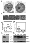
Figure 3. VSV infection activates autophagy independent of replication
A. VSV infection induces autophagosome formation as measured by electron microscopy. Representative images are shown for cells that are either uninfected or infected at an MOI=5, 20 hours post infection. B. Higher magnification images of autophagosomes in VSV-infected cells. C. Active and UV-inactivated virus induce autophagic vesicles. Cells were infected with UV-inactivated VSV-GFP as above. Autophagic bodies were counted for at least 20 cells from each treatment and presented as a notched box plot. The horizontal darkline represents the median for each category. The box represents the interquartile range and the whiskers encompass the most extreme data values. The notches represent a confidence interval for the median and nonoverlapping notches indicate different medians at the 5% significance level. There is a significant difference between uninfected and infected or UV-virus infected (* Wilcoxon test, p-value = 9.0e-5 and p-value =3.7e-3), respectively. However, there is no significant difference in the number of autophagosomes per cell between infected and UV-virus infected (Wilcoxon test, p-value = 0.22). D. The induction of autophagic vesicles by VSV is Atg5-dependent. S2 cells were pre-treated with either control (LacZ) or Atg5 dsRNA for 3 days and infected with VSV-GFP at an MOI=5 for 20 hours. Autophagic bodies were counted for 35 cells from each treatment and presented as a notched box plot. The horizontal black line represents the median for each category. Whiskers include the most extreme data value. Nonoverlapping notches indicate different medians at the 5% significance level. There is a significant difference between control and Atg5-depleted cells (* Wilcoxon test, p-value = 1.2e-07). These findings were observed in at least two independent experiments. E. Immunoblot analysis of cells treated with control dsRNA or dsRNA against Atg8 (Atg8a and Atg8b). Atg8 is observed in resting cells (Atg8-I, ~16kD). F. A smaller form of Atg8 is induced in cells that are serum starved, or infected with VSV, or UV inactivated VSV (Atg8-II, ~14kD). These data show representative experiments; similar findings were made in at least three repetitions.
Figure 4
Figure 4. VSV infection induces autophagy in Drosophila cells
VSV induces autophagosomes as measured by LC3 re-localization. A. Drosophila cells were transfected with a GFP-LC3 reporter (green). Cells were either uninfected or infected with wild type VSV or UV inactivated VSV for 24 hours, fixed, probed with anti-VSVM (Red) and with Hoechst 33342 (blue). A representative image is shown. Arrows indicate GFP-LC3+ puncta. B. The percentage of cells with punctae were counted for five experiments and fold change was graphed for cells that were uninfected (blue) infected with wild type VSV (red) or UV inactivated VSV (green); error bars indicate standard deviation; * p<0.01 student’s ttest. C. There is an increase in the number of punctae per cell upon infection with either VSV or UV inactivated VSV graphed as the number of punctae per cell normalized to 100% for each treatment (n>150 for each condition). D. The induction of GFP-LC3 punctae is cell-autonomous. The percentage of cells with punctae were counted for three experiments and fold change was graphed for cells that were either uninfected (blue), infected (red), or uninfected in a well where 30% of the cells were infected (orange). Only the infected cells have an increase in the GFP-LC3 punctae; error bars indicate standard deviation; * p<0.01 student’s ttest. E. There is no increase in the percentage of cells with punctae in cells that were transfected with viral RNA (red) compared to controls (blue). The percentage of cells with punctae was counted for three experiments and fold change was; error bars indicate standard deviation. F. There is no increase in the percentage of cells with punctae in cells that were transfected with viral RNP (red) compared to controls (blue). The percentage of cells with punctae was counted for three experiments and fold change was graphed; error bars indicate standard deviation. G. There is an increase in the percentage of cells with punctae in cells that were infected with VSV G+ VPs compared to blebs isolated from untransfected control cells. The average of three experiments is shown; error bars indicate standard deviation; * p<0.01 student’s ttest.
Figure 5
Figure 5. VSV infection induces autophagy in primary cells and in adult flies
A. Primary hemocytes expressing GFP-LC3 (Hml-gal4>UAS-eGFP-huLC3, green) were either uninfected or infected ex vivo for two hours, then fixed and stained with Hoescht 33342 to visualize nuclei (blue) by fluorescence microscopy. A representative image is shown. B. Percentage of cells with punctae were counted (n>40 for each condition). Error bars indicate range of two independent experiments. Increased punctae formation is observed in the infected cells; * p<.001, chi-square test in each experiment. C. Adult wild type flies were infected with VSV-GFP for three days. The flies were monitored for infection (GFP+) and autophagy (Lysotracker+) and counterstained with Hoescht 33342 to observe the nuclei. There is increased Lysotracker staining in the infected cells in vivo in the animal. White arrows indicate that cells that are Lysotracker+ are also GFP+. Blue arrows (merged image) show that the GFP cells throughout the tissue are not Lysotracker+. These data show representative experiments; similar findings were made in at least three repetitions.
Figure 6
Figure 6. Autophagy is antiviral in adult flies
A. Adult flies expressing ubiquitous and high level dsRNA against Atg18 (Actin-Gal4; UAS-Atg18IR) or sibling controls (+; UAS-Atg18IR) were challenged with 104 pfu VSV and morbitity was monitored as a function of time post-infection. Log rank test reveals that loss of Atg18 significantly increases susceptibility (p<0.001). A representative experiment is shown; similar findings were made in at least three experiments. B. Adult flies expressing ubiquitous and high level dsRNA against Atg18 (Actin-Gal4; UAS-Atg18IR) or their sibling controls (+; UAS-Atg18IR) were challenged with VSV and monitored over time for viral replication as measured by immunoblot against virally produced GFP and normalized to a cellular control protein. * indicates a non-specific band. C. Adult flies expressing ubiquitous and high level dsRNA against Atg18 (Actin-Gal4; UAS-Atg18IR, red bars) or their sibling controls (+; UAS-Atg18IR, blue bars) were challenged with VSV and monitored over time for viral replication as measured by viral titers. Plaque assays reveal a significant increase in viral titers in Atg18-depleted flies post infection. D. Adult flies expressing ubiquitous and high level dsRNA against Atg18 (Actin-Gal4; UAS-Atg18IR) or their sibling controls (+; UAS-Atg18IR) were challenged with VSV and monitored over time for viral replication as measured by Northern blot. There is a significant increase in the levels of GFP RNA in the Atg18-depleted animals. E. Adult flies expressing ubiquitous and high level dsRNA against Atg7 (Actin-Gal4; UAS-Atg7IR) or their respective sibling controls (+; UAS-Atg7IR) were challenged with VSV and monitored over time for viral replication as measured by Northern blot. There is a significant increase in the levels of GFP RNA in the Atg7-depleted animals. F. Adult flies expressing ubiquitous and high level dsRNA against Atg12 (Actin-Gal4; UAS-Atg12IR) or their respective sibling controls (+; UAS-Atg12IR) were challenged with VSV and monitored over time for viral replication as measured by Northern blot. There is a significant increase in the levels of GFP RNA in the Atg12-depleted animals.
Figure 7
Figure 7. The PI3K/Akt pathway controls autophagy and viral replication in adult flies
A. Flies carrying a heat-shock inducible Gal4 were crossed to UAS-PTEN, UAS-Δp60 or control at room temperature. Progeny were collected and heat shocked at 37°C for one hour on the day of infection and every two days following challenge. Viral replication was monitored over time for viral replication by immunoblot against virally produced GFP and normalized to cellular proteins. A significant decrease in viral replication was observed post infection. B. Flies carrying a heat-shock inducible PTEN were crossed to flies heterozygous for a mutant Atg1 allele at room temperature. Progeny were collected and heat shocked at 37°C for one hour on the day of infection and every two days following challenge. Viral replication was monitored over time for viral replication by immunoblot against virally produced GFP and normalized to a cellular control protein. A significant increase in viral replication was observed in the heterozygous animals post infection. C. Flies carrying a heat-shock inducible Gal4 were crossed to UAS-Akt IR, at room temperature. Progeny were collected and heat shocked at 37°C for one hour on the day of infection and every two days following challenge. Viral replication was monitored over time for viral replication by immunoblot against virally produced GFP and normalized to cellular proteins. A significant decrease in viral replication was observed post infection. D. Flies were injected with insulin in the presence or absence of VSV. Phospho-Akt and Total-Akt were monitored by immunoblot. A significant decrease in Phospho-Akt was observed post VSV infection. These data show representative experiments; similar findings were made in at least three repetitions.

Comment in

Similar articles

Cited by

References

    1. Abe A, Chen ST, Miyanohara A, Friedmann T. In vitro cell-free conversion of noninfectious Moloney retrovirus particles to an infectious form by the addition of the vesicular stomatitis virus surrogate envelope G protein. J Virol. 1998;72:6356–6361. - PMC - PubMed
    1. Andrade RM, Wessendarp M, Gubbels MJ, Striepen B, Subauste CS. CD40 induces macrophage anti-Toxoplasma gondii activity by triggering autophagy-dependent fusion of pathogen-containing vacuoles and lysosomes. J Clin Invest. 2006;116:2366–2377. - PMC - PubMed
    1. Arico S, Petiot A, Bauvy C, Dubbelhuis PF, Meijer AJ, Codogno P, Ogier-Denis E. The tumor suppressor PTEN positively regulates macroautophagy by inhibiting the phosphatidylinositol 3-kinase/protein kinase B pathway. J Biol Chem. 2001;276:35243–35246. - PubMed
    1. Bilen J, Bonini NM. Genome-wide screen for modifiers of ataxin-3 neurodegeneration in Drosophila. PLoS Genet. 2007;3:1950–1964. - PMC - PubMed
    1. Brand AH, Perrimon N. Targeted gene expression as a means of altering cell fates and generating dominant phenotypes. Development. 1993;118:401–415. - PubMed

Publication types