. 2009 Apr 17;30(4):556-65.
doi: 10.1016/j.immuni.2009.02.005. Epub 2009 Apr 9.

The NLRP3 inflammasome mediates in vivo innate immunity to influenza A virus through recognition of viral RNA

Affiliations

The NLRP3 inflammasome mediates in vivo innate immunity to influenza A virus through recognition of viral RNA

Irving C Allen et al. Immunity. .

Abstract

The nucleotide-binding domain and leucine-rich-repeat-containing (NLR) family of pattern-recognition molecules mediate host immunity to various pathogenic stimuli. However, in vivo evidence for the involvement of NLR proteins in viral sensing has not been widely investigated and remains controversial. As a test of the physiologic role of the NLR molecule NLRP3 during RNA viral infection, we explored the in vivo role of NLRP3 inflammasome components during influenza virus infection. Mice lacking Nlrp3, Pycard, or caspase-1, but not Nlrc4, exhibited dramatically increased mortality and a reduced immune response after exposure to the influenza virus. Utilizing analogs of dsRNA (poly(I:C)) and ssRNA (ssRNA40), we demonstrated that an NLRP3-mediated response could be activated by RNA species. Mechanistically, NLRP3 inflammasome activation by the influenza virus was dependent on lysosomal maturation and reactive oxygen species (ROS). Inhibition of ROS induction eliminated IL-1beta production in animals during influenza infection. Together, these data place the NLRP3 inflammasome as an essential component in host defense against influenza infection through the sensing of viral RNA.


Figures

Figure 1
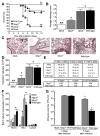
Figure 1. Characterization of influenza virus A/PR/8/34 pathogenicity and immune response in mice deficient in inflammasome signaling pathways
Wild type, MyD88−/−, ASC−/−and Caspase-1 (Casp1−/−) deficient mice were challenged intranasally (i.n.) with influenza A/PR/8/34 and survival was monitored 14 dpi. (A) ASC−/− and Casp1−/− mice demonstrated significantly increased mortality compared to the wild type animals (*p<0.05; log rank), while (B) MyD88−/− mice demonstrated modestly increased mortality. Mock-inoculated (n=7); influenza-infected wild type (n=19); MyD88−/− (n=11); ASC−/− (n=12); and Casp1−/− (n=7). (C–F) Lungs were harvested 3 dpi. from wild type, ASC−/−, Myd88−/− and mock-infected mice. Sections through the main bronchiole of the left lobe were stained with H&E. (C) No abnormal lung histopathology was observed in mock-challenged animals. (D) Wild type and (E) MyD88−/− influenza-challenged mice demonstrated increased airway inflammation. (F) ASC−/− mice demonstrated significantly reduced inflammation following influenza challenge. (G) Histological scoring of H&E stained lung sections demonstrated a significant increase in inflammation following influenza challenge in all mice. However, ASC−/− mice demonstrated a significant attenuation compared to MyD88−/− and wild type animals (*p<0.05; **p<0.01). Wild type (n=8); MyD88−/− (n=7); ASC−/− (n=6); wild type mock-infected (n=4). Results are representative of at least 2 independent experiments.
Figure 2
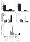
Figure 2. The Nlrp3 inflammasome is required for survival and mediates airway inflammation and viral clearance following pulmonary challenge with influenza virus
A Wild type, Nlrp3−/− and Nlrc4−/− mice were challenged i.n. with influenza A/PR/8/34 and survival was monitored 14 dpi. Nlrp3−/− mice demonstrated significantly increased mortality compared to wild type animals (**p<0.01; log rank). In contrast, loss of Nlrc4 did not have any effect on mouse survival. Wild type mock-infected (n=7); wild type infected (n=19); Nlrp3−/− infected (n=16); and Nlrc4−/− infected (n=7). All survival experiments were performed together with the mice studied in Figure 1, therefore, the wild type controls are identical to those shown in Figure 1. (B) Lungs from wild type, Nlrp3−/− and Nlrc4−/− mice were harvested for histology and scored. A significant increase in airway inflammation was observed following influenza challenge in wild type and Nlrc4−/− animals, while a significant attenuation was observed in Nlrp3−/− mice (*p<0.05; **p<0.01). (C) No abnormal lung histopathology was observed in the mock-challenged wild type animals. (D) Nlrp3−/− mice demonstrated significantly attenuated inflammation following influenza virus challenge. (E) Nlrc4−/− and (F) wild type influenza challenged mice demonstrated increased airway inflammation. (G) To profile the cell types involved in the host immune response to influenza infection, bronchoalveolar lavage (BAL) was performed with differential staining and FACS analysis, 3 dpi. Consistent with the histology data, significantly fewer total cells were recovered in the BAL fluid (BALF) from Nlrp3−/− animals (*p<0.05), compared with either Nlrc4−/− or wild type mice. (H) BALF cellularity indicated a significant influx of macrophages and neutrophils in all influenza-challenged animals, with no difference observed in the composition of cells present in the airways. Wild Type (n=8); Nlrp3−/− (n=5); Nlrc4−/− (n=3); ASC−/− (n=6); wild type mock-infected (n=4). Results are representative of 3 independent experiments. (I) While no differences were observed in BALF composition, differential staining revealed a significant decrease in the total number of monocytes and neutrophils present in the Nlrp3−/− mice (*p<0.05). Wild type (n=8); Nlrp3−/− (n=4); Nlrc4−/− (n=4); wild type mock-infected (n=5). (J) Lungs were weighed and homogenized in sterile PBS either 3 or 7 dpi. Cell free supernatants were titered by standard plaque assay. Nlrp3−/− mice demonstrated a significant defect in vial clearance by day 7 (*p<0.05). Wild type mock-infected, d3 (n=3); wild type, infected, d3 (n=9); Nlrp3−/− infected, d3 (n=9); wild type mock-infected d7 (n=4); wild type infected, d7 (n=6); Nlrp3−/− infected, d7 (n=7).
Figure 3
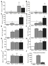
Figure 3. Nlrp3 inflammasome components are highly expressed in myeloid cells and in the lungs during influenza A virus infection
(A–B) ASC and Nlrp3 gene expression was assessed in a panel of cells considered important for viral infection. Primary mouse bone marrow derived macrophages (BM), B cells, T cells, mouse airway epithelial cells (MAECs) and mouse embryonic fibroblasts (MEFs) were assessed by real-time (rt) quantitative PCR with samples normalized to 18s and standardized to expression in MEFs. Each cell line was assessed in triplicate and data are representative of 3 independent experiments. (C–D) Primary mouse BM s and MAECs were differentiated and challenged with mouse adapted influenza A/PR/8/34 (MOI=10). Nlrp3, Nlrc4 and IL-1β transcripts were assessed by rtPCR normalized to 18s and standardized to naive levels. (C) A significant increase in IL-1β mRNA expression was observed in BM following influenza challenge (MOI=10); however, no significant change was observed in Nlrp3 or Nlrc4 expression (*p<0.05). (D) Influenza challenge (MOI=1) resulted in a significant increase in Nlrp3 expression and a modest decrease in Nlrc4 and IL-1β expression in the MAECs. (E) Whole lungs were removed from wild type mice either 3 dpi. or 7 dpi. and homogenized. Total RNA was extracted from tissue pellets and expression levels of Nlrp3, ASC, Il-1β and Caspase-1 (Casp1) were assessed by rtPCR normalized to the 18S house keeping gene and compared to the mock-infected lungs. Transcription levels are significantly elevated by day 3 following influenza inoculation and begin to decline by day 7 (*p<0.05). All experiments are representative of at least 2 independent experiments with 3–5 mice per group.
Figure 4
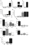
Figure 4. Mice lacking components of the Nlrp3 Inflammasome demonstrate significantly altered levels of proinflammatory cytokines following pulmonary challenge with influenza
Wild type, ASC−/−, Nlrp3−/− and Nlrc4−/− mice were challenged with influenza A/PR/8/34. Serum and cell free BALF were assessed 3 dpi., unless otherwise noted. (A–B) Significantly reduced levels of IL-1β(A) and IL-18 (B) were observed in the serum from Nlrp3−/− and Asc−/− mice (*p<0.05). (C–F) Nlrp3−/− and ASC−/− mice also demonstrated significantly reduced levels of BALF IL-1β (C) and MIP2α (D). KC was slightly decreased in Nlrp3−/− mice (E), while TNFα was reduced in ASC−/− mice (F) (*p<0.05). BALF levels of IL-18 were below the level of detection (data not shown). (G–I) All infected animals demonstrated a significant increase in IL-6 (G), IFNγ(H) and IL-12p40 (I) with no differences observed between genotypes. Mock, n=5; Nlrp3−/−, n=6; Nlrc4−/−, n=6; ASC−/−, n=9; Wild Type, n=5. (J) BALF from moribund Nlrp3−/− mice demonstrate significantly increased local levels of IL-6 compared to moribund wild type animals, 7 dpi.(*p<0.05). Mock, n=3; Nlrp3−/−, n=6; wild type, n=3. All data are representative of at least 2 independent experiments.
Figure 5
Figure 5. NLRP3 inflammasome activation in response to influenza virus is dependent upon lysosomal maturation and ROS production in human cells
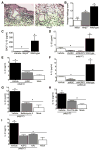
(A) Depiction of the primary ciliated human airway epithelial (HAE) cultures which were infected with the human pathogenic influenza virus A/Victoria/3/75 (H3N2; MOI = 1). Supernatants are collected from the apical and basolateral surfaces. (B) Robust viral replication was observed over the course of the HAE infection. (C) IL-1β levels were detected in the apical, but not basolateral, compartment 48 hours post-infection. (D) Human nasal airway epithelial cell lines (JME) were infected with A/Victoria/3/75 (MOI=1) and increased NLRP3 and ASC mRNA expression was observed. Data was normalized to 18s and compared to the expression in similarly treated human type II alveolar epithelial-like cell lines (A549). (E) Increased IL-1β protein was observed 24 hours post-infection in the cell free supernatant from the JMEs. (F) Human THP-1 monocyte cell lines were infected with A/Victoria/3/75 (MOI=5) and increased IL-1β levels were observed in the supernatant over a 24hr time course. (G–I) Influenza mediated IL-1β generation is dependent upon the NLRP3 inflammasome in human monocytes. (G) Human THP-1 cells were infected with lentivirus containing shRNA for ASC, NLRP3 or TUCAN (shASC, shNLRP3 or shTUCAN) or a mutated sh target sequence (shmut). Cell free supernatants were collected 24 hours post-infection (MOI=1). Influenza induced NLRP3- and ASC- dependent increase in IL-1β (*p<0.05), which was independent of TUCAN. (H) The general caspase inhibitor ZVAD-CHO and (I) the caspase-1 specific inhibitor Ac-YVAD-CHO, both inhibited IL-1β release in a dose-dependent manner (*p<0.05). (J–L) Influenza mediated IL-1β maturation requires lysosomal maturation and ROS production in THP-1. IL-1β release during influenza infection (MOI=5) was attenuated following treatment with (J) the lysosome inhibitor bafilomycin A (100–250nM), (K) the cathepsin B inhibitor CA-074-Me (50μM), and (L) the ROS inhibitors APDC (100μM) and NAc (50μM)(*p<0.05). (M) Treatment with the ROS inhibitor NAc (250mg/kg) inhibits influenza mediated IL-1β release in vivo (*p<0.05). NAc Mock, n=1; NAc Influenza, n=5, Vehicle Influenza, n=3.
Figure 6
Figure 6. The NLRP3 inflammasome is required for airway inflammation induced by nucleic acid analogs
Wild type and Nlrp3−/− mice received either 2 doses (50μg/dose) of poly(I:C) or vehicle on alternating days and were harvested 24hrs after the second dose was administered. (A–B) Poly(I:C) challenged animals demonstrated increased airway inflammation but less inflammatory cell influx was observed in Nlrp3−/− mice. (C) Histology scoring confirmed a significant attenuation in airway inflammation in the Nlrp3−/− mice (*p<0.05). (D) Significant decreases were observed in BALF IL-1β in the Nlrp3−/− mice compared to the wild type animals (*p<0.05). Vehicle challenged wild type (n=4); wild type, n=8; Nlrp3−/−, n=8. (E–G) IL-1β induction by viral RNA analogs in human monocytic cells is mediated by NLRP3 and ASC. (E) IL-1β secretion stimulated by the viral dsRNA analog poly(I:C) was significantly reduced in the shNLRP3 and shASC knockdown THP-1 cell lines and (F) following treatment with the caspase-1 specific inhibitor yVAD-CHO (*p<0.05). (G) IL-1β secretion stimulated by ssRNA40 was also significantly reduced in the shNLRP3 and shASC containing cells (*p<0.05). All knockdown studies are representative of at least 3 independent experiments. (H–J) Poly(I:C) mediated IL-1β maturation requires lysosomal maturation and ROS production in human monocytes. (H) The lysosome inhibitor bafilomycin A (100nM), (I) the cathepsin B inhibitor CA-074-Me (50μM), and (J) the ROS inhibitors APDC (100μM) and NAc (50μM) all significantly attenuated IL-1β production (*p<0.05).

Comment in

Similar articles

Cited by

References

    1. Adachi O, Kawai T, Takeda K, Matsumoto M, Tsutsui H, Sakagami M, Nakanishi K, Akira S. Targeted disruption of the MyD88 gene results in loss of IL-1- and IL-18-mediated function. Immunity. 1998;9:143–150. - PubMed
    1. Agostini L, Martinon F, Burns K, McDermott MF, Hawkins PN, Tschopp J. NALP3 forms an IL-1beta-processing inflammasome with increased activity in Muckle-Wells autoinflammatory disorder. Immunity. 2004;20:319–325. - PubMed
    1. Akira S, Uematsu S, Takeuchi O. Pathogen recognition and innate immunity. Cell. 2006;124:783–801. - PubMed
    1. Bartlett EJ, Hennessey M, Skiadopoulos MH, Schmidt AC, Collins PL, Murphy BR, Pickles RJ. The role of interferon in the replication of human parainfluenza virus type 1 wild type and mutant viruses in human ciliated airway epithelium. J Virol 2008 - PMC - PubMed
    1. Cottey R, Rowe CA, Bender BS. Influenza virus. Curr Protoc Immunol . 2001;Chapter 19(Unit 19):11. - PubMed

Publication types