. 2009 Apr;119(4):802-12.
doi: 10.1172/JCI36230. Epub 2009 Mar 2.

PICK1 deficiency causes male infertility in mice by disrupting acrosome formation

Affiliations

PICK1 deficiency causes male infertility in mice by disrupting acrosome formation

Nan Xiao et al. J Clin Invest. 2009 Apr.

Abstract

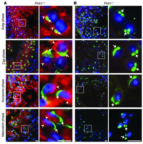
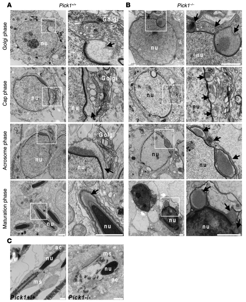
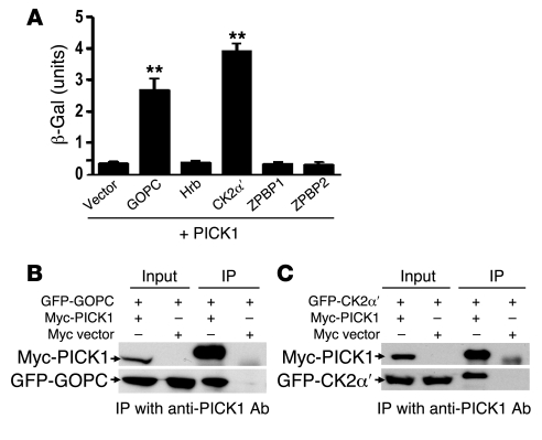
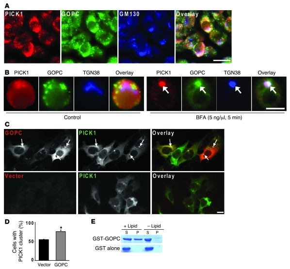
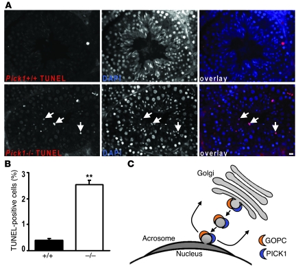
Protein interacting with C kinase 1 (PICK1) is a peripheral membrane protein involved in protein trafficking, a function that has been well characterized in neurons. Here, we report that male mice deficient in PICK1 are infertile and have a phenotype resembling the human disease globozoospermia. The primary defect in the testes of Pick1-knockout mice was fragmentation of acrosomes in the early stages of spermiogenesis. This fragmentation was followed by defects in nuclear elongation and mitochondrial sheath formation, leading to round-headed sperm, reduced sperm count, and severely impaired sperm motility. We found that PICK1 interacted with Golgi-associated PDZ- and coiled-coil motif-containing protein (GOPC) and the primary catalytic subunit of protein kinase 2 (CK2alpha'), proteins whose deficiencies lead to globozoospermia in mice. PICK1 was highly expressed in round spermatids and localized to Golgi-derived proacrosomal granules. GOPC colocalized with PICK1 in the Golgi region and facilitated formation of PICK1-positive clusters. Furthermore, there was an increase in apoptosis in the seminiferous tubules of Pick1-/- mice, a phenotype also seen in CK2alpha'-deficient mice. Our results suggest that PICK1 is involved in vesicle trafficking from the Golgi apparatus to the acrosome and cooperates with other proteins such as GOPC and CK2alpha' in acrosome biogenesis.


Figures

Figure 1
Figure 1. Decreased sperm number and abnormal sperm morphology in Pick1–/– mice.
(A) Total number of sperm from a single cauda epididymis: wild-type (Pick1+/+), 17.69 × 10⁶ ± 1.62 × 10⁶; Pick1+/–, 12.41 × 10⁶ ± 1.10 × 10⁶; Pick1–/–, 7.24 × 10⁶ ± 0.81 × 10⁶; mean ± SEM, n = 10, **P < 0.01. (B) Motile sperm number: Pick1+/+, 91.03 × 10⁵ ± 15.5 × 10⁵; Pick1+/–, 47.68 × 10⁵ ± 9.32 × 10⁵; Pick1–/–, 3.38 × 10⁵ ± 1.21 × 10⁵. (C) Linear motile sperm number: Pick1+/+, 58.80 × 10⁵ ± 13.59 × 10⁵; Pick1+/–, 22.13 × 10⁵ ± 5.55 × 10⁵; Pick1–/–, 0. (D) The percentage of globozoospermia-like sperm: Pick1+/+, 1.57% ± 0.81%; Pick1+/–, 2.26% ± 0.73%; Pick1–/–, 88.69% ± 9.57% (n = 3). (E) Morphology of unfixed sperm. Sperm from Pick1–/– mice lose the typical hook-shaped head of normal sperm; instead, they have round or irregular ball–like heads. In addition, defects in the tail can also be seen in sperm from Pick1–/– mice. (F) Immunostaining of acrosome matrix protein sp56 (red) and nucleus (nu, blue) in sperm. The acrosomes from Pick1–/– mice fail to acquire the crescent moon–shaped structure, are frequently fragmented, and are located in the wrong position. (G) Immunostaining of the mitochondrial sheath (ms, red) and nucleus in sperm. Mitochondrial sheaths in Pick–/– mice display various defects, including (left to right) abnormal sperm with aggregated mitochondrial sheaths, split mitochondrial sheaths, the mitochondrial sheath overlapping with the round nucleus, and the mitochondrial sheath wrapping around the round nucleus. Scale bars: 5 μm.
Figure 2
Figure 2. Abnormal spermiogenesis in Pick1–/– mice.
(A) H&E staining of the epididymis. The number of sperm in Pick1–/– mice is lower than that in Pick1+/+ mice in the cross sections of the cauda epididymis. Insets: Enlarged views of the heads of the sperm, showing that the sperm from Pick1–/– mice are round-headed. Scale bars: 10 μm. (B) The testes of Pick1–/– mice are smaller than those of Pick1+/+ or Pick1+/– mice. Scale bar: 2 mm. (C) Normalized testis weights (Testis weight (TW) ×1000 / BW) of Pick1–/– mice are significantly lower than those of the Pick1+/+ or Pick1+/– mice. Pick1+/+, 3.58 ± 0.11; Pick1+/–, 3.25 ± 0.09; Pick1–/–, 2.88 ± 0.19; n = 10. Data are presented as mean ± SEM; *P < 0.05, **P < 0.01. (D) The diameter of the seminiferous tubules in Pick1–/– mice is smaller than that of the Pick1+/+ and Pick1+/– mice. Pick1+/+, 203.7 ± 1.13 μm; Pick1+/–, 206.2 ± 5.2 μm; Pick1–/–, 181.3 ± 4.2 μm; n = 50. (E) H&E staining of testis. The lumens of seminiferous tubules in Pick1–/– mice are slightly larger than those in wild-type mice. The morphology of spermatogonia (white arrows), spermatocytes (white arrowheads), and round spermatids (black arrows) is similar in the 3 genotypes. However, there are fewer mature sperm in the Pick1–/– mice, and the heads of the sperm in Pick1–/– mice are round, in contrast to the hook-shaped heads found in Pick1+/+ and Pick1+/– mice (black arrowheads). The bottom row shows higher-magnification views of the boxed regions in the top panels. Scale bar: 10 μm.
Figure 3
Figure 3. PICK1 is highly expressed in spermatids and localized around the Golgi apparatus.
(A) DAB staining of testis sections from Pick1+/+ and Pick1–/– mice. PICK1 is highly expressed in round spermatids. The background staining of Pick1–/– mice is low. (B) Triple immunofluorescence staining of PICK1, β-tubulin, and DAPI, which marks nuclei. PICK1 is mainly concentrated in the perinuclear region of round spermatids. (C) PICK1 is partially colocalized with the Golgi marker GM130 in the spermatids in both the testis sections (upper panels) and testis smear stains (lower panels). Scale bars: 10 μm. (D) PICK1 is located on the Golgi-derived proacrosomal granules between the Golgi apparatus and the acrosome (ac). The arrowheads indicate gold particles labeling PICK1. The arrow indicates the nucleus envelope (ne). The right panel shows a higher-magnification view of the boxed region in the left panel. Scale bar: 200 nm.
Figure 4
Figure 4. Acrosome formation is disrupted in Pick1–/– mice.
Immunostaining of testis sections. PICK1 (red) and acrosome (green) were labeled by a guinea pig antibody against PICK1 and a mouse antibody against sp56. Nuclei were marked by DAPI (blue). Four phases of spermiogenesis are shown in progressive order. (A) Images from wild-type mice. PICK1 signals (arrowheads) are close to the acrosomes (arrows) in the Golgi phase and the cap phase but move to opposite ends of the nucleus in the acrosome and maturation phases. Acrosomes grow from single granules in the Golgi phase to caps covering the heads of the nuclei in the cap phase to the crescent moon-shaped structures at one pole of the nuclei in the acrosome and maturation phases. (B) Images from Pick1-knockout mice. PICK1 signals are notably missing in testes from Pick1–/– mice. There are multiple sp56-positive structures in spermatids of Pick1–/– mice throughout spermiogenesis. The morphology of the nuclei of the spermatids of Pick1+/+ and Pick1–/– mice was similar in the Golgi and cap phases. However, while the nuclei of Pick1+/+ mice become elongated in the acrosome and maturation phases, those of Pick1–/– mice remain round-shaped. Scale bars: 10 μm. Right panels show higher-magnification views of the boxed regions in the left panels.
Figure 5
Figure 5. TEM study of acrosome formation in Pick1–/– mice.
TEM images of spermatids showing 4 phases of spermiogenesis. (A) In the wild-type mice, a large number of Golgi-derived proacrosomal granules are present in the Golgi phase, when a single acrosome granule can be seen attached to the nuclear envelope. The acrosome flattens and grows to form a cap at one end of the nucleus in the cap phase. The acrosome becomes extended along the nuclear envelope in the subsequent acrosome and maturation phases. The nucleus starts to elongate in the acrosome phase and becomes hook-shaped in the maturation phase. (B) In Pick1–/– mice, multiple acrosomal vesicular structures are seen in all phases of spermatids, as indicated by the arrows. The nuclei of spermatids of Pick1–/– mice failed to elongate and remain round in the maturation phase. Right panels show higher-magnification views of the boxed regions in the left panels. (C) Sperm from the caput epididymis of Pick1+/+ and Pick1–/– mice. While mitochondria wrap around the axoneme in the midpiece of sperm from Pick1+/+ mice, they aggregate around the deformed nucleus in Pick1–/– mice. The deformed acrosome is also shown. Scale bars: 1 μm. ms, mitochondria sheath.
Figure 6
Figure 6. PICK1 interacts with GOPC and CK2α′, but not with Hrb, ZPBP1, or ZPBP2.
(A) Yeasts cotransformed with PICK1 and other cDNA as indicated were grown on liquid selective medium, and β-galactosidase activity was measured using ONPG as the substrate. Data are presented as mean ± SEM; n = 4; **P < 0.01 compared with vector control, as determined by Student’s t test. (B) GFP-GOPC transfected into HEK293T cells together with myc-PICK1 or the vector control. PICK1 was immunoprecipitated by an anti-PICK1 antibody. Anti-myc antibody (upper panel) or anti-GFP antibody (lower panel) was used for Western blotting. Expression of GFP-GOPC and myc-PICK1 in 293T cells is indicated in the lanes labeled “Input.” The immunoprecipitation products indicate that GFP-GOPC was pulled down in the presence only of PICK1 but not the vector control. (C) GFP-CK2α′ and myc-PICK1 or the empty myc vector were cotransfected into 293T cells and immunoprecipitated as described in B. Similarly, CK2α′ was coimmunoprecipitated with PICK1 specifically.
Figure 7
Figure 7. PICK1 and GOPC partially colocalize around the Golgi apparatus, and GOPC increases PICK1 clusters.
(A) Immunofluorescence staining showed that PICK1 (red) and GOPC (green) are both expressed in the perinuclear region on the round spermatids and they partially colocalize with the Golgi marker GM130 (blue). (B) In the rat testis smear, 5 ng/μl BFA treatment for 5 minutes leads to better colocalization of PICK1, GOPC, and TGN38, as indicated by the arrows. (C) GFP-PICK1 and myc-GOPC or the empty myc vector were cotransfected into 293T cells. GOPC increased the number of PICK1 clusters, and some of these clusters overlapped with the GOPC signal, as indicated by the arrows. (D) GOPC increases the percentage of HEK293T cells with PICK1 self-clusters to 76.1% ± 3.8% from 55.0% ± 0.8% in the vector control group (mean ± SEM; n = 3 experiments; *P < 0.05). (E) GST-GOPC was mixed with liposome and subjected to high-speed centrifugation. Equal amounts of pellet and supernatant were loaded and resolved by SDS-PAGE. GOPC was found to associate with liposomes and appeared in the pellet (top). In the control experiments, GOPC was not found in the pellet without liposomes, and GST itself did not bind to liposomes. Scale bars: 10 μm.
Figure 8
Figure 8. Increased apoptosis in the seminiferous tubules of Pick1–/– mice.
(A) TUNEL staining of the testis sections. Only a few apoptotic spermatogonia, labeled with red fluorescence, are seen in the seminiferous tubules of Pick1+/+ mice. In contrast, far more apoptotic germ cells, many of them spermatids, can be found in the seminiferous tubules of Pick1–/– mice, as indicated by the arrows. Scale bar: 10 μm. (B) Quantification revealed a significant increase in apoptotic cells in the seminiferous tubule of Pick1–/– mice. Data are presented as mean TUNEL-positive cells/100 cells ± SEM; n = 30 tubules; **P < 0.01. (C) Model illustrating PICK1’s role in acrosome formation. PICK1 and GOPC facilitate formation of trafficking vesicles from the Golgi apparatus to the acrosome. Both of them are removed from mature acrosomes and are possibly recycled for multiple rounds of vesicle trafficking.

Similar articles

Cited by

References

    1. Staudinger J., Zhou J., Burgess R., Elledge S.J., Olson E.N. PICK1: a perinuclear binding protein and substrate for protein kinase C isolated by the yeast two-hybrid system. J. Cell Biol. 1995;128:263–271. doi: 10.1083/jcb.128.3.263. - DOI - PMC - PubMed
    1. Xu J., Xia J. Structure and function of PICK1. Neurosignals. 2007;15:190–201. doi: 10.1159/000098482. - DOI - PubMed
    1. Torres R., et al. PDZ proteins bind, cluster, and synaptically colocalize with Eph receptors and their ephrin ligands. Neuron. 1998;21:1453–1463. doi: 10.1016/S0896-6273(00)80663-7. - DOI - PubMed
    1. Xia J., Zhang X., Staudinger J., Huganir R.L. Clustering of AMPA receptors by the synaptic PDZ domain-containing protein PICK1. Neuron. 1999;22:179–187. doi: 10.1016/S0896-6273(00)80689-3. - DOI - PubMed
    1. Dev K.K., Nishimune A., Henley J.M., Nakanishi S. The protein kinase C alpha binding protein PICK1 interacts with short but not long form alternative splice variants of AMPA receptor subunits. Neuropharmacology. 1999;38:635–644. doi: 10.1016/S0028-3908(98)00230-5. - DOI - PubMed

Publication types

MeSH terms