. 2009 Feb;20(3):963-72.
doi: 10.1091/mbc.e08-09-0979. Epub 2008 Nov 26.

Kinesin-8 from fission yeast: a heterodimeric, plus-end-directed motor that can couple microtubule depolymerization to cargo movement

Affiliations

Kinesin-8 from fission yeast: a heterodimeric, plus-end-directed motor that can couple microtubule depolymerization to cargo movement

Paula M Grissom et al. Mol Biol Cell. 2009 Feb.

Abstract

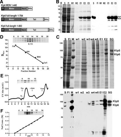
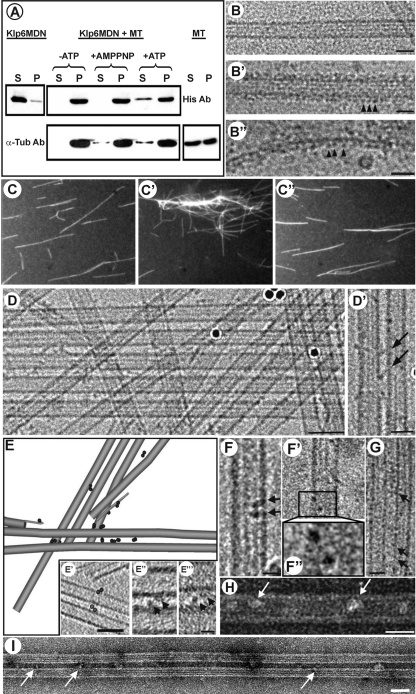
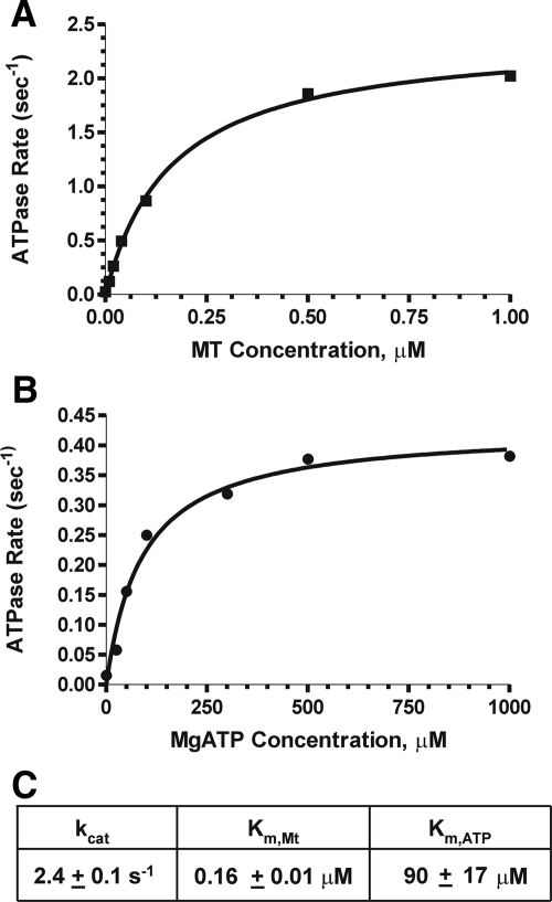
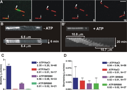
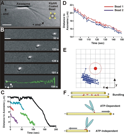
Fission yeast expresses two kinesin-8s, previously identified and characterized as products of the klp5(+) and klp6(+) genes. These polypeptides colocalize throughout the vegetative cell cycle as they bind cytoplasmic microtubules during interphase, spindle microtubules, and/or kinetochores during early mitosis, and the interpolar spindle as it elongates in anaphase B. Here, we describe in vitro properties of these motor proteins and some truncated versions expressed in either bacteria or Sf9 cells. The motor-plus-neck domain of Klp6p formed soluble dimers that cross-linked microtubules and showed both microtubule-activated ATPase and plus-end-directed motor activities. Full-length Klp5p and Klp6p, coexpressed in Sf9 cells, formed soluble heterodimers with the same activities. The latter recombinant protein could also couple microbeads to the ends of shortening microtubules and use energy from tubulin depolymerization to pull a load in the minus end direction. These results, together with the spindle localizations of these proteins in vivo and their requirement for cell viability in the absence of the Dam1/DASH kinetochore complex, support the hypothesis that fission yeast kinesin-8 contributes both to chromosome congression to the metaphase plate and to the coupling of spindle microtubules to kinetochores during anaphase A.


Figures

Figure 1.
Figure 1.
Preparation of Klp5/6 and its active fragment. (A) Constructs from klp5+ and klp6+ that were used to express the proteins studied here. Klp5pFL and Klp6pFL included the entire open reading frames of these genes (West et al., 2001), plus sequences to encode a 6-His C-terminal extension. Klp6pMDN included the motor domain (amino acids 1-403) plus the 36 amino acid putative α-helical segment. (B) Purification of Klp6MDN. Coomassie-stained SDS gel (left) and Western blot with penta-His antibodies (right) of samples collected during various stages of Klp6MDN purification on a Ni-NTA column. M, molecular weight standards; S, whole protein extract; Fl, flow-through; W1, W2, wash fractions (20 and 40 mM imidazole); and E1–E3, Klp6MDN elution fractions. (C) Purification of Klp5FL/Klp6FL. Coomassie-stained SDS gel (top) and Western blot with penta-His antibodies (bottom) of the purification of Klp5FL and Klp6FL after their coexpression from baculovirus in Sf9 cells. S, clarified supernatant from whole cell lysate; Fl, flow-through; M, molecular weight standards; w1–w5, wash-fractions; E1 and E2, Ni-NTA elution fractions; and SG, sucrose gradient-purified Klp5FL and Klp6FL. (D–F) Hydrodynamic characterization of Klp5FL/Klp6FL. (D) Sucrose gradient fractionation of protein standards and Klp5FL/Klp6FL Ni-NTA elution fraction E2 (as shown in C); blot of gradient fractions 6–20 (inset). (E) Gel filtration chromatography of Klp5FL/Klp6FL on a Superdex 200 column. Blot of fractions 7–12 (inset) showing the coelution of Klp5FL and Klp6FL. The elution positions of protein calibration standards are marked by arrows. (F) Ion exchange chromatography of Klp5FL/Klp6FL on a SP Sepharose XL cation exchange column with a blot of fractions 4–14 (inset). Klp5FL and Klp6FL coeluted during a 0–1 M NaCl gradient.
Figure 2.
Figure 2.
Evidence for Klp6MDN and Klp5/6FL cross-bridging of MTs. (A) Cosedimentation of Klp6MDN with MTs is ATP sensitive. Klp6MDN (2 μM) was incubated with Taxol-stabilized MTs (3 μM total tubulin) for 15 min at room temperature under various conditions: the absence of ATP, in the presence of 2 mM MgAMP-PNP, or in the presence of 2 mM MgATP. Samples were centrifuged for 30 min at 30,000 × g, and the pellets (P) were resuspended in SDS-buffer at a volume equal to the supernatants (S). Equal amounts of pellet and supernatant fractions were loaded on SDS gels and analyzed by Western blots. The top panel was probed with anti-His and the bottom panel with anti α-tubulin antibodies. (B) MT binding of Klp6MDN visualized by cryo-electron microscopy. (B) A rapidly frozen, ice-embedded, Taxol-stabilized MT. (B′) A similar MT after incubation with Klp6MDN for 10 min without ATP; motor-decorations (arrowheads) are evident on MT walls and on single protofilament extensions (B″). Bars, 25 nm. (C) Klp6MDN promotes ATP-sensitive MT bundling. Rhodamine-labeled MTs (C) were incubated with 0.5 μM Klp6MDN in the absence (C′) or presence (C″) of 1 mM MgATP. Samples were taken after 5 min, fixed with 0.2% glutaraldehyde, and then visualized by fluorescence light microscopy. (D) MT bundling in the presence of Klp6MDN visualized by cryo-EM. Without Klp6MDN most MTs were randomly distributed in the ice-layer (data not shown); in the presence of Klp6MDN, many MTs ran parallel to each other, forming bundles of up to ∼10 MTs (D). The distance between neighboring MTs within these bundles ranged from ∼10–13 nm. (D′) In some areas the gap between neighboring MTs was bridged by diagonal linkers (arrows). Dark circles are colloidal gold used for alignment of tilted views. Bars, 50 nm (D); 25 nm (D′). (E) Klp6MDN links between MTs were dimers when seen in 3D reconstructions. A 3D graphical model of a subvolume from a cryo-tomogram of ice-embedded MTs with Klp6MDN. The sample was imaged at 3° intervals over a tilt range of −63° to +72°. MTs and their protofilament extensions were modeled in gray, attached motors are shown as dark 4-nm discs. The majority of Klp6MDN particles occurred in pairs (circled in E′). E′–E‴ show 1-nm-thick tomographic slices of Klp6MDN dimers; E″ and E‴ show two motor heads (arrowheads) that bind adjacent MTs and are connected, most likely by their neck-domain. Bars, 50 nm (E′); 10 nm (E″ and E‴). (F–I) MT binding and bundling by Klp5/6FL visualized by cryo- (F and G) and negative staining (H and I) EM. Taxol-stabilized MTs were incubated with Klp5/6FL at a ratio of 1:2 plus 2 mM AMPPNP for 10 min. Klp5/6FL motors (arrows) bind to MTs (F–F″, H), often as dimers (F–F″). F″ is a 2× magnification of the two motor domains in the boxed area in (F′). Klp5/6FL also promotes MT bundle formation (G and I), whereby neighboring MTs seem to be connected by diagonal linkers (G, arrows). Bars, 50 nm (I); 25 nm (G and H); 12.5 nm (F and F′). Protein is black in B′–B″ and D–G and white in H and I due to different EM techniques used to prepare these images.
Figure 3.
Figure 3.
ATPase activities of Klp5/6FL. (A) Enzyme activity as a function of MT concentration: 100 nM Klp5/6FL, 1 mM MgATP, and Taxol-stabilized MTs (0–1 μM tubulin dimers). (B) Enzyme activity as a function of MgATP: 100 nM Klp5/6FL, 1 μM tubulin (polymerized into MTs), and 0–1 mM MgATP. The points on graphs A and B are an average of two experiments performed with the same protein preparation. (C) Steady-state ATPase values for Klp5/6FL.
Figure 4.
Figure 4.
Klp5/6FL is a plus end motor in vitro with no MT depolymerization activity. (A) Motility assay showing plus-end–directed motor activity of Klp5/6FL. Images of polarity marked MTs (green, Fl labeled seed with red, Rh extensions). First panel is a superposition of images taken through fluorescein isothiocyanate and Texas Red filter cubes; subsequent images show Texas Red images only with arrows marking the minus end of two MTs. Time between images in minutes. Bar, 5 μm. (B) Kymograph of polarity-marked MTs (Fl seed and Rh ends) visualized in Texas Red channel. MT with Klp5/6FL in the absence of ATP shows no motility or shortening; MT with Klp5/6FL plus 2 mM MgATP shows motility but no depolymerization (B′). (C) Summary bar graph of Klp5/6FL plus-end motility rates. Experiments were performed in the presence of NaCl/BRB80 buffer + ATP (blue bar); in NaCl/BRB80 buffer − ATP (red bar); in BRB80 buffer + ATP (purple bar); in BRB80 with no ATP (green bar). Error bars are standard deviations. (D) Summary bar graph showing the absence of MT shortening by Klp5/6FL. Experiments were performed in NaCl/BRB80 buffer + ATP (blue bar); in NaCl/BRB80 buffer − ATP (red bar); in BRB80 buffer + ATP (purple bar); BRB80 with no ATP (green bar). Error bars are standard deviations. Note difference in graph scales for C and D.
Figure 5.
Figure 5.
Klp5/6FL coated beads follow the end of a depolymerizing MT. (A) Experimental setup: Chlamydomonas axonemes (DIC image) have been elongated with tubulin plus GTP to form conventional MTs and then capped with Rh-tubulin in the presence of GMPCPP (arrows point to plus and minus MT ends). Tubulin was then washed out, and Klp5/6FL-coated beads were flowed into the chamber (note that bead is not drawn to scale). A given MT was uncapped by photobleaching its Rh-tubulin cap and images were recorded as the MT depolymerized. Bar, 2 μm. (B) Time-lapse images of a cluster of two or more Klp5/6FL-coated polystyrene beads. The beads move toward the axoneme (MT minus ends) during MT depolymerization (time in seconds). The final image (bottom) displays the trajectory of the entire movement. Bar, 2 μm. (C) Plots of distance versus time for moving Klp5/6FL-coated beads. Sometimes, beads detached from the shortening MTs before reaching the axonemes. Black line corresponds to the most processive example (shown in B). (D) Graph of distance to axoneme for the two attached beads traveling as one cluster. Selected images for this experiment are shown in B and one of these curves is shown in black in C. (E) Graph showing relative positions of the centers of two attached beads that traveled together. The motion of bead 2 (blue symbols) relative to the center of bead 1 (solid red circle) as they followed the shortening MT end. This graph is a view in the image plane with the x- and y-axes oriented as in B; the MT runs along the x-axis. Grid size is 0.2 μm, so the thin red line corresponds to the outer perimeter of bead 1. (F) Model of suggested Klp5/6FL motor activities functions in vivo. The enzyme (small red symbols) can bundle MTs, accomplish ATP-dependent transport of a cargo (drawn here as a chromosome) toward the MT plus end, and ATP-independent transport of a cargo (drawn here as a chromosome) toward the MT minus end as it follows tubulin depolymerization.

Similar articles

Cited by

References

    1. Cottingham F. R., Gheber L., Miller D. L., Hoyt M. A. Novel roles for saccharomyces cerevisiae mitotic spindle motors. J. Cell Biol. 1999;147:335–350. - PMC - PubMed
    1. Cottingham F. R., Hoyt M. A. Mitotic spindle positioning in Saccharomyces cerevisiae is accomplished by antagonistically acting microtubule motor proteins. J. Cell Biol. 1997;138:1041–1053. - PMC - PubMed
    1. DeZwaan T. M., Ellingson E., Pellman D., Roof D. M. Kinesin-related KIP3 of Saccharomyces cerevisiae is required for a distinct step in nuclear migration. J. Cell Biol. 1997;138:1023–1040. - PMC - PubMed
    1. Gandhi R., Bonaccorsi S., Wentworth D., Doxsey S., Gatti M., Pereira A. The Drosophila kinesin-like protein KLP67A is essential for mitotic and male meiotic spindle assembly. Mol. Biol. Cell. 2004;15:121–131. - PMC - PubMed
    1. Garcia M. A., Koonrugsa N., Toda T. Spindle-kinetochore attachment requires the combined action of Kin I-like Klp5/6 and Alp14/Dis1-MAPs in fission yeast. EMBO J. 2002a;21:6015–6024. - PMC - PubMed

Publication types

MeSH terms

LinkOut - more resources