

Glioblastoma microvesicles transport RNA and proteins that promote tumour growth and provide diagnostic biomarkers
- PMID: 19011622
- PMCID: PMC3423894
- DOI: 10.1038/ncb1800
Glioblastoma microvesicles transport RNA and proteins that promote tumour growth and provide diagnostic biomarkers
Abstract
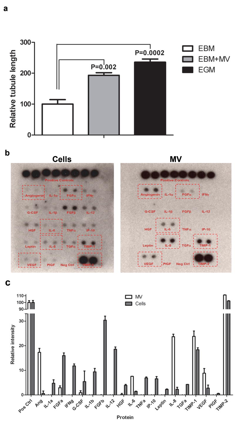
Glioblastoma tumour cells release microvesicles (exosomes) containing mRNA, miRNA and angiogenic proteins. These microvesicles are taken up by normal host cells, such as brain microvascular endothelial cells. By incorporating an mRNA for a reporter protein into these microvesicles, we demonstrate that messages delivered by microvesicles are translated by recipient cells. These microvesicles are also enriched in angiogenic proteins and stimulate tubule formation by endothelial cells. Tumour-derived microvesicles therefore serve as a means of delivering genetic information and proteins to recipient cells in the tumour environment. Glioblastoma microvesicles also stimulated proliferation of a human glioma cell line, indicating a self-promoting aspect. Messenger RNA mutant/variants and miRNAs characteristic of gliomas could be detected in serum microvesicles of glioblastoma patients. The tumour-specific EGFRvIII was detected in serum microvesicles from 7 out of 25 glioblastoma patients. Thus, tumour-derived microvesicles may provide diagnostic information and aid in therapeutic decisions for cancer patients through a blood test.
Figures
Comment in
-
Cancer stem cells as a prognostic indicator for glioblastoma multiforme.Biomark Med. 2010 Feb;4(1):127-8. doi: 10.2217/bmm.09.90. Biomark Med. 2010. PMID: 20387308 Free PMC article. No abstract available.
-
Glioblastoma produces tumor-promoting microvesicles.Nat Clin Pract Neurol. 2009 Mar;5(3):120-21. Nat Clin Pract Neurol. 2009. PMID: 20411603 No abstract available.
Similar articles
-
Glioblastoma microvesicles promote endothelial cell proliferation through Akt/beta-catenin pathway.Int J Clin Exp Pathol. 2014 Jul 15;7(8):4857-66. eCollection 2014. Int J Clin Exp Pathol. 2014. PMID: 25197356 Free PMC article.
-
Glioma microvesicles carry selectively packaged coding and non-coding RNAs which alter gene expression in recipient cells.RNA Biol. 2013 Aug;10(8):1333-44. doi: 10.4161/rna.25281. Epub 2013 Jun 17. RNA Biol. 2013. PMID: 23807490 Free PMC article.
-
From glioblastoma to endothelial cells through extracellular vesicles: messages for angiogenesis.Tumour Biol. 2016 Sep;37(9):12743-12753. doi: 10.1007/s13277-016-5165-0. Epub 2016 Jul 22. Tumour Biol. 2016. PMID: 27448307
-
Netrin-1 in Glioblastoma Neovascularization: The New Partner in Crime?Int J Mol Sci. 2021 Jul 31;22(15):8248. doi: 10.3390/ijms22158248. Int J Mol Sci. 2021. PMID: 34361013 Free PMC article. Review.
-
Role of extracellular vesicles in glioma progression.Mol Aspects Med. 2018 Apr;60:38-51. doi: 10.1016/j.mam.2017.12.003. Epub 2017 Dec 13. Mol Aspects Med. 2018. PMID: 29222067 Review.
Cited by
-
Extracellular Vesicles in Neurological Disorders.Subcell Biochem. 2021;97:411-436. doi: 10.1007/978-3-030-67171-6_16. Subcell Biochem. 2021. PMID: 33779926
-
Exosome and exosomal microRNA: trafficking, sorting, and function.Genomics Proteomics Bioinformatics. 2015 Feb;13(1):17-24. doi: 10.1016/j.gpb.2015.02.001. Epub 2015 Feb 24. Genomics Proteomics Bioinformatics. 2015. PMID: 25724326 Free PMC article. Review.
-
Analysis of exosome release as a cellular response to MAPK pathway inhibition.Langmuir. 2015 May 19;31(19):5440-8. doi: 10.1021/acs.langmuir.5b00095. Epub 2015 May 5. Langmuir. 2015. PMID: 25915504 Free PMC article.
-
The multifaceted exosome: biogenesis, role in normal and aberrant cellular function, and frontiers for pharmacological and biomarker opportunities.Biochem Pharmacol. 2012 Jun 1;83(11):1484-94. doi: 10.1016/j.bcp.2011.12.037. Epub 2011 Dec 31. Biochem Pharmacol. 2012. PMID: 22230477 Free PMC article. Review.
-
Extracellular vesicles: masters of intercellular communication and potential clinical interventions.J Clin Invest. 2016 Apr 1;126(4):1139-43. doi: 10.1172/JCI87316. Epub 2016 Apr 1. J Clin Invest. 2016. PMID: 27035805 Free PMC article. Review.
References
-
- Stupp R, et al. Radiotherapy plus concomitant and adjuvant temozolomide for glioblastoma. The New England journal of medicine. 2005;352:987–996. - PubMed
-
- Mazzocca A, et al. Cancer research. 2005;65:4728–4738. - PubMed
-
- Muerkoster S, et al. Tumor stroma interactions induce chemoresistance in pancreatic ductal carcinoma cells involving increased secretion and paracrine effects of nitric oxide and interleukin-1beta. Cancer research. 2004;64:1331–1337. - PubMed
-
- Singer CF, et al. Differential gene expression profile in breast cancer-derived stromal fibroblasts. Breast Cancer Res Treat. 2007 - PubMed
-
- Carmeliet P, Jain RK. Angiogenesis in cancer and other diseases. Nature. 2000;407:249–257. - PubMed
Publication types
MeSH terms
Substances
Grants and funding
LinkOut - more resources
Full Text Sources
Other Literature Sources
Molecular Biology Databases
