

T cells in rheumatoid arthritis
- PMID: 19007421
- PMCID: PMC2582813
- DOI: 10.1186/ar2412
T cells in rheumatoid arthritis
Abstract
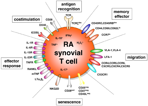
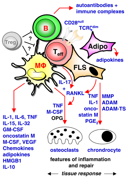
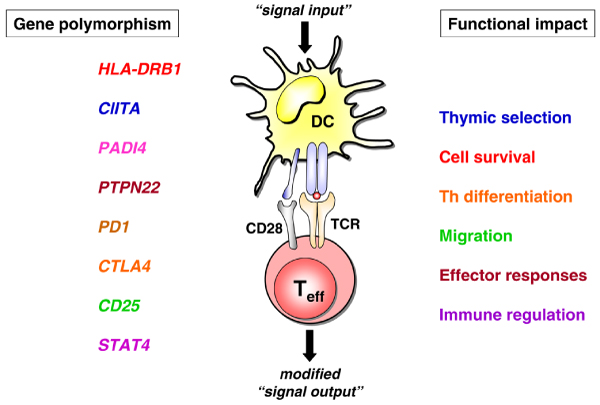
Over the past decade and a half, advances in our understanding of the pathogenesis of immune-mediated diseases such as rheumatoid arthritis (RA) have translated directly into benefit for patients. Much of this benefit has arisen through the introduction of targeted biological therapies. At the same time, technological advances have made it possible to define, at the cellular and molecular levels, the key pathways that influence the initiation and persistence of chronic inflammatory autoimmune reactions. As our understanding grows, it is likely that this knowledge will be translated into a second generation of biological therapies that are tailor-made for the patient. This review summarizes current perspectives on RA disease pathogenesis, with particular emphasis on what RA T cells look like, what they are likely to see, and how they contribute to persistence of the chronic inflammatory response.
Figures
Similar articles
-
The central role of T cells in rheumatoid arthritis.Clin Exp Rheumatol. 2007 Sep-Oct;25(5 Suppl 46):S4-11. Clin Exp Rheumatol. 2007. PMID: 17977483 Review.
-
New insights in the pathogenesis of rheumatoid arthritis.J Rheumatol Suppl. 1998 Jul;53:3-7. J Rheumatol Suppl. 1998. PMID: 9666411 Review.
-
Pathogenesis of rheumatoid arthritis.Med Clin North Am. 1997 Jan;81(1):29-55. doi: 10.1016/s0025-7125(05)70504-6. Med Clin North Am. 1997. PMID: 9012754
-
Immunological therapies for rheumatoid arthritis.Br Med Bull. 2005 Sep 20;73-74:71-82. doi: 10.1093/bmb/ldh051. Print 2005. Br Med Bull. 2005. PMID: 16174791 Review.
-
Therapeutic T-cell manipulation in rheumatoid arthritis: past, present and future.Rheumatology (Oxford). 2008 Oct;47(10):1461-8. doi: 10.1093/rheumatology/ken163. Epub 2008 May 25. Rheumatology (Oxford). 2008. PMID: 18503092 Review.
Cited by
-
Host-derived CD4+ T cells attenuate stem cell-mediated transfer of autoimmune arthritis in lethally irradiated C57BL/6.g7 mice.Arthritis Rheum. 2013 Mar;65(3):681-92. doi: 10.1002/art.37800. Arthritis Rheum. 2013. PMID: 23233229 Free PMC article.
-
Adenosine metabolic signature in circulating CD4+ T cells predicts remission in rheumatoid arthritis.RMD Open. 2024 Feb 17;10(1):e003858. doi: 10.1136/rmdopen-2023-003858. RMD Open. 2024. PMID: 38367982 Free PMC article.
-
Impaired activation-induced cell death promotes spontaneous arthritis in antigen (cartilage proteoglycan)-specific T cell receptor-transgenic mice.Arthritis Rheum. 2010 Oct;62(10):2984-94. doi: 10.1002/art.27614. Arthritis Rheum. 2010. PMID: 20564001 Free PMC article.
-
Expansion of human bone marrow-derived mesenchymal stromal cells with enhanced immunomodulatory properties.Stem Cell Res Ther. 2023 Sep 19;14(1):259. doi: 10.1186/s13287-023-03481-7. Stem Cell Res Ther. 2023. PMID: 37726837 Free PMC article.
-
Vaccines prevent reinduction of rheumatoid arthritis symptoms in collagen-induced arthritis mouse model.Drug Deliv Transl Res. 2023 Jul;13(7):1925-1935. doi: 10.1007/s13346-023-01333-8. Epub 2023 Mar 27. Drug Deliv Transl Res. 2023. PMID: 36971998 Free PMC article.
References
-
- Cope AP. Rheumatoid arthritis. In: Rich RR, Fleischer TA, Shearer WT, Schroeder HW, Frew AJ, Weyard CM, editor. Clinical Immunology. 3. New York, NY: Elsevier; 2008. pp. 52.1–52.23.
-
- Furst DE, Breedveld FC, Kalden JR, Smolen JS, Burmester GR, Sieper J, Emery P, Keystone EC, Schiff MH, Mease P, van Riel PL, Fleischmann R, Weisman MH, Weinblatt ME. Updated consensus statement on biological agents for the treatment of rheumatic diseases, 2007. Ann Rheum Dis. 2007;66(suppl):2–22. doi: 10.1136/ard.2007.081430. - DOI - PMC - PubMed
Publication types
MeSH terms
Grants and funding
LinkOut - more resources
Full Text Sources
Medical
Research Materials
Miscellaneous
