

Rhesus macaque rhadinovirus-associated non-Hodgkin lymphoma: animal model for KSHV-associated malignancies
- PMID: 18757778
- PMCID: PMC2582004
- DOI: 10.1182/blood-2008-04-151498
Rhesus macaque rhadinovirus-associated non-Hodgkin lymphoma: animal model for KSHV-associated malignancies
Abstract
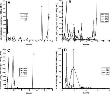
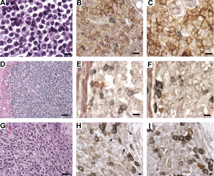
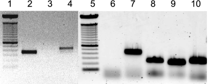
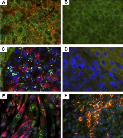
Rhesus macaque rhadinovirus (RRV) is closely related to Kaposi sarcoma-associated herpesvirus (KSHV) and is associated with the development of B-cell hyperplasia and persistent lymphadenopathy resembling multicentric Castleman disease in rhesus macaques (RMs) coinfected with simian immunodeficiency virus (SIV). Here we investigated whether RMs experimentally infected with SIV and RRV can develop other disease manifestations observed in HIV- and KSHV-infected patients. As reported earlier, inoculation of SIV-infected RMs with RRV results in persistent RRV infection, whereas immunocompetent animals infected with RRV exhibit viremia 2 weeks after infection, followed by a period of no virus detection until they are subsequently made immunodeficient by SIV infection. A subset of animals developed abnormal cellular proliferations characterized as extranodal lymphoma and a proliferative mesenchymal lesion. In situ hybridization and immunohistochemistry analysis indicate RRV is present in both malignancies, and DNA microarray analysis detected viral interleukin-6 (vIL-6) and viral FLICE-like inhibitory protein (vFLIP) transcripts. Reverse-transcriptase polymerase chain reaction analysis confirmed vIL-6 and vFLIP expression, and that of RRV open reading frames 72 and 73, homologs of KSHV open reading frames shown to be expressed in primary effusion lymphoma. These data support the utility of the RRV-/SIV-infected RM as an excellent animal model to investigate KSHV-like pathogenesis.
Figures
Similar articles
-
Viral interleukin-6 encoded by rhesus macaque rhadinovirus is associated with lymphoproliferative disorder (LPD).J Med Primatol. 2009 Oct;38 Suppl 1(Suppl 1):2-7. doi: 10.1111/j.1600-0684.2009.00369.x. J Med Primatol. 2009. PMID: 19863672 Free PMC article.
-
Induction of B cell hyperplasia in simian immunodeficiency virus-infected rhesus macaques with the simian homologue of Kaposi's sarcoma-associated herpesvirus.J Exp Med. 1999 Sep 20;190(6):827-40. doi: 10.1084/jem.190.6.827. J Exp Med. 1999. PMID: 10499921 Free PMC article.
-
Experimental infection of rhesus and pig-tailed macaques with macaque rhadinoviruses.J Virol. 1999 Dec;73(12):10320-8. doi: 10.1128/JVI.73.12.10320-10328.1999. J Virol. 1999. PMID: 10559350 Free PMC article.
-
Rhesus macaque rhadinovirus-associated disease.Curr Opin Virol. 2013 Jun;3(3):245-50. doi: 10.1016/j.coviro.2013.05.016. Epub 2013 Jun 6. Curr Opin Virol. 2013. PMID: 23747119 Free PMC article. Review.
-
Rhesus monkey rhadinovirus: a model for the study of KSHV.Curr Top Microbiol Immunol. 2007;312:43-69. doi: 10.1007/978-3-540-34344-8_2. Curr Top Microbiol Immunol. 2007. PMID: 17089793 Review.
Cited by
-
Non-human primate model of Kaposi's sarcoma-associated herpesvirus infection.PLoS Pathog. 2009 Oct;5(10):e1000606. doi: 10.1371/journal.ppat.1000606. Epub 2009 Oct 2. PLoS Pathog. 2009. PMID: 19798430 Free PMC article.
-
Animal Models for Gammaherpesvirus Infections: Recent Development in the Analysis of Virus-Induced Pathogenesis.Pathogens. 2020 Feb 12;9(2):116. doi: 10.3390/pathogens9020116. Pathogens. 2020. PMID: 32059472 Free PMC article. Review.
-
Genomic characterization of Japanese macaque rhadinovirus, a novel herpesvirus isolated from a nonhuman primate with a spontaneous inflammatory demyelinating disease.J Virol. 2013 Jan;87(1):512-23. doi: 10.1128/JVI.02194-12. Epub 2012 Oct 24. J Virol. 2013. PMID: 23097433 Free PMC article.
-
Viral interleukin-6 encoded by rhesus macaque rhadinovirus is associated with lymphoproliferative disorder (LPD).J Med Primatol. 2009 Oct;38 Suppl 1(Suppl 1):2-7. doi: 10.1111/j.1600-0684.2009.00369.x. J Med Primatol. 2009. PMID: 19863672 Free PMC article.
-
Disruption of LANA in rhesus rhadinovirus generates a highly lytic recombinant virus.J Virol. 2009 Oct;83(19):9786-802. doi: 10.1128/JVI.00704-09. Epub 2009 Jul 8. J Virol. 2009. PMID: 19587030 Free PMC article.
References
-
- Chang Y, Cesarman E, Pessin MS, et al. Identification of herpesvirus-like DNA sequences in AIDS-associated Kaposi's sarcoma. Science. 1994;265:1865–1869. - PubMed
-
- Cesarman E, Chang Y, Moore PS, Said JW, Knowles DM. Kaposi's sarcoma-associated herpesvirus-like DNA sequences in AIDS-related body-cavity-based lymphomas. N Engl J Med. 1995;332:1186–1191. - PubMed
-
- Soulier J, Grollet L, Oksenhendler E, et al. Kaposi's sarcoma-associated herpesvirus-like DNA sequences in multicentric Castleman's disease. Blood. 1995;86:1276–1280. - PubMed
-
- Oksenhendler E, Boulanger E, Galicier L, et al. High incidence of Kaposi sarcoma-associated herpesvirus-related non-Hodgkin lymphoma in patients with HIV infection and multicentric Castleman disease. Blood. 2002;99:2331–2336. - PubMed
Publication types
MeSH terms
Substances
Grants and funding
LinkOut - more resources
Full Text Sources
Other Literature Sources
Medical
Molecular Biology Databases
Research Materials
