. 2008 Jun;118(6):2088-97.
doi: 10.1172/JCI33392.

RelA/p65 promotes osteoclast differentiation by blocking a RANKL-induced apoptotic JNK pathway in mice

Affiliations

RelA/p65 promotes osteoclast differentiation by blocking a RANKL-induced apoptotic JNK pathway in mice

Sergio Vaira et al. J Clin Invest. 2008 Jun.

Abstract

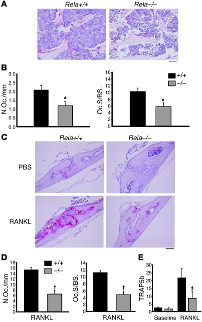
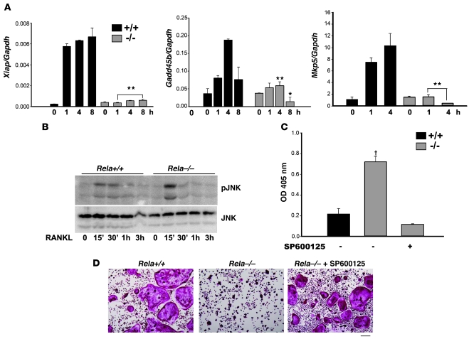
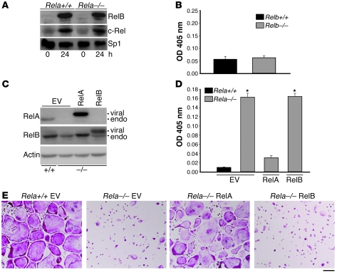
Osteoclasts (OCs) function to reabsorb bone and are responsible for the bone loss associated with inflammatory arthritis and osteoporosis. OC numbers are elevated in most disorders of accelerated bone destruction, reflecting altered rates of precursor differentiation and apoptosis. Both of these processes are regulated by the JNK family of MAP kinases. In this study, we have demonstrated that the NF-kappaB subunit RelA/p65 inhibits JNK-mediated apoptosis during a critical period of commitment to the OC phenotype in response to the cytokine RANKL. This RelA/p65-mediated arrest of cell death led to enhanced OC differentiation. Hence, Rela-/- OC precursors displayed prolonged JNK activation in response to RANKL, and this was accompanied by an increase in cell death that prevented efficient differentiation. Although complete blockade of JNK activity inhibits osteoclastogenesis, both short-term blockade in RelA-deficient cultures and suppression of the downstream mediator, Bid rescued apoptosis and differentiation. These antiapoptotic effects were RelA specific, as overexpression of RelA, but not RelB, blocked apoptosis and rescued differentiation in Rela-/- precursors. Thus, RelA blocks a RANKL-induced, apoptotic JNK-Bid pathway, thereby promoting OC differentiation. Consistent with this, mice lacking RelA/p65 in the hematopoietic compartment were shown to have a deficient osteoclastogenic response to RANKL and were protected from arthritis-induced osteolysis.


Figures

Figure 1
Figure 1. RelA is important for basal and stimulated OC formation in vivo.
(A) TRAP-stained sections of vertebrae from Rela+/+ and Rela–/– RCs 4 months after bone marrow reconstitution. Scale bar: 100 μm. (B) Histomorphometric analysis was performed on TRAP-stained sections to determine the OC number (N.Oc./mm) and OC surface (Oc.S./BS.). *P < 0.05; n = 8/group. (C) Rela+/+ and Rela–/– RCs were injected with 100-μg RANKL or PBS daily for 5 days and sacrificed on the sixth day. TRAP-stained coronal sections of calvaria at the sagittal suture show a consistent increase in OCs along sutures and sinusoids only in Rela+/+ RANKL-treated animals. Scale bar: 200 μm. (D) Quantification of the N.Oc./mm and Oc.S./BS. (percentage of bone surface covered by OCs) along calvarial surfaces of mice in C confirms the significant difference in response of Rela+/+ and Rela–/– RCs to RANKL injection. In the PBS-injected animals, only rare OCs are detected in either genotype. P < 0.005; n = 5/group. (E) Serum levels of TRAP5b were determined 1 day prior to first RANKL injection (baseline) and at sacrifice on day 6 (RANKL). P = 0.01; n = 5/group.
Figure 2
Figure 2. Rela–/– RCs are protected from arthritis-induced bone loss.
(A) Arthritis was induced in Rela+/+ and Rela–/– RCs 5 weeks after marrow transplantation by injection of K/BxN serum at days 0, 2, and 7, with LPS on day 2. Histological sections of ankle joints at day 14 stained with H&E show an extensive and similar inflammatory infiltrate in the joint spaces. Scale bar: 500 μm. (B) The inflammatory response was also evaluated by measurement of hind paw thickness (sum of both paws) during induction of arthritis. n = 8/group. The experiment was repeated twice, with similar results. (C) TRAP-stained sections of metatarsals demonstrate OCs both on the outer bone surface adjacent to inflammatory pannus (arrowheads) and in marrow spaces. Scale bar: 100 μm. (D) Histomorphometric analysis of N.Oc./mm and Oc.S./BS. on the outer bone surface demonstrate significantly more OCs in Rela+/+ RCs compared with Rela–/– RCs. *P < 0.001; n = 8/group. (E) Serum CTX was measured at day 0 and 14 of serum transfer arthritis, and the fold change for each RC was plotted, demonstrating a significant rise only in the Rela+/+ group. P < 0.05; n = 6/group.
Figure 3
Figure 3. Rela–/– BMMs are sensitive to RANKL-induced cell death.
(A) Rela+/+ or Rela–/– BMMs were cultured in M-CSF and RANKL or (B) cultured with wild-type calvarial osteoblasts in the presence of 1,25 vitamin D3, then fixed and TRAP stained. Scale bar: 500 μm. The number of OCs per field (mean ± SEM) is shown, demonstrating fewer OCs in the absence of RelA. (C) Cell number was assessed by MTT assay in BMMs cultured with M-CSF and RANKL for 3–48 hours, showing fewer cells after 48 hours in Rela–/– cultures relative to Rela+/+ (*P < 0.00001; n = 4). (D) Apoptosis was measured by DNA fragmentation assay after 18 or 36 hours in the presence of M-CSF, with (+) or without (–) RANKL. After 36 hours, Rela–/– BMMs cultured with M-CSF and RANKL, but not with M-CSF alone, had increased cell death. ^P < 0.0001 compared with Rela+/+ without RANKL at 36 hours; P < 0.00001 relative to Rela+/+ with RANKL at 36 hours. (E) Caspase-3 (casp 3) activation was measured in Rela+/+ and Rela–/– BMMs cultured in M-CSF/RANKL for 36 and 48 hours, with or without ZVAD, and fold induction relative to the same cells in M-CSF alone was plotted. #P < 0.05 relative to Rela+/+ 36 hours; P < 0.01, ΧP < 0.00001 relative to Rela+/+ at 36 or 48 hours. (F) OCs were generated with or without ZVAD during the first 48 hours. Scale bar: 500 μm. (G) OCs were generated as in F on cortical bone slices. Resorption pits (brown) were stained with horseradish peroxidase-conjugated wheat germ agglutinin. Scale bar: 100 μm.
Figure 4
Figure 4. The absence of RelA leads to enhanced JNK activation.
(A) Pre-OCs (BMMs cultured for 2 days with M-CSF and RANKL) generated from Rela+/+ (black bars) and Rela–/– (gray bars) mice were then starved of serum and cytokines and restimulated with RANKL for the indicated times prior to RNA extraction and real-time quantitative PCR for xiap, Gadd45b, and Mkp5. Rela–/– pre-OCs show significantly less induction of xiap, Gadd45b and Mkp5. *P < 0.5, **P < 0.01 compared to Rela+/+ at the same time point. (B) Rela+/+ and Rela–/– pre-OCs were starved and restimulated with RANKL for the indicated times, and total lysates were analyzed by immunoblot for pJNK, demonstrating enhanced JNK activation in Rela–/– cultures. A parallel blot with equal loading was used to assess total JNK protein. (C) Rela+/+ and Rela–/– BMMs were cultured in RANKL, with or without the JNK inhibitor SP600125 (1 μm), for 36 hours Apoptosis was then assessed by DNA fragmentation assay. RANKL-mediated apoptosis was abrogated by the JNK inhibitor in Rela–/– cells. The data are representative of 3 independent experiments. P < 0.05 Rela–/– versus Rela+/+ or Rela–/– with SP600125. (D) Rela+/+ and Rela–/– BMMs were cultured in osteoclastogenic conditions for 8 days, with or without SP600125 for the first 36 hours, showing rescue of differentiation with short-term JNK inhibition. Scale bar: 200 μm.
Figure 5
Figure 5. Knockdown of Bid rescues Rela–/– osteoclastogenesis.
(A) Rela+/+ and Rela–/– BMMs were stimulated with RANKL at the indicated times, and total cell lysates were examined by immunoblot for expression of Bid. β-actin was used as loading control. (B) Rela–/– BMMs were transduced with retrovirus expressing 2 different siRNAs for Bid (siBid1 and siBid2) or luciferase (siLuc). Following puromycin selection, levels of Bid were examined by immunoblot, demonstrating reduction of Bid by the specific siRNAs. β-actin was used as loading control. (C) Rela+/+ and Rela–/– BMMs transduced with siLuc or siBid1 or -2 were cultured with RANKL for 36 hours and DNA fragmentation was measured. The data are representative of 3 independent experiments. *P < 0.05 versus Rela+/+ siLuc. (D) The same BMMs in C were cultured in osteoclastogenic conditions for 6 days, then stained for TRAP, showing rescue of differentiation with knockdown of Bid. Scale bar: 200 μm. (E) BMMs cultured on cortical bone slices were stained with horseradish peroxidase-conjugated wheat germ agglutinin to demonstrate resorption. Scale bar: 200 μm. (F) Pits in E were quantified, showing that reduction in apoptosis correlates with differentiation and bone resorption. Black bars indicate Rela+/+; gray bars indicate Rela–/–; *P < 0.05 versus Rela+/+ siLuc.
Figure 6
Figure 6. Enhanced RANKL-induced apoptosis is specific for RelA.
(A) BMMs from Rela+/+ and Rela–/– mice were cultured in the presence of M-CSF and RANKL for 0 or 24 hours. Equal amounts of nuclear extracts were incubated with biotinylated κB oligos bound to streptavidin-coated beads. Beads were washed to isolate κB-bound proteins, and these were analyzed by immunoblot for RelB or c-Rel. Unbound nuclear proteins were analyzed by immunoblot for Sp1 as loading control. (B) BMMs from Relb+/+ and Relb–/– mice were cultured with M-CSF and RANKL for 48 hours, and apoptosis was assessed by DNA fragmentation assay, showing no effect of RelB on apoptosis. (C) Rela–/– BMMs retrovirally transduced with empty vector (EV), RelA, or RelB, and Rela+/+ BMMs transduced with EV were selected in blastocydin, then analyzed by immunoblot. Viral-encoded proteins (both RelA and RelB), migrate more slowly than their endogenous (endo) counterparts, and are expressed at 2- to 3-fold over endogenous levels. (D) BMMs transduced in C were cultured with RANKL for 48 hours, and apoptosis was assessed by DNA fragmentation assay. While the EV control and RelB vectors do not abrogate the RANKL-induced cell death in Rela–/– cultures, RelA reduces apoptosis to the same level as Rela+/+ transduced with EV. *P < 0.001 Rela+/+ EV. (E) Transduced BMMs were grown in osteoclastogenic conditions for 6 days and then were fixed and stained for TRAP. Reexpression of RelA restores Rela–/– OC differentiation, while overexpression of RelB has no effect. Scale bar: 200 μm.

Similar articles

Cited by

References

    1. Teitelbaum S.L. Osteoclasts: what do they do and how do they do it? Am. J. Pathol. 2007;170:427–435. doi: 10.2353/ajpath.2007.060834. - DOI - PMC - PubMed
    1. Perkins N.D. Integrating cell-signalling pathways with NF-κB and IKK function. Nat. Rev. Mol. Cell Biol. 2007;8:49–62. doi: 10.1038/nrm2083. - DOI - PubMed
    1. Franzoso G., et al. Requirement for NF-κB in osteoclast and B-cell development. Genes Dev. 1997;11:3482–3496. doi: 10.1101/gad.11.24.3482. - DOI - PMC - PubMed
    1. Iotsova V., et al. Osteopetrosis in mice lacking NF-κB1 and NF-κB2. Nat. Med. 1997;3:1285–1289. doi: 10.1038/nm1197-1285. - DOI - PubMed
    1. Ruocco M.G., et al. IκB kinase (IKK)β, but not IKKα, is a critical mediator of osteoclast survival and is required for inflammation-induced bone loss. J. Exp. Med. 2005;201:1677–1687. doi: 10.1084/jem.20042081. - DOI - PMC - PubMed

Publication types

MeSH terms